

Ultrasound vs CT vs MRI: When Doctors Choose Sound Waves and When Another Scan Is Needed

Key Takeaways
- Ultrasound uses sound waves and no radiation, which is why it is the standard first scan in pregnancy, in children and for repeated monitoring over time.
- CT reads tissue density and completes its imaging in seconds, making it the preferred emergency tool for bleeding, fractures, lung problems and kidney stones.
- MRI maps the behavior of water in tissue, giving it unmatched detail of the brain, spinal cord, cartilage and ligaments without any ionizing radiation.
- Sound cannot pass through bone or gas, so ultrasound is effectively blind to the adult brain, the lungs and much of the bowel.
- A single abdominal CT delivers roughly the equivalent of several years of natural background radiation, a small risk that becomes meaningful mainly with repeated scanning.
- Most metal implants and dental work are compatible with MRI after individual screening; only specific devices and fragments rule it out.
Doctors usually choose ultrasound first when the question involves soft tissue near the surface, fluid, the pelvis, the pregnant uterus or moving blood, because it uses sound waves with no radiation. CT is chosen when speed, bone, lung, or bleeding detail matters, while MRI is reserved for the brain, spinal cord, joints and organs where fine tissue contrast counts. The treating team decides based on the specific question.
A woman sits on the edge of an exam table with a folded referral form in her hand. Her right side has ached for a week, and the doctor has just said the word “scan.” Her first question is not about her gallbladder. It is: which scan, and why that one?
The ultrasound vs CT vs MRI decision looks like a menu from the outside, as if the pricier or more powerful option must be the better one. Inside a radiology department it works more like choosing a tool from a drawer. A sound-wave probe, an X-ray ring and a giant magnet each answer a different kind of question, and the wrong tool can produce a beautiful image of the wrong thing.
This explainer walks through how each machine actually sees, what each one misses, and the reasoning that leads a clinician to say “ultrasound today” or “this needs an MRI.” The final choice always belongs to the team looking after you, but understanding the logic makes that conversation far less mysterious.
Why ultrasound is often the first scan doctors reach for
Ultrasound is the imaging equivalent of tapping a wall to find the stud behind the plaster. A handheld probe called a transducer sends pulses of sound far above the range of human hearing into the body and listens for the echoes that bounce back. Different tissues reflect sound differently: fluid lets it pass almost silently, solid organs scatter it, and bone or air stop it nearly cold. A computer turns the timing and strength of those echoes into a live, moving picture on the screen.
That word “live” is the whole point. A sonographer, the trained professional who performs the scan, can tilt the probe, ask you to breathe in, press gently on a tender spot and watch what happens in real time. No other common scan lets a clinician interact with the anatomy while looking at it. Doppler ultrasound, a setting that measures the shift in echo frequency from moving blood, adds color to show which way blood is flowing and how fast.
The ultrasound vs CT vs MRI question therefore often starts here for practical reasons. Ultrasound involves no ionizing radiation, which is why it is the standard method for checking a pregnancy (MedlinePlus). It needs no injection for most exams, can be wheeled to a bedside, and a typical appointment lasts about 15 to 45 minutes according to the NHS. When the clinical question is “is there fluid, a stone, a cyst, or a blocked vessel in something I can reach with sound,” ultrasound is frequently the first and sometimes the only step.
Its limits are equally physical. Sound cannot pass through bone or through gas, so the skull, the lungs and a bowel full of air act like closed doors. Those doors are exactly where the other two machines come in.
How a CT scan works: X-rays taken in slices
Picture a loaf of bread. A plain X-ray photographs the whole loaf from one side, so every slice overlaps in a single flat image. Computed tomography, usually shortened to CT, spins a thin X-ray beam around the body and records how much of it passes through from hundreds of angles. A computer then reconstructs the data into individual cross-sectional slices, and stacks them to build a three-dimensional model that can be turned and viewed from any direction.

The scanner itself looks like a large doughnut. You lie on a table that slides through the ring while the X-ray tube rotates inside the housing. The scanning portion is quick; the NHS notes that a CT scan usually takes about 10 to 20 minutes, and the actual image acquisition within that window is often a matter of seconds. That speed is the reason emergency departments lean on CT so heavily. A patient who cannot lie still for long, or who may be bleeding internally, needs answers measured in minutes rather than the better part of an hour.
CT reads density. Bone appears bright white because it blocks X-rays, air appears black because it blocks almost none, and soft organs fall into shades of gray between them. Fresh bleeding shows up well, as do fractures, lung changes and kidney stones. An iodine-based contrast dye, a liquid injected into a vein or swallowed to make blood vessels and certain organs stand out, can be added when the question involves circulation or subtle organ detail (Mayo Clinic).
The trade-off is the radiation. CT uses far more X-ray exposure than a single chest film, and every scan carries a small theoretical addition to lifetime cancer risk. How small is a fair question, and it gets its own section below.
How an MRI works: magnets, radio waves and the water in your tissues
Magnetic resonance imaging does not use X-rays or sound. It uses the fact that the body is mostly water, and that the hydrogen atoms in water behave like tiny compass needles. Inside the scanner’s very strong magnetic field those needles line up. Brief pulses of radio waves knock them out of alignment, and as they swing back they release a faint signal. Tissues release that signal at different rates depending on how much water they hold and how tightly it is bound, and a computer maps those differences into images (NHS).
The result is unmatched contrast between soft tissues that look nearly identical on CT. Gray matter and white matter in the brain, the discs between the vertebrae, cartilage inside a knee, the fibers of a tendon, the layers of the uterine wall: MRI separates them with a clarity that X-ray density simply cannot match. Radiologists, the physicians who interpret scans, can also switch between different pulse sequences to highlight fluid, fat, inflammation or old versus new bleeding within the same appointment.
Clarity costs time. An MRI examination typically lasts anywhere from 15 to 90 minutes depending on the body part and the number of sequences, according to the NHS, and the machine makes loud knocking and buzzing sounds as its coils switch on and off. You lie inside a tunnel that is narrower and deeper than a CT ring, and movement blurs the picture, so staying still matters more than in any other scan.
A gadolinium-based contrast agent, a metal-containing liquid injected into a vein, is sometimes used to make tumors, inflammation or blood vessels more visible. Because MRI involves no ionizing radiation, the main safety questions revolve around the magnet itself and around metal in or on the body, which we cover under eligibility.
Ultrasound vs CT scan: what sound waves see that X-rays miss
Ask a radiologist what an ultrasound can see that a CT cannot and the answer usually comes in three parts: motion, flow and texture in real time.
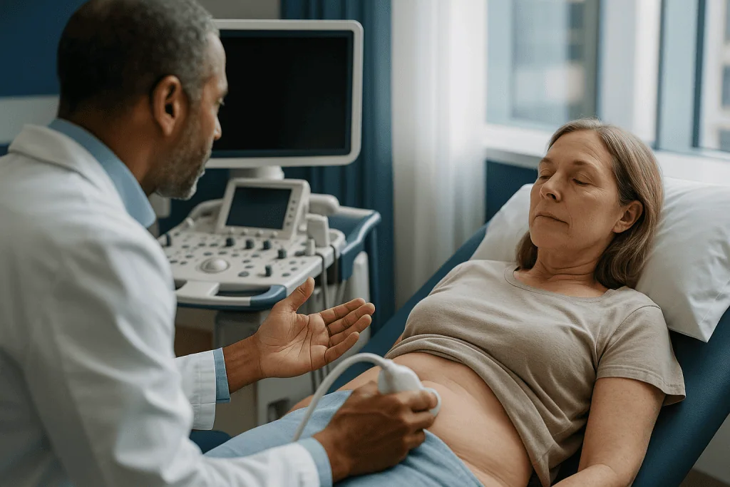
Motion first. A CT is a frozen moment. An ultrasound is a video. Watching a heart valve open and close, seeing whether a fetus moves, or observing how a gallbladder wall behaves when the probe presses on it all require a live picture. Echocardiography, the ultrasound of the heart, gives cardiologists a moving view of chamber size and valve function that a single CT snapshot cannot replicate.
Flow second. Doppler settings show blood direction and speed without any dye. That makes ultrasound the usual starting test for a suspected clot in a leg vein, for narrowing of the carotid arteries in the neck, and for checking blood supply to a transplanted kidney. CT can image vessels superbly, but it needs injected iodine contrast and a radiation dose to do so.
Texture third. A simple fluid-filled cyst and a solid nodule can look similar on CT yet differ sharply on ultrasound, where fluid appears black and echo-free while solid tissue scatters sound. That distinction matters in the thyroid, the breast, the ovaries and the testicles, all superficial organs where ultrasound excels. A sonographer can also guide a needle in real time for a biopsy or fluid drainage, watching the tip move toward the target.
Where CT wins the ultrasound vs CT scan comparison is depth and obstruction. Sound weakens as it travels, so deep structures in a larger body may be hard to see, and anything behind bone or bowel gas disappears. Lungs, the skull interior, most of the bowel wall and subtle fractures belong to CT. Neither machine is “better”; each is blind where the other sees.
What can an MRI see that an ultrasound cannot?
The honest answer is: almost everything ultrasound cannot reach, plus finer detail in the places both can reach.
Ultrasound’s reach ends at bone and air. MRI has no such barrier. The brain and spinal cord, sealed inside the skull and vertebral canal, are essentially invisible to sound in adults yet are MRI’s home territory. Multiple sclerosis plaques, small strokes, pituitary abnormalities, disc herniations pressing on nerve roots and spinal cord inflammation are typically MRI diagnoses. In infants, whose skull bones have not yet fused, ultrasound through the soft spot can show the brain, which is one of the few exceptions.
Inside joints, ultrasound sees tendons and fluid near the surface well, but it cannot look inside the knee to assess the menisci and cruciate ligaments, or into the hip socket for cartilage damage. MRI shows these structures along with bone marrow, where early stress fractures and bone bruises appear before any change is visible on X-ray or CT.
Depth and consistency also favor MRI. Ultrasound quality depends heavily on the operator’s skill, the patient’s body habitus and the angle of approach. MRI produces standardized images of the whole region regardless of who positions the coil, which makes it more reliable for follow-up comparisons over months and years. For the liver, pancreas, adrenal glands and pelvis, MRI can characterize a lesion first spotted on ultrasound: is it a benign fatty deposit, a fluid-filled cyst, a blood-filled hemangioma or something that needs a biopsy?
What MRI cannot do is match ultrasound’s convenience. It cannot be done at a bedside, cannot show real-time needle guidance as easily, takes far longer, and cannot be used freely in someone with certain implanted devices. That is why clinicians so often sequence them: ultrasound to ask the first question, MRI to answer the harder one.
MRI vs CT scan differences: what MRI shows that CT cannot
People often assume CT and MRI are interchangeable big machines. They overlap in body coverage but not in what they measure, and that difference explains most of the MRI vs CT scan differences a doctor weighs.
CT measures density. MRI measures water behavior. Structures with very similar density but different water content look alike on CT and distinct on MRI. The clearest example is the brain: a small ischemic stroke, an area where blood supply has been cut off, may be invisible on CT in the first hours yet show clearly on a diffusion MRI sequence, which detects the way water movement changes inside injured cells (Mayo Clinic). Conversely, a fresh hemorrhage shows brilliantly on CT within minutes, which is why CT is done first when a stroke is suspected: it rapidly separates bleeding from clot and guides urgent treatment decisions.
Soft tissue detail is the other divide. Cartilage, ligaments, tendons, nerves and the spinal cord itself have almost no density contrast from surrounding tissue on CT. MRI separates them cleanly. Tumors inside the liver, prostate, uterus or muscles are often characterized on MRI after CT has shown that “something is there.”
CT keeps the advantage where bone, calcium and air are involved. Complex fractures, lung nodules, bowel obstruction, kidney stones and calcified arteries are CT questions. CT is also the workhorse for staging many cancers because it surveys the whole chest, abdomen and pelvis in one short pass.
Then there is the practical layer. CT is faster, more widely available around the clock, tolerates movement better and has no magnet-related exclusions. MRI has no radiation and superior tissue contrast but demands time, stillness and metal screening. When a doctor says “we need the MRI, not the CT,” it is nearly always because the question lives in soft tissue detail rather than density.
Why do doctors use ultrasound instead of MRI?
Patients sometimes leave an appointment quietly wondering whether they were given the lesser test. The reasoning is usually the opposite: ultrasound was chosen because it answers the question with the least burden.
Safety comes first. Ultrasound carries no ionizing radiation and no magnetic field. That makes it the default in pregnancy, in young children, and in anyone who needs repeated imaging over time, such as monitoring a known cyst or a fetus across several visits (MedlinePlus).
Speed and access come next. An ultrasound machine can be wheeled to a bedside, an emergency bay or an intensive care unit. A patient who is unstable, in pain, or unable to lie still for the duration of an MRI can still be examined. There is no screening questionnaire about pacemakers or metal fragments, no confined tunnel and no loud noise.
Suitability is the quiet third reason. For many common questions, ultrasound is not a compromise but the best-matched tool. Gallstones, kidney swelling, thyroid nodules, testicular pain, breast lumps in younger women, early pregnancy, leg vein clots and heart valve motion are all areas where guidelines place ultrasound first. Ordering an MRI for a straightforward gallstone question would be like using a telescope to read a book.
Where ultrasound falls short, the plan changes. If the sonographer cannot see the whole organ because of bowel gas or body size, if the result is unclear, or if the finding needs characterization, the radiologist’s report will typically recommend CT or MRI as the next step. That sequencing is not indecision. It is a deliberate ladder designed to reach the answer with the fewest scans, the least radiation and the shortest wait, and each rung is chosen by the treating team based on what the previous one showed.
Ultrasound vs CT vs MRI at a glance: a side-by-side table
The table below distills the practical differences most people want to know before an appointment. Time ranges are typical figures published by the NHS and vary by body region and the number of images required; they are not promises for any individual scan.
| Feature | Ultrasound | CT scan | MRI |
|---|---|---|---|
| How it sees | Echoes of high-frequency sound | X-ray absorption from many angles | Radio signals from hydrogen in a magnetic field |
| Ionizing radiation | None | Yes | None |
| Typical appointment length | About 15–45 minutes (NHS) | About 10–20 minutes (NHS) | About 15–90 minutes (NHS) |
| Strengths | Fluid, superficial organs, pregnancy, blood flow, real-time guidance | Bone, lung, fresh bleeding, kidney stones, trauma, speed | Brain, spinal cord, joints, soft-tissue detail, lesion characterization |
| Blind spots | Behind bone or gas; deep tissue in larger bodies | Subtle soft-tissue contrast; early stroke | Calcium, fine bone detail, patients who cannot stay still |
| Contrast used | Rarely; microbubble agents in select cases | Iodine-based, sometimes | Gadolinium-based, sometimes |
| Main eligibility issues | Very few | Pregnancy, iodine allergy, kidney function | Certain implants, metal fragments, claustrophobia, kidney function for contrast |
| Portable | Yes | No | No |
Read the table as a map of questions rather than a ranking of machines. A clinician who suspects a blood clot in the leg will not glance at the MRI column at all, and one who suspects a torn knee ligament will not send you for a CT. The right scan is the one whose strengths line up with the specific uncertainty in your case.
One more nuance the table cannot show: these scans are often used in sequence rather than in competition. Ultrasound may find a liver lesion, CT may stage it, and MRI may characterize it. Each step narrows the question before the next machine is asked.
Who is usually offered which scan, and who is asked to wait
Eligibility is rarely about age or fitness. It is about a handful of specific circumstances that push the decision toward one machine or delay it.
Pregnancy steers toward ultrasound and, when more detail is needed, toward MRI without contrast. CT is generally avoided during pregnancy unless the benefit clearly outweighs the radiation risk to the fetus, a judgment the treating team makes case by case (NHS). Gadolinium contrast is also usually avoided in pregnancy because it crosses the placenta.
Metal changes the MRI conversation. Some older pacemakers, certain cochlear implants, aneurysm clips, metal fragments in the eye from grinding work and some infusion pumps are unsafe in a strong magnetic field. Many modern devices are labeled MRI-conditional, meaning they can be scanned under specific settings, but every patient completes a detailed screening form and the department checks device documentation before proceeding. If MRI cannot be done safely, CT or ultrasound becomes the alternative (Mayo Clinic).
Kidney function matters for contrast dye in both CT and MRI. Iodine contrast can strain already weakened kidneys, and gadolinium is used cautiously in advanced kidney disease. A blood test for kidney function is often requested beforehand, and a non-contrast scan or a different modality may be chosen instead.
Body size and stillness shape the choice too. Sound weakens with depth, so ultrasound may be limited in a larger patient. MRI demands that you stay motionless for long stretches; very young children, people in severe pain, and those with significant claustrophobia may need sedation, an open-design scanner, or a CT instead.
Finally, some people are simply asked to wait. A vague symptom with reassuring examination findings may warrant a review in a few weeks before any scan, because imaging too early can turn up incidental findings, unexpected abnormalities of no clinical importance, that then trigger further tests and worry.
Radiation, contrast dye and other risks in plain numbers
Risk conversations about scans tend to swing between dismissal and alarm. The evidence sits in between.
Ultrasound has no known harmful effects at the energy levels used in diagnostic imaging (MedlinePlus). Its risks are practical rather than physical: an inconclusive result, or an operator-dependent miss.
CT delivers ionizing radiation, which can damage DNA. Harvard Health notes that a CT scan of the abdomen delivers roughly the equivalent of several years of natural background radiation, the low-level exposure everyone receives from soil, rocks and cosmic rays. The added lifetime cancer risk from a single scan is small and difficult to measure directly, but it is not zero, and it accumulates with repeated scans. That is why radiologists use the lowest dose that still answers the question and why pediatric protocols are adjusted for body size. A scan that changes management is worth its dose; a scan ordered for reassurance alone may not be.
Contrast agents carry their own small risks. Iodine-based CT contrast can cause a warm flush, a metallic taste and, rarely, an allergic-type reaction ranging from hives to a serious response requiring treatment. It can also affect kidney function in people whose kidneys are already impaired, so a kidney blood test is often checked first (Mayo Clinic). Gadolinium for MRI is generally well tolerated; serious reactions are rare, and a scarring condition of the skin and organs called nephrogenic systemic fibrosis has been linked to older gadolinium agents in patients with severe kidney disease, which is why kidney function is screened.
MRI’s remaining risks relate to the magnet: loose metal objects becoming projectiles, heating of some implants and the intense noise, for which ear protection is standard. Claustrophobia and anxiety are real for many people and can usually be discussed in advance.
What actually happens on the day, and what the following days look like
Preparation differs more than people expect. For an abdominal ultrasound you may be asked to fast for several hours so the gallbladder is full and bowel gas is reduced; for a pelvic ultrasound you may need a full bladder to lift the bowel out of the way. A cool gel is spread on the skin to eliminate air between probe and body, and the sonographer moves the probe while watching the screen. Some pelvic or early-pregnancy scans use a slim internal probe, which is discussed and consented beforehand.
For CT you change into a gown, remove metal, and may receive contrast through a small cannula in a vein. The table moves through the ring, a voice asks you to hold your breath for a few seconds, and it is over quickly. If contrast was given you are typically asked to drink normally afterward to help clear it (NHS).
For MRI the screening form comes first, then earplugs or headphones, then positioning inside the tunnel with a plastic coil placed over the body part. Sequences run for several minutes each, with pauses in between; you can speak to the operator through an intercom and hold a call button throughout.
The days afterward are usually uneventful. Contrast leaves the body through the kidneys within a day or so in people with normal kidney function. Mild bruising at the cannula site is common. Any rash, swelling or breathing difficulty after contrast warrants urgent medical attention.
Results follow a separate timeline. A radiologist reviews the images and writes a report to the referring clinician; the interval varies by urgency and by service, and emergency findings are communicated the same day. Ask at the appointment how and when you should expect to hear, and who will explain the report to you.
What people often get wrong about ultrasound, CT and MRI
Several myths travel from waiting room to waiting room. Correcting them makes the imaging conversation clearer.
“MRI is the best scan, so it should find everything.” MRI is the best scan for soft-tissue detail. It is a poor scan for lung parenchyma, small calcifications and fine bone architecture, and it can miss fresh bleeding that CT shows instantly. Best depends entirely on the question.
“Ultrasound is only for pregnancy.” Obstetric scanning is one application among dozens. Heart, thyroid, breast, liver, kidney, vascular, musculoskeletal and emergency trauma scanning all rely on it daily.
“One CT will give me cancer.” A single CT adds a small, hard-to-measure increment to lifetime risk. Harvard Health frames the exposure as equivalent to a few years of natural background radiation, not a dangerous dose. The concern is cumulative and unnecessary scanning, which is why the ordering decision matters more than the machine.
“MRI uses radiation too.” It does not. MRI uses magnetic fields and radio waves; there is no ionizing radiation involved (NHS).
“A normal scan means nothing is wrong.” Each modality has blind spots, and some conditions, such as many functional bowel disorders or early nerve problems, produce no visible change on any scan. A clear image rules out the things that scan can see; it does not rule out illness.
“Contrast dye is dangerous and should be refused.” Contrast carries small risks that are screened for in advance. Refusing it can leave a scan unable to answer the question, prompting another test. The safer path is to raise allergy and kidney history openly and let the team choose.
“If I have a metal filling or a hip replacement I can never have an MRI.” Most dental work and orthopedic implants are MRI-compatible. Screening determines this individually.
Questions to ask your care team before the scan
A good imaging conversation is short and specific. These questions tend to produce the most useful answers, and none of them will surprise a clinician who orders scans regularly.
- What exact question is this scan meant to answer, and how will the result change what we do next?
- Why this modality rather than the others? Is there a reason ultrasound is not enough, or a reason MRI is needed instead of CT?
- Will contrast dye be used, and do I need a kidney blood test or an allergy review first?
- Do I need to fast, arrive with a full bladder, or stop any medicine beforehand? (Never stop a prescribed medicine on your own; the team will tell you if anything needs adjusting.)
- I have an implant, a pacemaker, metal fragments, or I might be pregnant. Does that change the plan?
- If the scan is unclear or incomplete, what is the likely next step?
- How and when will I get the results, and who will explain them to me?
- If this is one of several scans over time, is there a way to limit my total radiation exposure?
Bring a list of previous scans if you have had any. Radiologists compare new images against old ones whenever possible, and an earlier ultrasound or CT from another service can sometimes answer the current question without a new scan at all.
Ask, too, about comfort. Departments routinely accommodate claustrophobia, hearing sensitivity, mobility limitations and the need for a companion in the room during ultrasound. Sedation for MRI is possible in selected cases and is arranged in advance rather than on the day. Raising these needs early avoids a cancelled appointment and a longer wait.
Finally, if you do not understand why a scan has been recommended, say so. The decision rests with your treating team, and a clear explanation of the reasoning is part of good care, not an imposition.
When to call your doctor: red-flag signs that should not wait for a scheduled scan
Most imaging is planned, and most symptoms that lead to a scan can safely wait for the appointment. Some cannot. The signs below call for same-day medical assessment, usually through emergency services, regardless of whether a scan is already booked. The team will decide which imaging, if any, is needed and how urgently.
- Sudden severe headache described as the worst of your life, especially with a stiff neck, confusion, or loss of consciousness.
- Sudden weakness or numbness on one side of the body, facial droop, slurred speech, or loss of vision: these are stroke warning signs, and rapid CT is time-critical.
- Chest pain with breathlessness, sweating or pain spreading to the arm or jaw.
- Sudden shortness of breath with a swollen, painful leg, which may indicate a clot traveling to the lung.
- Severe abdominal pain with a rigid abdomen, persistent vomiting, or vomiting blood.
- Pregnancy with heavy bleeding or severe one-sided pelvic pain.
- A head injury followed by drowsiness, repeated vomiting or worsening confusion.
- Loss of bladder or bowel control with new leg weakness or numbness in the saddle area.
After a scan, contact your care team promptly if you develop a rash, facial swelling, wheezing or difficulty breathing following contrast, or if the cannula site becomes hot, red and increasingly painful. Also call if you have not heard about results within the timeframe you were given; reports occasionally need chasing, and you are entitled to ask.
Everything else, from mild persistent discomfort to a finding you want explained, belongs in a normal appointment. Imaging is a powerful tool, but it works best when it is asked a clear question by a clinician who knows your history. That partnership, not the machine, is what turns a picture into a plan.
Frequently asked questions
What can an ultrasound see that a CT scan cannot?
Ultrasound shows motion and blood flow in real time, which a CT snapshot cannot. It watches heart valves open, tracks blood direction with Doppler and separates fluid-filled cysts from solid nodules by their echo pattern. It also guides needles live during biopsies. CT still wins for anything behind bone or gas, and for deep structures in larger bodies.
What can an MRI see that an ultrasound cannot?
MRI sees through bone and air, so it images the brain, spinal cord and the inside of joints, all of which are largely hidden from sound. It also distinguishes tissues with similar echo patterns, such as cartilage, ligaments, bone marrow and different types of liver lesion. Ultrasound remains quicker, portable and free of magnet-related restrictions.
Why do doctors use ultrasound instead of MRI?
Because for many questions it answers the problem fastest with the least burden. Ultrasound has no radiation, no magnet screening, no tunnel, and can be done at a bedside in about 15 to 45 minutes. Gallstones, thyroid nodules, leg clots and pregnancy are examples where guidelines place it first. MRI is added when the finding needs finer characterization.
What can an MRI show that a CT scan cannot?
MRI shows soft-tissue contrast that CT lacks: early ischemic stroke on diffusion sequences, spinal cord inflammation, torn knee ligaments, tendon tears, bone marrow changes and the internal character of tumors. CT excels instead at bone detail, calcification, lung tissue and fresh bleeding, which is why stroke pathways run CT first to rule out hemorrhage.
What are the main mri vs ct scan differences in safety?
CT uses ionizing radiation; MRI does not. MRI instead involves a strong magnet, so people with certain implants or metal fragments must be screened, and the scan is loud and lengthy. Both may use contrast dye with small allergy and kidney-related risks. Neither is unsafe when used appropriately; the risk profile simply differs.
Is an ultrasound vs ct scan decision affected by pregnancy?
Yes. Ultrasound is the default in pregnancy because it uses no radiation and has no known harmful effects at diagnostic levels. CT is generally avoided unless the benefit clearly outweighs the fetal radiation risk, a decision made by the treating team. MRI without contrast is often the alternative when more detail is required.
How long does each scan take?
According to the NHS, an ultrasound typically lasts about 15 to 45 minutes, a CT appointment about 10 to 20 minutes, and an MRI anywhere from 15 to 90 minutes depending on the body area and sequences. These are typical ranges, not guarantees; complex studies or contrast injections can lengthen any of them.
Can I have an MRI with a metal implant?
Often yes. Most orthopedic implants, dental fillings and surgical staples are MRI-compatible or MRI-conditional. Certain older pacemakers, some cochlear implants, aneurysm clips and metal fragments in the eye can be unsafe. Every patient completes a screening form, and the department checks device documentation before scanning. If MRI is unsafe, CT or ultrasound is used instead.
Why did my doctor order a CT after my ultrasound was normal?
A normal ultrasound rules out what ultrasound can see, not everything. If your symptoms point toward structures hidden behind bone or bowel gas, such as the lungs, deep abdomen or bones, CT can look where sound cannot. It may also be chosen to survey a wider area quickly. Ask your clinician what specific question the second scan is meant to answer.
What does contrast dye do and is it necessary?
Contrast makes blood vessels and certain tissues stand out by changing how they absorb X-rays (iodine for CT) or emit signal (gadolinium for MRI). It is used when the question involves circulation, inflammation or tumor characterization. Kidney function and allergy history are checked beforehand. Whether it is needed depends on the clinical question, and the radiology team decides.
References
This article is for general information only and is not a substitute for professional medical advice. Please consult a qualified doctor about your individual situation.

More from the Blog

What a CT Scan Shows, and Why Doctors Order One
A CT scan shows detailed cross-sectional images of bones, organs, blood vessels and soft tissue by combining many X-ray views into slices. Doctors most…

Can an Ultrasound Detect Cancer? What It Can Show, Its Limits and the Next Steps
An ultrasound can detect abnormal lumps, masses and changes in organs that may be cancer, and it is especially good at telling a fluid-filled…
What a PET-CT Scan Shows That a CT Alone Cannot: Metabolic Activity in Cancer Staging
A PET-CT scan shows how actively tissues are using energy, mapped onto a detailed anatomical picture. The PET part tracks a mildly radioactive sugar…
When Does a Brain Lesion Need a Stereotactic Biopsy Instead of Open Surgery?
A brain lesion is usually sampled with a stereotactic needle biopsy when the tissue diagnosis will change treatment but removing the lesion is not…

Stereotactic Brain Biopsy Step by Step: Planning Scans, Coordinates and the Needle Path
A stereotactic brain biopsy uses a detailed MRI or CT scan, a three-dimensional coordinate system and a thin needle to remove a tiny sample…
What Happens During an EMG Test? Nerve Conduction Study, Needle Electrodes and Timing
During an EMG test, a clinician first performs a nerve conduction study, placing sticky surface electrodes on the skin and delivering brief electrical pulses…