

What a Cataract Examination Shows: Lens Grading, Vision Testing and Lens Measurements
Key Takeaways
- A cataract eye exam combines vision testing, slit-lamp lens grading and, when surgery is considered, biometry measurements of the eye's length and corneal curve.
- Glare and contrast tests can reveal real-world vision loss that the standard letter chart misses, particularly with cortical and posterior subcapsular cataracts.
- The three main cataract types are nuclear, cortical and posterior subcapsular, each named for the layer of the lens that clouds first and each affecting sight differently.
- Dilating drops take about 20 to 30 minutes to work and leave vision blurred and light-sensitive for roughly four to six hours, according to the National Eye Institute.
- Guidelines frame the decision to operate around how a cataract affects daily life, not around a specific grade or chart score.
- The NHS puts full recovery after cataract surgery at around four to six weeks, with a new glasses prescription usually issued only once the eye has healed.
A cataract eye exam typically combines three kinds of information: vision testing (how sharply you see, often with glare and contrast checks), a slit-lamp examination that grades how cloudy each part of the lens has become, and, if surgery is being considered, biometry measurements of the eye's length and corneal curve used to plan an artificial lens. Together these show whether the lens explains your symptoms and whether surgery is likely to help.
The first clue is often small. A man notices the candle on a restaurant table has grown a soft halo. A woman turns her phone brightness all the way up and still squints at the crossword. Someone else fails the vision line at the driver’s license counter and feels a jolt of embarrassment, then worry. By the time they sit in the exam chair, most people have one question: is it a cataract, and how bad is it?
A cataract eye exam answers that question in layers. It is not a single test with a yes-or-no result. It is a set of measurements, each looking at a different part of the eye, that an ophthalmologist or optometrist assembles into a picture of what the lens is doing to your sight.
This explainer walks through those layers in the order they usually happen, what each one reveals, and what the numbers do and do not decide about treatment.
Why the measurements matter more than the word "cataract"
Here is the honest starting point: almost everyone who lives long enough develops some lens clouding. The National Eye Institute reports that more than half of all Americans aged 80 or older either have a cataract or have already had one removed. Being told you have “a bit of cataract” is closer to being told you have a few gray hairs than to being handed a diagnosis that demands action.
What matters is the gap between what the lens looks like and what you can actually do. A dense-looking lens in someone who reads comfortably and drives without trouble may not need anything but a follow-up appointment. A milder-looking one in a night-shift driver who is blinded by oncoming headlights might be limiting daily life far more. The exam exists to measure that gap with numbers rather than impressions.
Three sets of numbers do most of the work. Vision testing tells the team how the eye performs, including under conditions that mimic real life such as glare. Lens grading tells them how much of the performance loss the cataract can plausibly explain. Biometry, the measurement of the eye’s dimensions, tells them what artificial lens would be needed if surgery is chosen. Each answers a different question, and none of them alone settles the decision.
The opinion this article takes is a simple one, and it is grounded in how guidelines from bodies such as the NHS frame the decision: the exam should be judged by how well it connects findings to your day-to-day sight, not by how impressive the equipment is. A good report reads like a story about your vision, with the lens as one character among several.
How do eye doctors test for cataracts?
The short version, drawn from the Mayo Clinic’s description of cataract diagnosis, is that clinicians rely on your history, a vision chart, a slit-lamp examination and a dilated look at the back of the eye. Everything else refines those four steps.

History comes first and is more useful than people expect. The clinician wants to know which tasks have become harder, whether one eye seems worse, whether bright light or dim light causes more trouble, and whether your glasses prescription has drifted. A pattern of needing frequent changes to reading glasses, or of near vision briefly improving while distance vision worsens, is a recognized feature of certain cataract types, which is why the question gets asked.
Vision testing follows. This is the familiar letter chart, but read with your current glasses and then with lenses the clinician adjusts, so the team can separate blur that new glasses would fix from blur they cannot. Many practices add a glare test, which repeats the chart with a bright light shining toward the eye, because some cataracts drop performance dramatically only under those conditions.
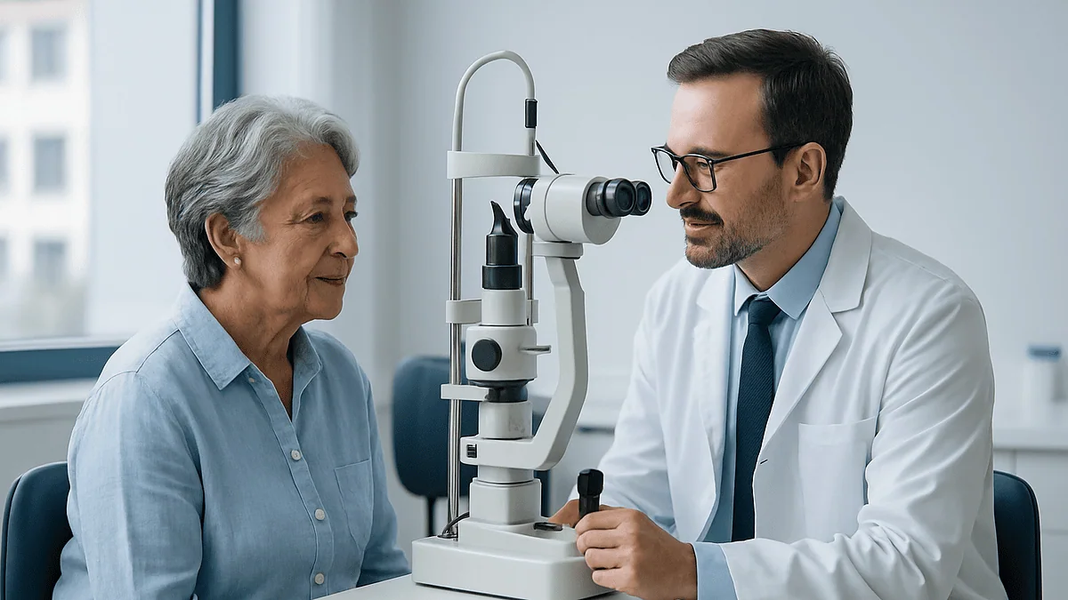
The slit lamp is the centerpiece. A slit lamp is a microscope with a bright, adjustable beam of light that lets the examiner look at the front of the eye in cross-section, almost like slicing a tomato with light instead of a knife. Through it, the lens can be inspected layer by layer and its cloudiness graded.
The dilated examination closes the loop. Drops widen the pupil so the retina and optic nerve can be seen, ruling in or out other causes of poor sight. Only when those parts look healthy can the team confidently attribute your symptoms to the lens, which is the whole point of a cataract eye exam.
What actually happens during a cataract eye exam, step by step
Most visits follow a predictable order, with the drops placed early so they have time to work while other tests run. The table below summarizes what each step feels like from the chair and what it tells the team.
| Step | What you experience | What it shows |
|---|---|---|
| History and symptoms | Conversation about tasks, lighting, glasses changes | Which eye and which situations are most affected |
| Visual acuity and refraction | Reading letters through changing lenses | Best corrected sight; blur that glasses cannot fix |
| Glare and contrast testing | Chart read with a bright light or faded letters | Real-world disability the standard chart misses |
| Eye pressure check | Brief puff of air or a gentle probe after numbing drops | Screens for glaucoma before any surgery planning |
| Slit-lamp exam and lens grading | Chin on a rest, bright beam, a few minutes | Type, location and density of lens clouding |
| Dilated retinal exam | Drops, then a bright light and a lens held near the eye | Health of retina, macula and optic nerve |
| Biometry (if surgery considered) | Looking at a target inside a machine | Eye length and corneal curve for lens planning |
Two details help people relax. The air-puff or probe used to check eye pressure, called tonometry, does not hurt; numbing drops are used when the probe touches the eye. And the bright light of the slit lamp is uncomfortable for a moment but causes no harm, because the beam is designed for exactly this purpose.
According to the National Eye Institute, dilating drops need about 20 to 30 minutes to take full effect, and vision stays blurry and light-sensitive for roughly four to six hours afterward. That waiting window is often when the biometry and chart tests are done, so the visit feels busier than the list suggests. Sunglasses for the trip home and a driver, if one is available, make the afternoon far easier.
Vision testing: why a 20/40 line does not tell the whole story
Visual acuity, the standard letter-chart score, is the number everyone remembers. “20/40” means you read at 20 feet what a person with typical sight could read at 40. It is a useful yardstick and it feeds into driving standards, but it is measured under nearly ideal conditions: a black letter on a white background, in a well-lit room, with no headlights coming toward you.
Cataracts are unfair to that test. A lens with clouding at its back surface, near the center, can let a patient read 20/30 on the chart while making sunny sidewalks or night driving genuinely difficult. The scattered light that produces halos and glare simply does not show up when the target is high-contrast and the room is calm.
That is why many clinics add two supplementary tests. Glare testing repeats the chart while a bright light shines toward the eye, imitating a low sun or approaching car; a sharp drop in the line you can read is a sign the cataract is scattering light. Contrast sensitivity testing uses letters that fade from black to pale gray and measures the faintest shade you can still make out, which tracks tasks like reading a menu in a dim restaurant or spotting a curb at dusk.
Refraction, the process of trying different lenses in front of your eye, is the other half of vision testing. Its job is subtraction. If new lenses bring the eye back to clear sight, glasses may be a reasonable path for now. If the best possible lenses still leave the chart blurry, the team knows the limitation sits inside the eye rather than in your spectacles. The Mayo Clinic lists this step alongside the chart as a core part of diagnosis for precisely that reason.
Ask for these results in plain terms: the best corrected line for each eye, and whether glare or contrast made it worse. Those two facts often matter more than any single grade the lens receives.
Lens grading on the slit lamp: nuclear, cortical and posterior subcapsular
The natural lens sits just behind the pupil and is built like an onion, with a firm center called the nucleus, softer surrounding layers called the cortex, and a thin outer bag called the capsule. Cataracts are named by which layer clouds first, and the Mayo Clinic describes three main patterns.
A nuclear cataract forms in the center and typically turns the lens yellow, then brown. Because the central lens hardens and changes its focusing power, distance vision blurs gradually and colors lose their vividness. Some people notice they can briefly read without glasses again, a shift sometimes called second sight, before the clouding overtakes the benefit.
A cortical cataract begins as wedge-shaped streaks at the outer edge of the lens that creep toward the center like spokes on a wheel. Light hitting those spokes scatters, so glare is often the leading complaint even when the chart score looks respectable.
A posterior subcapsular cataract is a small, dense patch at the back of the lens, directly in the path of light. It tends to progress faster than the other types, interferes with reading and bright-light vision early, and is more often seen in people who have used corticosteroid medicines or who have diabetes, according to the National Eye Institute.
Grading puts a number on each of these. Clinicians commonly use a scale from trace to 4+, or a standardized photographic reference set in which the examiner matches what they see to a series of images of increasing density. The grade is a description, not a verdict: a 2+ nuclear cataract in one person may cause more disability than a 3+ in another, depending on pupil size, the other eye, and the tasks that fill the day. Grading exists so that change can be tracked honestly from one visit to the next and so that the team can judge whether the lens findings are enough to explain the vision loss you report.
The dilated exam: checking the parts a new lens cannot fix
Removing a cataract replaces one component of the eye’s optical system. It does nothing for the film at the back, the retina, or the cable that carries the picture to the brain, the optic nerve. If either of those is damaged, surgery may clear the view without restoring the sight a person hoped for. This is why every thorough cataract eye exam includes a dilated look at the back of the eye, a step the Mayo Clinic and MedlinePlus both list as standard.
Dilation drops relax the muscle that narrows the pupil, opening a wider window. Through that window the clinician examines the macula, the small central area of the retina responsible for reading and face recognition, along with the surrounding retina and the optic nerve head. Age-related macular degeneration, diabetic changes in the retinal blood vessels, and glaucoma damage to the nerve are the conditions most often uncovered, and each changes the conversation.
A dense cataract can make this view hazy, and in some cases the examiner cannot see the macula well enough to judge it. Some practices then use an imaging scan called optical coherence tomography, which builds a cross-sectional picture of the retina using reflected light, or an ultrasound scan when the lens is too cloudy for light to pass. These tools are not about the cataract itself; they are about making sure nothing behind it is being missed.
Eye pressure is measured during the same visit. Pressure that is too high can point to glaucoma, a condition in which the optic nerve is damaged over time, and it can also influence how a surgical plan is organized. None of these findings means a cataract cannot be treated. They do mean the team can give you a realistic account of what surgery might change and what it cannot, which is a kinder outcome than a surprise after the fact.
Lens measurements explained: how biometry sizes an artificial lens
If cataract surgery is being considered, a further set of measurements enters the picture. Biometry is the measurement of the eye’s physical dimensions, and its purpose is to calculate the focusing power of the artificial lens, called an intraocular lens or IOL, that will replace the cloudy one.
Two numbers do most of the work. Axial length is the distance from the front surface of the cornea to the retina, in effect the depth of the eyeball. Keratometry measures the curvature of the cornea, the clear dome at the front of the eye, which contributes a large share of the eye’s total focusing power. A longer eye or a flatter cornea needs a different lens power than a shorter eye or a steeper one, in the same way that a projector must be refocused when it is moved closer to or farther from the wall.
Most modern devices gather these figures optically. You rest your chin on a support and look at a target while a beam of low-energy light measures the distances by timing its reflections. The process is painless and typically takes a few minutes per eye. When the cataract is too dense for light to pass through, an ultrasound probe, applied after numbing drops, can measure axial length instead.
Additional measurements often join the set. Corneal topography maps the surface shape in detail and can reveal astigmatism, an unevenness of the cornea that blurs images at all distances. Anterior chamber depth, the space between the cornea and the lens, and white-to-white distance, the width of the cornea, help refine where the new lens will sit.
These figures are then run through mathematical formulas that predict the lens power most likely to produce the target focus. Formulas differ in how they weight each measurement, and surgeons often compare several. Biometry is precise but not perfect, which is why a good team frames the result as a plan with a small margin rather than a promise of a specific prescription.
What the measurements can and cannot decide about your new lens
Biometry produces a power. It does not, by itself, choose a lens design or a target focus. Those are decisions, and they belong to you and the surgeon together.
The first decision is where to aim. A standard monofocal intraocular lens provides one clear focal distance. Many people aim for distance and plan to wear reading glasses; others, especially lifelong near-sighted readers, prefer to keep near focus and wear glasses for driving. Some teams offer a strategy in which one eye is set for distance and the other for near, an approach the brain can adapt to for some people and not for others. The NHS notes that most people still need glasses for at least some tasks after cataract surgery, and that a new prescription is usually issued only once the eye has healed.
The second decision concerns lens design. Toric lenses correct astigmatism found on corneal measurements. Multifocal and extended-depth-of-focus lenses split or stretch light to provide more than one focal distance; they reduce dependence on glasses for some people but can add halos and reduce contrast, particularly at night. Whether any of these options suits you depends on the measurements, the health of the macula, your visual tasks and your tolerance for trade-offs, and the surgeon will explain what the findings support.
The third decision is what surgery cannot deliver. Measurements do not predict how the retina will perform once the cataract is gone, and a lens cannot correct nerve or macular disease. Prior refractive surgery on the cornea also makes biometry less predictable, because the formulas were built around unaltered corneas; if you have had laser vision correction, tell the team, since it changes the formula they use.
A useful way to hear the numbers is as a set of constraints. They narrow the field of sensible choices. The final choice remains a conversation.
How long does a cataract eye exam take, and how should you prepare?
Plan for a long appointment rather than a quick check. The dilating drops alone need about 20 to 30 minutes to open the pupil fully, according to the National Eye Institute, and the remaining tests, history taking and discussion fit around that window. When biometry and retinal imaging are added ahead of possible surgery, a visit can stretch to the better part of a morning or afternoon. Clinics vary, so it is fair to ask when you book.
The after-effects last longer than the visit. The National Eye Institute puts blurred near vision and light sensitivity from dilation at roughly four to six hours. Reading and screen work are difficult in that period and driving is not advised, so arranging a ride, or planning for public transport with sunglasses, removes a source of stress.
Preparation is mostly about information. Bring your current glasses, including any separate pairs for reading or computer work, since refraction starts from what you already wear. A list of medicines matters more than people realize: some drugs used for prostate symptoms can affect how the pupil behaves during surgery, corticosteroids are linked to a particular cataract pattern, and blood-thinning medicines are relevant to surgical planning. Do not stop or change anything before the visit; simply bring the list so the prescribing clinician and the eye team can coordinate.
Contact lens wearers are often asked to leave their lenses out for a period before biometry, because lenses can temporarily reshape the cornea and skew the keratometry readings. The length of that break depends on lens type and is set by the clinic.
Finally, write down the tasks that trouble you most. “Headlights at night” or “reading labels in the pharmacy” gives the examiner something concrete to match against the grades and the glare test, and it keeps the conversation about your life rather than only about your lens.
Who is usually offered cataract surgery, and who is asked to wait
Guidelines in the UK and the US converge on the same principle: surgery is offered when a cataract affects a person’s quality of life or ability to carry out daily activities, not when the lens reaches a particular grade. The NHS puts it plainly, describing the decision as based on whether your eyesight is affecting your day-to-day life, and stating that surgery should not be withheld on the basis of a chart score alone.
People who commonly move toward surgery include those whose best corrected vision no longer meets driving standards, those whose glare disability makes night driving unsafe even with a passable chart line, and those whose reading, work or hobbies have narrowed because of blur that glasses cannot fix. Surgery is also sometimes recommended when a cataract prevents the team from monitoring or treating another condition at the back of the eye, such as diabetic retinal disease, because the cloudy lens blocks the view.
People who are often asked to wait fall into a few groups. Someone whose symptoms are mild and whose vision is corrected well by an updated glasses prescription may be offered monitoring, with repeat grading at intervals to track change. Someone with active inflammation or infection on the surface of the eye is usually treated for that first. Someone whose retinal or nerve disease would limit the benefit may be counseled about realistic expectations before proceeding, and may reasonably decide not to.
Waiting is not a rejection. Cataracts generally progress slowly, and unlike some eye conditions, delaying surgery does not usually harm the eye itself, though very dense, long-standing cataracts can become technically harder to remove. The Mayo Clinic frames the timing as a shared judgment: when the benefits to your daily function outweigh the risks for you, and not before. That decision always rests with you and your treating team.
What the days and weeks after cataract surgery usually look like
People often arrive at the exam already wondering how much downtime surgery involves, so it is worth describing the typical arc, with the caveat that your surgeon’s instructions take precedence over any general account.
Cataract surgery is almost always a day procedure. The NHS describes the operation itself as taking around 30 to 45 minutes, usually under local anesthetic drops or an injection near the eye, with the patient awake and going home the same day. A protective shield is commonly worn for the first night and sometimes while sleeping for a few days afterward.
In the first days, the NHS lists gritty sensations, watering, blurred or double vision and a red or bloodshot appearance as normal, with these effects usually easing within a few days. Prescribed drops, typically an anti-inflammatory and an antibiotic, are used for a period set by the surgeon; how often and for how long is a decision for the prescribing team, and the schedule matters, so follow the written instructions exactly.
Full recovery is longer than the visible healing. The NHS advises that it can take four to six weeks to recover fully, and it recommends avoiding strenuous exercise, swimming, eye rubbing and getting soap or water in the eye during the early period, again for durations set by your team. Driving is off the table until your surgeon confirms you meet the standard. Many people return to reading, television and light activities within days, but heavy lifting and dusty or dirty environments are usually restricted for longer.
Glasses come last. Because the eye’s focus settles as it heals, the NHS notes that a new spectacle prescription is generally issued only after healing is complete, often around six weeks. If both eyes need surgery, the NHS describes them as usually done as separate operations, with the second scheduled once the first has settled. A follow-up examination, often repeating parts of the original exam, checks pressure, the lens position and the retina.
Risks and alternatives, described in plain clinical language
A fair explainer describes the downside as clearly as the upside. Cataract surgery is one of the most frequently performed operations, and serious complications are uncommon, but they exist and a good consultation names them.
The NHS lists possible complications including infection inside the eye, bleeding, swelling of the retina or cornea, retinal detachment, damage to the capsule that holds the lens, and, rarely, permanent loss of vision. It also describes posterior capsule opacification, a clouding of the thin membrane behind the new lens that can develop months or years later and cause blur similar to the original cataract; this is usually treated with a brief outpatient laser procedure. Some people notice increased glare, halos or a change in how colors appear, particularly with lenses that provide more than one focus.
Alternatives are limited, and it helps to hear that directly. No medicine, drop, diet or exercise has been shown in mainstream evidence to reverse a cataract, a point MedlinePlus and the National Eye Institute both make. The non-surgical options are practical: an updated glasses prescription, anti-glare coatings, brighter task lighting, magnifiers for reading, and sunglasses with brimmed hats for outdoor glare. For many people at an early stage these measures are entirely reasonable, and monitoring with repeat grading is a legitimate plan rather than a failure to act.
Risk also varies with the individual eye. Very dense cataracts, very short or very long eyes, small pupils, prior eye surgery, and certain medicines that affect the iris can make the operation more complex, and the biometry and slit-lamp findings are part of how the surgeon anticipates that. Ask how your particular measurements affect the plan.
Nothing here should be read as encouragement toward or away from surgery. The risks and alternatives are laid out so that the decision, which sits with you and your treating team, is made with the full picture in view.
What people often get wrong about cataracts and the exam
Misconceptions cluster around a few themes, and correcting them makes the exam results easier to interpret.
The first is that a cataract is a film growing over the eye. It is not. The clouding happens inside the lens, behind the pupil, as proteins in the lens break down and clump, according to the National Eye Institute. Nothing is peeled off; the whole lens is removed and replaced.
The second is that a cataract must be “ripe” before it can be removed. This idea dates from older surgical techniques. Modern surgery uses ultrasound to break up and remove the lens through a small incision, and the timing is now driven by how your sight affects your life, as the NHS and Mayo Clinic both describe, not by density.
The third is that cataracts can be dissolved with drops, supplements or diet. Mainstream evidence does not support any such treatment, and MedlinePlus is explicit that surgery is the only effective way to remove a cataract. Protecting the eyes from ultraviolet light, not smoking and managing diabetes are linked to lower risk of developing cataracts, but they do not clear one that has formed.
The fourth is that a cataract can grow back. The natural lens, once removed, does not regenerate. The blur some people experience later is usually the capsule behind the artificial lens becoming cloudy, which is a different problem with a different, simpler treatment.
The fifth is that a passing chart score means there is nothing to discuss. As the glare and contrast tests show, a lens can pass the chart and still fail the road at night. Conversely, a dramatic-sounding grade does not obligate anyone to operate.
A final misconception concerns the exam itself: that it is only about the lens. Roughly half of the visit is spent confirming that the retina, nerve and eye pressure are healthy, because those are the findings that most change what surgery can realistically offer.
Questions to ask your care team after a cataract eye exam
The most valuable ten minutes of the visit are the ones after the tests, when the findings are on the screen and the clinician is in the room. Having questions ready turns a summary into a conversation. These are the ones that tend to unlock the most useful answers.
- What is my best corrected vision in each eye, and did the glare or contrast testing change it?
- Which type of cataract do I have, what grade did you record, and how does that compare with my last visit?
- Does the cataract fully explain my symptoms, or did you find anything in the retina, macula or optic nerve that also matters?
- What did the biometry show, and is there anything unusual about my eye measurements, such as significant astigmatism or a very long or short eye?
- If I chose surgery, what focus would you suggest aiming for, and what would I most likely still need glasses to do?
- What are the realistic risks for my particular eye, given these findings and my medical history?
- Do any of my current medicines affect the plan, and who should I speak to about them?
- If I decide to wait, what should I watch for, and when would you want to repeat the grading?
- Would you operate on one eye first, and what usually determines the timing of the second?
- Who do I contact, and how quickly, if something changes before my next appointment?
Notice that several of these ask the clinician to connect the numbers to your life rather than to recite them. That framing is deliberate. A grade of 2+ nuclear sclerosis means little on its own; the same grade alongside “and it accounts for the drop under glare that is making your night driving unsafe” is a finding you can act on.
Write the answers down, or ask whether a copy of the report can be shared with you. Many people find that reviewing the figures at home, away from the bright lights and the dilated blur, is when the plan finally makes sense.
When to call your doctor
Cataracts themselves rarely cause emergencies, but the eye can develop other problems at the same time, and some symptoms after an examination or surgery need prompt attention rather than a wait for the next scheduled visit.
Contact your eye care team the same day, or seek urgent care, if you experience any of the following: a sudden loss or sharp drop in vision in either eye; a new curtain, shadow or veil moving across part of your visual field; a sudden shower of new floaters or flashes of light, which can signal a retinal tear or detachment; severe eye pain, especially with headache, nausea or a red eye, which can indicate a rapid rise in eye pressure; or a sudden onset of double vision that persists.
After cataract surgery, the NHS advises contacting the surgical team urgently for increasing pain rather than the mild discomfort expected in the first days, worsening rather than improving redness, vision that becomes worse after initially clearing, a sticky or pus-like discharge, or a sudden increase in floaters or flashes. Infection inside the eye is rare but is treated as an emergency because early treatment matters greatly.
Less urgent, but still worth a call rather than waiting for the next annual visit: a noticeable change in vision between appointments, new difficulty meeting driving standards, or glasses that no longer help within months of being updated. These can indicate that a cataract is progressing faster than expected and may prompt earlier repeat grading.
If you are unsure whether a symptom is urgent, err on the side of calling. Eye teams would far rather hear about a false alarm than see someone who waited. And whatever the finding, the decision about what to do next, including whether and when to consider surgery, rests with you and the clinicians who have examined your eyes directly.
Frequently asked questions
How do eye doctors test for cataracts?
Clinicians rely on your symptom history, a letter chart read through adjustable lenses, a slit-lamp examination that inspects the lens in cross-section, and a dilated look at the retina and optic nerve. Many practices add glare and contrast testing to capture real-world difficulty. If surgery is being considered, biometry measures the eye’s dimensions to plan an artificial lens. The combination shows whether the lens explains your vision loss and what treatment could realistically change.
What are the first signs of cataracts that people notice?
Early changes are usually gradual: mild blur that new glasses do not fully fix, halos around lights at night, increased glare from sunlight or headlights, colors that look faded or yellowed, and frequent changes in glasses prescription. Some people briefly find reading easier without glasses. None of these confirms a cataract on their own, since other conditions cause similar changes, so new or worsening symptoms should be assessed with a dilated eye examination.
How long does a cataract eye exam take?
Set aside a long appointment. Dilating drops need about 20 to 30 minutes to take full effect according to the National Eye Institute, and the remaining tests, discussion and any biometry fit around that window, so a full pre-surgical assessment can occupy much of a morning or afternoon. Blurred near vision and light sensitivity from the drops last roughly four to six hours afterward, so plan not to drive and bring sunglasses.
Can a routine eye exam detect a cataract?
Yes. Any comprehensive eye examination that includes a slit-lamp check of the lens will detect cataract changes, and many people first learn of early clouding this way before they have noticed symptoms. A routine exam may not include glare testing or biometry, which are added when the findings or your symptoms suggest surgery might be worth discussing. Ask your clinician what the lens looked like and whether any grade was recorded for future comparison.
What does a cataract grade like 2+ actually mean?
It is a description of density, not a verdict. Clinicians typically grade each part of the lens on a scale from trace to 4+, or match what they see against a standardized set of reference photographs. A 2+ nuclear cataract describes moderate central clouding, but how much it limits your sight depends on pupil size, the other eye and the tasks you rely on. Grades matter most for tracking change between visits.
What is cataract surgery recovery time and how many days of rest are needed?
The NHS describes gritty sensations, watering, redness and blur as normal for the first few days, with full recovery taking around four to six weeks. Many people return to light activities such as reading and television within days, while strenuous exercise, swimming and dusty environments are restricted for longer periods set by the surgeon. Driving resumes only when your surgeon confirms you meet the legal standard. Your own team’s instructions override any general timeline.
Why do I need my eyes dilated for a cataract exam?
Dilation widens the pupil so the clinician can examine the retina, macula and optic nerve behind the lens. Cataract surgery replaces only the lens, so the team needs to know whether those structures are healthy before predicting what surgery could achieve. Dilation also gives a fuller view of the lens itself, including the outer cortex where some cataracts begin. The drops cause temporary blur and light sensitivity but no lasting effect.
What are the lens measurements before cataract surgery for?
Biometry measures the eye’s axial length, from the cornea to the retina, and the curvature of the cornea, then feeds those numbers into formulas that calculate the power of the artificial lens. Additional measurements map astigmatism and the space where the new lens will sit. The results narrow the sensible lens options, but choices about focus target and lens design remain a shared decision between you and the surgeon.
Will I still need glasses after cataract surgery?
Most people do, at least for some tasks, according to the NHS. A standard monofocal lens provides one clear distance, so people who aim for distance vision usually need reading glasses, and those who aim for near vision need glasses for driving. Lenses that provide more than one focus reduce that dependence for some people but can add halos or reduce contrast. A new prescription is usually issued once healing is complete.
Can cataracts be treated with eye drops or supplements instead of surgery?
No treatment other than surgery has been shown in mainstream evidence to remove or reverse a cataract, a point MedlinePlus and the National Eye Institute both make. Non-surgical approaches can help you manage symptoms for a time: updated glasses, anti-glare coatings, brighter task lighting, magnifiers and sunglasses. Protecting the eyes from ultraviolet light, not smoking and managing diabetes are linked to lower risk, but they do not clear an existing cataract.
References
- NHS: Cataract surgery
- National Eye Institute (NIH): Cataracts
- National Eye Institute (NIH): Get a Dilated Eye Exam
- MedlinePlus: Cataract
This article is for general information only and is not a substitute for professional medical advice. Please consult a qualified doctor about your individual situation.

More from the Blog

How DCR Surgery Creates a New Tear Drainage Path: Endoscopic and External Approaches
DCR surgery (dacryocystorhinostomy) treats a blocked tear duct by creating a new opening between the tear sac at the inner corner of the eye…

Living With Glaucoma: Drop Routines, Regular Pressure Checks and Everyday Habits That Help
Living with glaucoma means protecting the sight you still have, because vision already lost to the disease does not return. In practice that comes…

Blood Thinners, Prostate Medicines and Cataract Treatment: What Your Surgeon Checks First
Most people do not need to stop blood thinners for routine cataract surgery, because the operation uses a small incision and usually topical anesthetic,…
How Eye Angiography Is Done: Dilating Drops, the Dye Injection and the Photo Sequence
Eye angiography is done in three stages. First, drops widen the pupils so the camera can see the back of the eye. Next, a…

What Makes Super LASIK Personalized: Wavefront Maps and Custom Corneal Reshaping
Super LASIK is a marketing label some providers use for wavefront-guided LASIK. An instrument called an aberrometer measures how light is distorted as it…

How Long Does LASIK Take? The Flap, the Laser Seconds and the Waiting Room Before
LASIK usually takes about 30 minutes or less for both eyes, and the excimer laser reshapes each cornea in well under a minute. Most…