

What Are the Risks of Scoliosis Surgery? Nerve Monitoring, Infection and Implant Issues
Key Takeaways
- Intraoperative neuromonitoring records sensory and motor signals through the spinal cord continuously, letting the team pause and adjust within minutes if signals drop.
- The rods and screws are a temporary internal splint; once bone graft fuses the vertebrae, the bone carries the load and a broken rod in a healed fusion often causes no symptoms.
- Surgeons typically consider fusion for curves beyond roughly 45 to 50 degrees, according to the Mayo Clinic, while moderate curves in growing children are usually braced or observed.
- Reported complication rates vary widely between studies depending on patient group, with adolescents with idiopathic curves generally at the low end and older adults with long fusions at the high end.
- Deep infection around implants can appear months or years after surgery, sometimes with only vague back pain, so late new symptoms deserve imaging rather than waiting.
- The NHS notes that contact sports are generally avoided for about a year after fusion while the bone matures, even though school and most activities resume much sooner.
Scoliosis surgery risks include nerve or spinal cord injury, surgical site infection, bleeding that may need a transfusion, anesthetic complications, and implant problems such as rod breakage, screw loosening or failure of the fusion to knit. Serious complications are uncommon and teams use intraoperative nerve monitoring, antibiotics and careful implant planning to lower them, but no operation is risk-free. The decision always rests with the treating team.
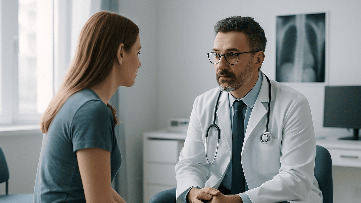
The X-ray is clipped to the light box, and the curve on it looks like a question mark. A fifteen-year-old sits on the exam table swinging her feet while her father asks the surgeon the only thing he really wants to know: what could go wrong? It is the right question, and it deserves a better answer than either “nothing, don’t worry” or the alarming lists that live on the internet.
Scoliosis surgery risks are real, measurable and, for the most part, well understood. Some are the ordinary hazards of any large operation under general anesthesia. Others belong specifically to work done a few millimeters from the spinal cord, with metal anchored into growing or aging bone. A handful are rare but serious enough that surgical teams have built entire systems, from continuous nerve monitoring to sterile-field protocols, to keep them rare.
This explainer walks through those risks honestly: what they are, how often they tend to show up, what is done to prevent them and what patients and families can watch for afterward.
What do scoliosis surgery risks actually cover?
When surgeons talk about risk, they are not describing a single hazard. They are grouping several very different things under one word. Sorting them helps.
First come the general surgical risks that accompany any long operation under general anesthesia: bleeding, blood clots in the legs or lungs, reactions to anesthetic drugs, and lung complications such as pneumonia. Scoliosis surgery often runs for several hours and involves the patient lying face down, so these risks are taken seriously even in healthy teenagers.
Second are the risks specific to the spine. The operation takes place alongside the spinal cord and the nerve roots that branch from it. Injury to those structures can cause numbness, weakness or, very rarely, paralysis. This is the complication patients fear most, and it is the reason nerve monitoring has become such a central part of the operation.
Third are the risks that come from implanting metal and asking bone to heal around it. Screws can loosen, rods can break, and the fusion, the process in which bone graft knits two or more vertebrae into a single solid segment, can fail to unite. Infection can settle around the hardware and be stubborn to clear.
Finally there are the longer-term trade-offs: a fused section of spine no longer bends, and the segments above and below take on more work over the years.
Mainstream sources such as the Mayo Clinic and the NHS describe all of these as recognized complications, while stressing that most people who have the operation do not experience the serious ones. That balance, real risk but usually good outcomes, is the honest frame for everything that follows.
How scoliosis surgery works: what actually happens in the operating room
The most common operation for scoliosis is a posterior spinal fusion. The patient lies face down under general anesthesia. The surgeon makes an incision along the middle of the back and moves the long muscles aside to expose the bony arches of the vertebrae.
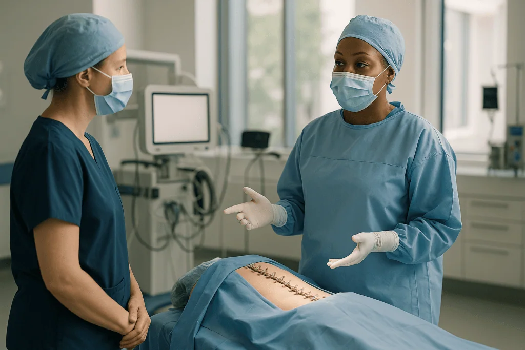
Small anchors, usually screws placed into the thick pillars of bone on each vertebra called pedicles, are inserted along the curved section. Two contoured metal rods are then attached to those anchors. By rotating and levering the rods, the surgeon gradually straightens and de-rotates the spine. The correction is never to perfectly straight; the aim is a balanced, stable spine, not a ruler.
Bone graft, either the patient’s own bone shaved from the vertebrae, donor bone, or a synthetic substitute, is packed along the treated segments. Over the following months this graft heals into a solid bridge of bone. The rods and screws act as an internal splint while that happens; once the fusion is solid, the bone, not the metal, does the work. MedlinePlus notes that people typically stay in the hospital for about three to four days after a spinal fusion, though the exact stay depends on age and how much of the spine was treated.
Some patients have an anterior approach through the side of the chest or abdomen, or a combined front-and-back procedure. Young children whose spines are still growing may instead have growing rods, which are lengthened periodically, or vertebral body tethering, a flexible cord that guides growth without fusing bone. The Mayo Clinic lists these as options for selected children, each with its own risk profile.
Throughout, an anesthesiologist manages blood pressure and fluids, and a neuromonitoring technician watches the electrical signals traveling through the spinal cord in real time.
Who is usually offered surgery, and who is usually asked to wait?
Surgery is not the default treatment for scoliosis. The NHS describes it as an option generally reserved for severe or progressing curves once other approaches are unlikely to help. According to the Mayo Clinic, surgeons typically begin to consider fusion when a curve measures beyond roughly 45 to 50 degrees, because larger curves tend to keep worsening and can eventually affect breathing or cause persistent pain.
The measurement in question is the Cobb angle, the angle between the most tilted vertebrae at the top and bottom of the curve on a standing X-ray. It is the shared language of scoliosis care, but it is not the only factor.
Growing adolescents with moderate curves are usually asked to wait and are often offered a brace instead. The Mayo Clinic notes that bracing is considered for moderate curves in children whose bones are still growing, because it can help stop a curve from getting bigger even though it does not reverse it. Adults with mild, stable curves and little pain are commonly monitored rather than operated on.
Who tends to move toward surgery? People whose curves are large or clearly progressing on serial X-rays, children whose curves are advancing despite bracing, adults whose scoliosis is producing nerve symptoms or pain that has not settled with non-surgical care, and patients with neuromuscular conditions where the curve threatens sitting balance or lung function.
Who is usually asked to pause? Patients with untreated infection, poorly controlled medical conditions, very low bone density that would not hold screws well, or those who smoke heavily, since smoking is associated with poorer bone healing. None of these are absolute bars; they are reasons to optimize first.
The decision is individual, made between the patient, the family and the surgical team, and it is never a race.
Nerve monitoring: how the team watches the spinal cord during the operation
The spinal cord cannot tell the surgeon it is in trouble while the patient is asleep. Intraoperative neuromonitoring gives it a voice.
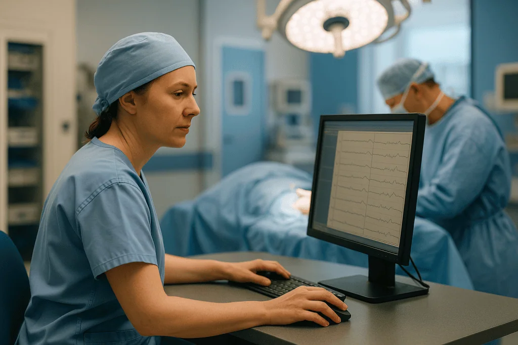
The technique works by sending tiny electrical pulses through the nervous system and measuring how they arrive at the other end. Somatosensory evoked potentials, often shortened to SSEPs, stimulate a nerve in the leg and record the signal reaching the scalp, testing the sensory pathways. Motor evoked potentials, or MEPs, do the reverse: they stimulate the brain’s motor area and record the response in leg muscles, checking the pathways that control movement. Electrodes are placed before the first incision and readings continue throughout.
If the signals drop in size or slow down beyond a set threshold, the team pauses. They raise the blood pressure to improve blood flow to the cord, check the position of any recently placed screw, and may partially release the correction. Most alerts resolve with these steps. If they do not, the surgeon can reduce the correction or, rarely, remove hardware and stage the operation for another day.
Before continuous monitoring became widely used, surgeons relied on the wake-up test: lightening the anesthetic enough for the patient to move their feet on command, then deepening it again. Some teams still keep it available as a backup.
Monitoring is not a guarantee. It can miss injuries that develop after the operation, for example from swelling or a blood clot pressing on the cord, and it can occasionally give false alarms. What it does is convert a silent, invisible risk into something that can be seen and acted on within minutes. Johns Hopkins describes neuromonitoring as part of standard practice in modern scoliosis surgery, and most spine centers treat it as non-negotiable for deformity correction.
Infection after scoliosis surgery: why it happens and how teams prevent it
Any incision can become infected, but scoliosis surgery has features that make infection a particular concern: a long wound, a long operation, and metal implants that bacteria can cling to and shield themselves on.
Surgical site infections come in two broad types. Superficial infections involve the skin and the tissue just beneath it. They usually appear within the first few weeks, with redness, warmth, spreading pain or fluid leaking from the wound. Deep infections reach the muscle layer and the implants themselves. These can show up early or, less commonly, months to years later, sometimes with only vague back pain and fatigue. MedlinePlus lists infection among the recognized risks of scoliosis surgery in children, alongside bleeding and nerve injury.
Prevention starts before the operation. Patients are often screened and treated for skin bacteria, asked to wash with an antiseptic before admission, and encouraged to stop smoking and optimize nutrition. In the operating room, an antibiotic is given intravenously shortly before the incision so that tissue levels are high when the skin is opened; the specific drug and timing are decided by the anesthetic and surgical team. Sterile technique, limited traffic in the room and careful wound closure round out the routine. Some surgeons also place antibiotic powder directly into the wound, a practice still being evaluated in research.
Treatment depends on depth. A superficial infection may settle with wound care and antibiotics chosen by the treating team. A deep infection around the hardware usually needs a return to the operating room to wash out the wound, sometimes more than once, followed by a longer course of antibiotics. Removing the implants is generally avoided until the fusion has healed, because the spine still needs the internal splint.
Children with neuromuscular scoliosis, people with diabetes and those with poor nutrition carry a higher infection risk, which is why their preparation is often more intensive.
Implant issues: what can go wrong with rods and screws
The hardware in a scoliosis operation is a scaffold, not a permanent replacement for bone. Understanding that clarifies most implant problems.
Screw malposition happens when a pedicle screw strays outside the bony channel it was meant to occupy. The pedicles in a curved, rotated spine can be narrow and oddly angled, especially at the top of the chest. A screw that breaches the inner wall can irritate a nerve root or, rarely, the spinal cord; one that breaches the outer wall may sit close to blood vessels or the lung lining. Surgeons use X-ray or navigation during placement and check each screw afterward, and neuromonitoring provides a further safety net.
Loosening occurs when the bone around a screw fails to grip. It is more common in osteoporotic bone, in very long constructs and in patients whose fusion has not fully healed.
Rod breakage sounds dramatic but has a simple mechanism: metal that flexes millions of times eventually fatigues. If the fusion has healed, a broken rod often causes no symptoms and may be found on a routine X-ray. If the fusion has not healed, the rod has been doing bone’s job for too long, and breakage is a signal that a pseudarthrosis, a failed fusion, may be present.
Prominence is when a screw head or rod end can be felt or seen under the skin, particularly in slim patients. It is uncomfortable rather than dangerous and can be revised once the fusion is solid.
Metal sensitivity is uncommon; most modern implants are titanium or cobalt-chrome alloys chosen partly for their tolerance in the body.
The Cleveland Clinic and Mayo Clinic both list hardware failure among possible complications. The typical remedy is a revision operation, planned rather than emergency, and often smaller than the original.
Bleeding, transfusion and anesthesia: the risks that are not about the spine
The vertebrae are rich in blood supply, and exposing many of them at once means steady blood loss over several hours. Managing that loss is one of the anesthesiologist’s main tasks.
Several tools are used. Blood pressure may be deliberately kept a little lower during exposure to reduce oozing, then raised during correction to protect the spinal cord. Cell salvage collects blood lost during surgery, filters it and returns it to the patient. Antifibrinolytic medicines, drugs that slow the breakdown of blood clots at the wound surface, are commonly given during spine deformity surgery; the decision to use them, and how, belongs to the anesthetic team. Even with these measures, some patients need a transfusion of donated blood, and consent discussions usually cover that possibility in advance.
General anesthesia itself carries small risks: allergic reactions, nausea, sore throat from the breathing tube, and, very rarely, more serious cardiac or respiratory events. Lying face down for hours can cause pressure on the eyes, face, chest and knees, so the team pads and repositions carefully. Vision problems after prone spine surgery are extremely rare but documented, which is why positioning receives so much attention.
Blood clots in the legs can form during long periods of immobility and, if a piece travels to the lungs, cause a pulmonary embolism. Early walking, compression devices on the calves and, in selected adults, clot-preventing medication are the standard countermeasures. Children have a lower baseline clot risk than adults but are still encouraged up and moving quickly.
A rarer post-operative problem is a slowing of the bowel called ileus, which usually resolves within days. Chest infections can follow shallow breathing from pain, which is one reason good pain control and breathing exercises matter in the first days.
Is scoliosis surgery dangerous? What the complication numbers show
People search this question in stark terms, so the answer deserves to be precise rather than reassuring or alarming.
A systematic review published in the journal Scoliosis and indexed by the NIH examined the reported rate of complications in scoliosis surgery across the medical literature. Its central finding was uncomfortable but useful: reported rates vary enormously from study to study, depending on how complications were defined, how long patients were followed and whether the population was adolescents with idiopathic curves or adults and children with neuromuscular disease. The authors concluded that patients deserve a frank discussion of this uncertainty rather than a single tidy percentage.
Some patterns are consistent across sources. Adolescents with idiopathic scoliosis, meaning curves with no identifiable underlying cause, tend to have the lowest complication rates. They are young, their bones heal well and they rarely have other medical conditions. Adults, particularly older adults with degenerative curves and long fusions extending to the pelvis, have higher rates of infection, implant failure and medical complications. Children with cerebral palsy, muscular dystrophy or spina bifida have higher infection and respiratory complication rates because of their underlying conditions.
Death is a rare event in scoliosis surgery. It is documented, and honest consent discussions acknowledge it, but it belongs at the far tail of the risk distribution, most often linked to major medical events in patients with significant heart or lung disease.
Permanent spinal cord injury is uncommon and has become less so with routine neuromonitoring. Temporary nerve root symptoms such as numbness or a patch of weakness are somewhat more frequent and usually improve.
So: dangerous compared with a filling at the dentist, yes. Dangerous compared with leaving a large, progressing curve untreated for decades? That is the calculation each patient makes with their team, and it is different for every spine.
Scoliosis surgery side effects in the long term: stiffness, adjacent segments and non-union
The most certain consequence of a spinal fusion is not a complication at all. It is the intended result: the fused segment no longer moves.
How much that matters depends on where the fusion ends. The Cleveland Clinic notes that fusion limits motion in the treated part of the spine. A fusion confined to the upper and middle back costs little flexibility, because those segments contribute relatively little to bending. A fusion that extends into the lower lumbar spine reduces the ability to bend and twist noticeably, and fusions to the pelvis change how a person sits and lifts. Surgeons work to end the fusion as high as safely possible for exactly this reason.
Adjacent segment disease describes accelerated wear in the discs and joints just above or below a fusion, which now absorb motion the fused segment used to share. It develops over years to decades, is more of a concern with long fusions into the lower back, and does not affect everyone. Whether it reflects the fusion itself or the natural aging of a spine that was already abnormal is still debated in the research literature.
Pseudarthrosis, the failure of the bone graft to fuse solidly, may cause persistent pain, loss of correction or rod breakage. Smoking, poor nutrition, certain anti-inflammatory medicines and long fusions raise the risk. It is usually diagnosed on CT scanning and treated with a revision that adds fresh graft and stronger fixation.
Flat-back syndrome, a loss of the normal inward curve of the lower back, was more common with older implant designs and leaves people leaning forward and fatiguing quickly. Modern rod contouring has made it less frequent.
Some people report chronic back pain years after fusion; others report far less pain than before. The evidence on long-term pain is mixed, and the honest summary is that fusion changes the spine’s mechanics permanently, for better and for worse.
Scoliosis surgery risks at a glance
The table below groups the main risks by when they tend to appear and what the team does about them. Timing is typical, not guaranteed; a deep infection can surface late, and a rod can break years after the operation.
| Risk | Typical timing | Main prevention | What patients might notice |
|---|---|---|---|
| Nerve or spinal cord injury | During surgery or first days | Neuromonitoring, blood pressure management, careful screw placement | New numbness, weakness, bladder or bowel change |
| Bleeding needing transfusion | During surgery | Cell salvage, antifibrinolytic medicines, positioning | Usually none; disclosed in consent |
| Surgical site infection | Days to weeks; occasionally months or years | Pre-op antibiotics, skin preparation, sterile technique | Wound redness, drainage, fever, rising pain |
| Blood clot | First one to two weeks | Early walking, calf compression, medication in selected adults | Calf swelling or pain, sudden breathlessness |
| Screw loosening or malposition | Early to months | Imaging or navigation, checking each screw | Focal pain, radiating leg or trunk symptoms |
| Pseudarthrosis (non-union) | Six months onward | Good graft, stable fixation, no smoking | Persistent pain, loss of correction |
| Rod breakage | Months to years | Solid fusion, appropriate rod material | Often none; sometimes a click or new ache |
| Adjacent segment wear | Years to decades | Ending fusion as high as safe, core conditioning | Gradual stiffness or pain above or below fusion |
Two things stand out from this list. Most early risks are actively watched for, and most have a planned response. The later risks are slower, quieter and depend heavily on the length of the fusion, which is one reason surgeons think so carefully about where to stop.
Spinal fusion for scoliosis recovery: what the first days and weeks usually look like
Knowing the shape of a normal recovery helps a family tell the ordinary aches from the warning signs.
The first day is spent mostly in bed, with pain controlled through a combination of medicines chosen by the anesthetic and surgical team. Nurses check sensation and movement in the legs every few hours; this is the routine screening for late nerve problems, not a sign of concern. A urinary catheter is common and is usually removed within a day or two.
Walking often begins on the first or second day, with a physical therapist alongside. It feels precarious at first because the trunk has been straightened and lengthened, sometimes by a few centimeters, and the body’s sense of its own center has to recalibrate. Deep breathing exercises are encouraged to keep the lungs clear.
MedlinePlus describes a hospital stay of roughly three to four days after spinal fusion, longer for adults with extensive fusions or for children with neuromuscular conditions. Most adolescents go home without a brace; some adults and children wear one for a period.
According to the NHS, it is usually possible to return to school after a few weeks and to take part in most sports after a few months, with contact sports generally avoided for about a year while the fusion matures. Bending, lifting and twisting are restricted during the early months to protect the healing bone.
Fatigue is the symptom families are least prepared for. Even a healthy teenager may nap in the afternoons for weeks. Appetite and bowel function can take a week or more to settle. Numbness around the incision is common and often partially permanent.
Follow-up X-rays at intervals over the first year or two confirm that the correction is holding and the fusion is knitting. Recovery timelines are typical ranges, not promises; each surgeon sets restrictions based on what was done and how the bone is healing.
Can I live with scoliosis? The alternatives to surgery and their trade-offs
Yes, and most people do. Scoliosis is common, most curves are mild, and the majority never need an operation. The NHS notes that many people with scoliosis live full, active lives without surgery.
Observation is the first alternative. For small curves in children, that means a standing X-ray every several months during growth to check whether the curve is changing. For adults with stable curves, it may simply mean periodic review if symptoms change. The trade-off is uncertainty: a curve can progress, and the window for less invasive options can narrow.
Bracing is the main non-surgical treatment for growing children with moderate curves. The Mayo Clinic explains that a brace does not straighten the spine but can prevent a curve from worsening while the skeleton matures. Braces are worn for many hours a day, which is a genuine burden for a teenager, and they stop being useful once growth ends.
Physical therapy and exercise help with pain, posture and core strength at any age. Specific scoliosis-focused exercise programs exist; the evidence that they change curve size is limited and mixed, and mainstream guidance presents them as supportive rather than corrective.
For adults, pain management, activity modification and, where a nerve root is compressed, targeted injections can control symptoms without addressing the curve itself.
Living with an untreated large curve carries its own risks: continued progression, cosmetic distress, pain, and, in very large thoracic curves, reduced lung capacity. Those risks accumulate slowly, which is precisely why the surgical decision can be hard: it trades a concentrated, front-loaded set of risks for a diffuse, long-term set.
Neither path is wrong in the abstract. The treating team’s job is to lay both sets of trade-offs side by side for the particular spine in front of them.
What people often get wrong about scoliosis surgery complications
Myths cluster around this operation, partly because it is dramatic and partly because online accounts skew toward the unusual. A few corrections.
“The rods hold you together for life.” They do not. The implants are a temporary internal splint. Once the fusion heals, bone bears the load; the metal simply stays because removing it would mean another operation. A broken rod in a healed fusion is often an incidental finding.
“Any tingling after surgery means paralysis is coming.” Transient numbness around the incision, in the chest wall or down a thigh is common and usually reflects small sensory nerves cut or stretched during exposure. New weakness or changes in bladder or bowel control are different and warrant urgent contact, but ordinary skin numbness is expected.
“Fusion means you can never bend again.” Most fusions for adolescent scoliosis stop above the lower lumbar segments, which do the majority of bending. Many people return to dance, gymnastics, cycling and swimming. Range of motion is reduced, not abolished.
“Surgery straightens the spine completely.” Surgeons aim for a balanced spine with the curve substantially reduced. Pushing for a perfectly straight line raises the risk of nerve injury and hardware strain, so a residual curve is normal and intended.
“Complication rates are the same everywhere.” The systematic review indexed by the NIH found wide variation in reported rates depending on patient group and study design. Adolescents with idiopathic curves and older adults with degenerative curves are not comparable populations.
“If you are going to have surgery, sooner is always better.” Timing is individual. Operating on a very young child risks the crankshaft phenomenon, where continued growth of the front of the spine twists a posterior fusion, which is why growing rods and tethers exist for that age group.
Questions to ask your care team before scoliosis surgery
A good consultation is a conversation, not a briefing. These questions tend to surface the information that matters most, and a well-prepared team will welcome them.
- Which levels of my spine will be fused, and why does the fusion stop where it does? How much bending and twisting will I lose?
- Will nerve monitoring be used throughout the operation? What happens if the signals change?
- What is your team’s approach to reducing blood loss, and how likely is a transfusion in my case?
- What steps will be taken before and during surgery to lower my infection risk? Are there things I can do in the weeks beforehand?
- Which implant material will be used, and what are the plans if a screw loosens or a rod breaks years from now?
- How will I know if the fusion has healed, and when will that be checked?
- What does a typical recovery look like for someone my age with my curve, and what restrictions will apply in the first months?
- What are the specific risks that apply to me because of my age, bone density, other conditions or the size of my curve?
- What happens if we choose to wait? How will the curve be monitored, and what would prompt us to revisit the decision?
- Who do I call, at any hour, if something worries me after I go home?
Writing the answers down during the visit helps; so does bringing a second person to listen. For adolescents, it is worth asking that at least part of the conversation be addressed to them directly. They are the ones who will live with the spine, and their questions about sport, appearance and school are as legitimate as any adult’s about complication rates.
No question on this list has a universally correct answer. Their value is in hearing how your particular team reasons about your particular case.
When to call your doctor after scoliosis surgery
Most post-operative discomfort is expected: incision soreness, muscle aches, fatigue, patches of skin numbness and stiffness that eases with gentle walking. Certain signs are different and should prompt a call to the surgical team the same day, or a visit to emergency care if the team cannot be reached.
Seek care urgently for any new weakness in the legs, difficulty walking that is worse than the day before, new numbness spreading beyond the incision, or any change in bladder or bowel control, including difficulty passing urine or unexpected incontinence. These can signal pressure on the spinal cord or nerves from swelling or a blood collection and are treated as emergencies.
Call promptly if the wound becomes increasingly red, hot or swollen, if it begins to leak fluid or pus, if the edges separate, or if a fever develops, particularly when combined with rising pain at the surgical site. Infection is easier to manage the earlier it is recognized.
Sudden shortness of breath, chest pain, coughing up blood, or a swollen, painful calf may indicate a blood clot in the leg or lung and warrant emergency assessment.
Also contact the team for pain that escalates rather than settles, pain that is not controlled by the plan you were sent home with, a sudden change in the shape of your back, a clicking or grinding sensation, persistent vomiting, or an inability to keep fluids down.
Months or years later, new or worsening back pain, a visible prominence under the skin, or a noticeable loss of the correction should prompt a review with a spine specialist rather than watchful waiting. Late infections and rod breakage can be quiet, and an X-ray is often all it takes to reassure or to plan the next step.
When in doubt, call. Surgical teams would far rather hear about a false alarm than miss a real one.
Frequently asked questions
Is scoliosis surgery worth the risk?
That depends on the size and behavior of the curve, the person’s age and health, and how much the curve is affecting daily life, which is why the decision belongs to the patient and the treating team together. Surgery concentrates a defined set of risks into one event, while a large, progressing curve carries slower long-term risks of pain, deformity and, in severe cases, reduced lung capacity. Weighing those two very different risk profiles is the heart of the consultation.
What are the downsides of scoliosis surgery?
The certain downside is permanent loss of motion in the fused segment, which matters more the lower in the back the fusion extends. Possible downsides include infection, nerve injury, bleeding, implant loosening or breakage, failure of the fusion to heal, and accelerated wear in the segments above or below over the years. Recovery also takes months, with activity restrictions and fatigue that many families underestimate. Most people do not experience the serious complications, but all are recognized possibilities.
Is scoliosis surgery dangerous compared with other operations?
It is a major operation on a structure millimeters from the spinal cord, so it carries more risk than minor surgery, but serious complications are uncommon and modern safeguards such as neuromonitoring have reduced them. Systematic reviews show reported complication rates vary widely by patient group. Adolescents with idiopathic curves generally do well; older adults with degenerative curves and long fusions face higher rates of infection and implant problems. Your team can describe the risk profile specific to your case.
What is the survival rate of scoliosis surgery?
Death after scoliosis surgery is rare, and honest consent discussions acknowledge it without quoting a single universal figure, because risk depends heavily on age and underlying health. The rare deaths that are reported are most often linked to major heart or lung events in patients with significant medical conditions rather than to the spinal work itself. Healthy adolescents sit at the very low end of this risk. Ask your surgical team how the figures apply to your situation.
Can I live with scoliosis without surgery?
Most people with scoliosis do exactly that, because most curves are mild and never require an operation. Observation with periodic X-rays, bracing for moderate curves in growing children, exercise and pain management for adults are all mainstream approaches. The trade-off is that a large or progressing curve can worsen over time and, in severe cases, affect breathing or cause persistent pain. Regular review with a spine specialist helps keep that decision under watch.
What are the most common scoliosis surgery complications?
Across mainstream sources, the most frequently listed complications are surgical site infection, bleeding that may need a transfusion, implant problems such as screw loosening or rod breakage, failure of the fusion to knit, and temporary nerve symptoms such as numbness. Permanent spinal cord injury is uncommon. Longer term, loss of motion in the fused area is universal by design, and wear in adjacent segments can develop over years. Rates differ substantially between adolescents and adults.
How does nerve monitoring during scoliosis surgery actually work?
Electrodes placed before the incision send tiny electrical pulses through the nervous system and record how they arrive at the other end. Somatosensory evoked potentials test the sensory pathways by stimulating a leg nerve and recording at the scalp; motor evoked potentials stimulate the brain and record muscle responses in the legs. If signals shrink or slow beyond a threshold, the team pauses, raises blood pressure, checks recently placed hardware and may ease the correction.
What are the long-term scoliosis surgery side effects?
The fused segment stays permanently stiff, which is more noticeable with fusions into the lower back. Over years, the discs and joints just above and below the fusion may wear faster, a process called adjacent segment disease that does not affect everyone. Rods can fatigue and break, usually without symptoms if the fusion is solid. Some people report chronic back pain decades later while others report far less pain than before surgery; the long-term evidence is mixed.
How long does spinal fusion for scoliosis recovery take?
MedlinePlus describes a hospital stay of roughly three to four days after spinal fusion, and the NHS notes that returning to school usually takes a few weeks, most sports a few months, and contact sports about a year while the bone matures. Fatigue can last for weeks even in healthy teenagers. The fusion itself takes many months to become fully solid, which is why bending, lifting and twisting are restricted early on. Your surgeon sets the specific timeline.
Can the rods and screws be removed after scoliosis surgery?
They can, but they usually are not, because removal means another operation and the healed fusion no longer needs them. Implants are typically taken out only for a specific reason such as a deep infection that will not clear, a screw head that is painfully prominent, or a suspected metal sensitivity. Removal is generally delayed until imaging confirms the fusion is solid, since the spine still relies on the hardware as an internal splint until then.
References
- MedlinePlus – Scoliosis surgery – child
- MedlinePlus – Spinal fusion
- NIH PMC – Rate of complications in scoliosis surgery: a systematic review of the PubMed literature
This article is for general information only and is not a substitute for professional medical advice. Please consult a qualified doctor about your individual situation.

More from the Blog

What Happens in a Schroth Method Session: Elongation, Rotational Breathing and Corrections
A Schroth method session is a one-to-one physical therapy appointment for scoliosis built around three steps: elongation (lengthening the spine), rotational angular breathing (sending…

How Nucleoplasty Works: The Needle, X-Ray Guidance and Radiofrequency Inside the Disc
Nucleoplasty is a minimally invasive, needle-based procedure for selected contained disc herniations. Guided by live X-ray (fluoroscopy), a clinician passes a thin probe into…
How ACDF Surgery Is Performed: The Front-of-Neck Approach, Disc Removal and the Fusion
ACDF surgery is done through a small incision at the front of the neck. The surgeon gently moves the windpipe, food pipe and blood…
When Does Juvenile Scoliosis Move From Monitoring to Bracing or Surgery? The Decision Points
Juvenile scoliosis is usually monitored while the curve stays below about 20 to 25 degrees, braced when a still-growing child's curve reaches roughly 25…
Which Spine Operation Fits Which Problem? Decompression, Fusion and Disc Surgery Compared
Decompression, fusion and disc surgery treat different mechanical problems. Decompression removes bone or ligament that is squeezing nerves, usually for spinal stenosis. Disc surgery…
Which Symptoms After Spine Surgery Need a Same-Day Call? Wound, Bladder and Leg Changes
Call your surgical team the same day if you notice new or worsening leg weakness, numbness spreading between the legs or around the genitals,…