

When Do Glaucoma Drops Give Way to Laser or Surgery? How Treatment Usually Escalates

Key Takeaways
- Every mainstream glaucoma treatment, from drops to surgery, works by lowering eye pressure; none currently restores nerve fibers or vision already lost.
- In the LiGHT randomized trial, about three-quarters of people who started with selective laser trabeculoplasty were at target pressure without drops at three years, which is why guidelines now allow laser as a first option in early open-angle glaucoma.
- The effect of laser trabeculoplasty builds over several weeks and can fade over years, so drops are usually continued until the follow-up check and the procedure may need repeating.
- Escalation is usually triggered by pressure staying above an individual target, by visual-field or OCT scans showing progression, or by an inability to use drops reliably, not by a single high reading.
- Minimally invasive glaucoma surgery lowers pressure less than trabeculectomy or tube shunts but recovers faster, which places it in mild to moderate disease rather than advanced cases.
- After trabeculectomy or tube surgery, increasing redness, pain, discharge or a sudden drop in vision can indicate infection at any point in later years and warrants same-day contact with the eye team.
Glaucoma drops usually give way to laser or surgery when eye pressure stays above the target the eye specialist has set, when visual-field or optic-nerve scans show damage progressing despite treatment, or when drops cannot be tolerated or used reliably. Laser is often the middle step; surgery is generally reserved for advanced or fast-moving disease. The ophthalmologist decides based on each person's pressure, damage, and daily life.
The bottle sits on the bathroom shelf next to the toothpaste, and every night for six years the routine has been the same: tilt the head, one drop, blink, done. Then the optometrist frowns at a printout, the referral letter arrives, and a new word appears in the conversation. Laser. Sometimes a heavier one. Surgery.
For most people with glaucoma, that moment feels like a failure, as if the drops were a test they somehow did not pass. It rarely is. Glaucoma is a slow, chronic condition, and the way glaucoma treatment options are arranged is closer to a staircase than a cliff: each step is added when the one below it stops doing enough to protect the optic nerve.
This explainer walks through that staircase honestly, including what the evidence shows about laser before drops, why some people wait and others are moved forward quickly, and which questions are worth asking before anyone picks up an instrument.
What are the main glaucoma treatment options, in plain language?
Glaucoma is a group of eye conditions in which the optic nerve, the cable carrying images from the eye to the brain, is gradually damaged, most often because the pressure inside the eye is too high for that particular nerve to tolerate. Every treatment currently in mainstream use works on the same lever: lowering that pressure. Nothing yet in routine care repairs nerve fibers already lost, which is why the emphasis falls on catching the condition and protecting what remains.
The options sort into three broad tiers. Medicated eye drops either reduce how much fluid the eye produces or improve how that fluid drains. Laser procedures use focused light to adjust the eye’s own drainage tissue or, in some cases, to reduce fluid production. Surgery creates a new drainage route, either with a tiny implanted device or by fashioning a channel in the wall of the eye. The NHS and Mayo Clinic both describe this same ladder, with drops as the usual starting point and surgery for cases where pressure remains too high or the disease keeps advancing.
Two things shape which rung a person stands on. The first is the type of glaucoma. Open-angle glaucoma, the most common form in the United States, progresses over years and is usually managed stepwise. Angle-closure glaucoma, where the drainage angle is physically narrowed or blocked, often calls for laser early because the anatomy itself is the problem. The second is how much damage already exists and how quickly it is moving. According to the National Eye Institute, about three million Americans have glaucoma, and roughly half do not know it, which means many people meet the condition for the first time already partway up the staircase.
How does lowering eye pressure protect the optic nerve?
The front of the eye is constantly filled with a clear fluid called aqueous humor, which nourishes the lens and cornea and then drains away through a sieve-like meshwork tucked into the angle where the iris meets the cornea. Picture a sink with the tap always running. When the drain is efficient, the water level holds steady. When it clogs or narrows, the level rises. In the eye, that rising level is intraocular pressure.

The optic nerve leaves the back of the eye through a small opening, and the delicate nerve fibers passing through it are vulnerable to sustained pressure. Over time they thin and die, producing the characteristic pattern of vision loss that begins at the edges and, if unchecked, creeps toward the center. Because the brain quietly fills in missing peripheral information, people rarely notice until a significant amount is gone. Johns Hopkins Medicine notes that this is why glaucoma is often called a silent thief of sight.
Pressure is not the whole story. Some people develop nerve damage at pressures that would be considered normal in others, a pattern called normal-tension glaucoma. Others live for years with elevated pressure and no damage at all, a state described as ocular hypertension. What matters clinically is the relationship between a given eye’s pressure and a given nerve’s tolerance. This is why eye specialists set an individual target pressure rather than aiming for a single universal number.
Every escalation in treatment is a bid to bring pressure down to that target and keep it there. Drops turn the tap down or widen the drain modestly. Laser reshapes or stimulates the drain. Surgery installs a bypass. The mechanism is the same throughout; only the force applied changes.
Why do eye drops come first for most people with glaucoma?
Drops earn their place at the bottom of the staircase because they are reversible, adjustable, and carry no surgical risk. If a particular drop causes stinging or a rash, it can be swapped. If pressure falls further than needed, the prescriber can pull back. That flexibility is hard to match once tissue has been altered by a laser or a scalpel.
Several classes exist. Prostaglandin analogs improve fluid outflow through a secondary drainage route and are usually taken once daily, which the NHS identifies as the most common first choice. Beta-blockers reduce fluid production. Alpha agonists and carbonic anhydrase inhibitors do a little of both. Some people end up on combination drops that put two mechanisms in one bottle. Which class is chosen, and in what order, is a decision for the prescribing ophthalmologist based on the type of glaucoma, other health conditions, and how the first choice is tolerated.
The catch is adherence. A drop only works when it lands in the eye, at roughly the same time, day after day, for the rest of a person’s life. Mayo Clinic flags that missed doses are a common reason pressure control slips, and the barriers are ordinary human ones: arthritis making the bottle hard to squeeze, a shift-work schedule, side effects such as redness or dryness, or simply not feeling any different, since glaucoma rarely produces symptoms until late.
None of this means drops are weak. For many people they hold pressure at target for years, and some never move beyond them. It does mean that when an eye specialist raises the idea of laser, the question in the room is often not “Have the drops stopped working?” but “Is there a way to protect this nerve that relies less on a bottle?”
How does a specialist know the eye drops are not working well enough?
Two things are tracked at every glaucoma review: the pressure itself and the state of the nerve. Pressure is measured with tonometry, a quick test in which a puff of air or a gentle probe assesses how firm the eye is. The nerve is monitored two ways. A visual field test maps the edges of vision by asking the person to click when faint lights appear, revealing blind spots they might never notice in daily life. Optical coherence tomography, or OCT, is a painless scan that measures the thickness of the nerve fiber layer at the back of the eye to a fraction of a millimeter.

Escalation is usually considered when one of three patterns appears. Pressure remains above the individual target despite drops being used correctly. The visual field or OCT shows the damage advancing even when pressure looks acceptable, which suggests the target itself needs to be lower. Or the drops are working but the person cannot keep using them, because of side effects, cost, dexterity, memory, or a lifestyle that makes a rigid schedule unrealistic.
Before recommending laser or surgery, most specialists first check the basics. Is the drop being instilled correctly? Are two drops being used within a minute of each other, so the second washes out the first? Has a second or third drop class been tried? The NHS describes this sequence: adjusting or adding drops before moving to procedures.
The pace also matters. A person in their eighties with mild, stable damage might be watched for years on a single drop. Someone in their forties whose field test worsened noticeably between two visits has decades of vision to protect and less margin for slow experimentation. The same pressure reading can mean very different things in those two rooms.
What does laser treatment for glaucoma actually involve?
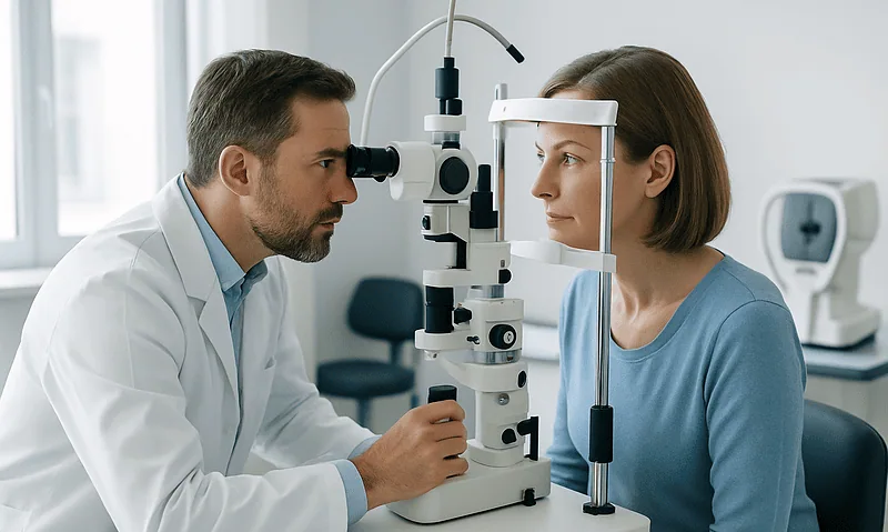
Laser treatment for glaucoma is an outpatient procedure done with the person seated at a slit lamp, the same chin-rest microscope used for routine eye exams. The eye is numbed with anesthetic drops, a small lens is placed against the cornea to aim the beam, and the treatment itself typically lasts a few minutes. Most people describe a series of clicks and flashes rather than pain, though some feel pressure or a brief ache.
Three laser procedures cover most situations. Selective laser trabeculoplasty, usually shortened to SLT, delivers low-energy pulses to the drainage meshwork in open-angle glaucoma. It does not burn a hole; it appears to trigger a biological response that improves how well the tissue drains. Laser peripheral iridotomy is used for angle-closure glaucoma and creates a tiny opening in the outer iris so fluid can flow around a blocked angle. Cyclophotocoagulation targets the tissue that produces fluid and is generally reserved for advanced or hard-to-control disease, according to Mayo Clinic.
The effect of SLT is not instant. Mayo Clinic notes it can take several weeks for the full pressure-lowering effect to appear, so drops are usually continued in the interim and reviewed afterward. The NHS points out that the effect can fade over time and the procedure may need repeating, or drops may eventually be reintroduced.
Afterward, the eye may be slightly red or light-sensitive for a day or so, and a short course of anti-inflammatory drops is sometimes prescribed. Vision is usually clear enough to go home the same day, though most people arrange for someone else to drive because pupils are often dilated for the procedure.
Laser vs drops for glaucoma: is laser sometimes offered first?
For decades the order was fixed: drops first, laser when drops faltered. That assumption was tested directly in a large randomized trial called LiGHT, published in The Lancet and indexed on PubMed, which assigned people with newly diagnosed open-angle glaucoma or ocular hypertension to either SLT or eye drops as their first treatment. At three years, about three-quarters of those who started with laser were at their target pressure without needing any drops at all, and eyes in the laser-first group were less likely to have needed surgery.
The result shifted guideline thinking. The NHS now lists laser trabeculoplasty as an option that may be offered instead of drops as initial treatment for some people with open-angle glaucoma or ocular hypertension, and Mayo Clinic describes it as a possible first step. This is a genuine change in how glaucoma treatment options are sequenced, not a marketing flourish.
It does not mean laser is universally better. The trial population was people with early disease and no prior treatment. SLT is not appropriate for every drainage angle, its effect can wear off and need repeating, and some people simply prefer the control of a medicine they can stop. Others reach the opposite conclusion: a few minutes in a chair once every few years feels preferable to a nightly ritual for life.
The honest summary is that for early open-angle glaucoma, laser and drops are both reasonable first choices, and which is offered depends on the anatomy of the angle, the person’s preferences, and the specialist’s judgment. Someone already on drops who is struggling with them can reasonably ask whether SLT might reduce their reliance on the bottle. Whether it is suitable is for the ophthalmologist to assess.
When is glaucoma surgery needed, and who is usually asked to wait?
Surgery moves up the list when pressure cannot be held at target with drops and laser combined, when damage keeps progressing despite an apparently acceptable pressure, or when the disease is already advanced at diagnosis and there is little nerve left to lose. The NHS frames surgery as the option when other treatments have not worked, and Mayo Clinic describes it as a step taken when medicine and laser are insufficient or when the eye specialist judges that a much lower pressure is needed than those approaches can deliver.
Certain situations push people forward sooner. Very high pressures that threaten rapid damage, angle-closure that has not responded to iridotomy, secondary glaucoma caused by inflammation or prior eye injury, and childhood glaucoma, where the anatomy of the drainage system itself is abnormal, often lead to surgical discussion early. A person whose visual field is already constricted enough to affect reading or mobility has less room to wait and see.
Others are usually asked to hold. Someone with mild damage, a pressure only slightly above target, and a stable field over several visits is generally a candidate for another drop or a laser rather than an operation. The same is true when adherence problems have not yet been addressed, since surgery cannot fix a bottle that is not being used, but a different bottle or a laser might. Advanced age is not by itself a reason to operate or to refrain; the relevant question is how many years of vision are at stake relative to the risks of the procedure.
Every one of these judgments is individual. The specialist weighs the speed of decline, the remaining nerve, the other eye, general health, and what the person themselves is prepared to accept. The staircase is the same for everyone; the pace at which someone climbs it is not.
Trabeculectomy, tube shunts and minimally invasive glaucoma surgery explained
Glaucoma surgery falls into three families. Trabeculectomy, the longest-established, creates a small flap in the white of the eye beneath the upper eyelid so fluid can seep out into a tiny reservoir under the conjunctiva called a bleb. The surgeon controls how freely fluid escapes with fine stitches that can be adjusted afterward. It is typically done under local anesthetic with sedation and remains the procedure many specialists turn to when a substantially lower pressure is needed.
Tube shunt surgery, sometimes called a drainage implant, places a slender silicone tube inside the eye connected to a small plate positioned under the conjunctiva toward the back of the eyeball. Fluid drains along the tube to the plate and is absorbed there. Tubes are often chosen when scarring from previous surgery, inflammation, or certain secondary glaucomas make a trabeculectomy less likely to keep working, as Cleveland Clinic explains.
Minimally invasive glaucoma surgery, abbreviated MIGS, is a newer group of procedures that use microscopic devices or small incisions to improve drainage through the eye’s existing pathways. They are frequently combined with cataract surgery, since the surgeon is already inside the eye. MIGS generally lowers pressure by a smaller amount than trabeculectomy or tubes but with faster recovery and a lower rate of serious complications, which makes it a middle option for mild to moderate disease rather than a replacement for traditional surgery in advanced cases.
The words “minimally invasive” deserve a caveat. They describe the size of the incision and the recovery, not the seriousness of the decision. Any operation inside the eye carries risk, and which family of surgery is suitable depends on the type and severity of glaucoma, the state of the conjunctiva, and whether a cataract is also present.
Comparing glaucoma treatment options at a glance
Laying the tiers side by side helps explain why an eye specialist might reach for one rather than another. The table below summarizes the general character of each option as described by the NHS, Mayo Clinic and Cleveland Clinic. It is a map, not a menu; suitability is always individual.
| Option | How it lowers pressure | Typical role | Common trade-offs |
|---|---|---|---|
| Eye drops | Reduce fluid production or improve outflow | Usual first step; can be added or combined | Daily lifelong use; redness, stinging, dryness; adherence |
| Selective laser trabeculoplasty | Stimulates the drainage meshwork | First step or after drops in open-angle glaucoma | Effect can fade over years; may need repeating; short-lived redness |
| Laser iridotomy | Opens a path around a narrowed angle | Angle-closure and narrow angles | Glare or a light streak in some people; does not treat open-angle disease |
| MIGS | Micro-devices or small incisions improve existing drainage | Mild to moderate disease, often with cataract surgery | Smaller pressure reduction than traditional surgery |
| Trabeculectomy | Creates a new drainage flap and bleb | Moderate to advanced disease needing low pressure | Longer recovery; infection, scarring, over- or under-drainage |
| Tube shunt | Implanted tube drains fluid to a plate | Complex or previously operated eyes | Device-related complications; longer recovery |
Reading down the table, two patterns emerge. Pressure-lowering power tends to increase toward the bottom, and so do recovery time and the seriousness of possible complications. That is the trade the staircase represents. A person with early disease can usually afford to accept a smaller effect in exchange for lower risk; a person losing field quickly may need to accept more risk to gain more protection. No row is “best.” Each is best for a particular eye at a particular moment, and the specialist’s job is to match the two.
What do the days and weeks after laser or glaucoma surgery usually look like?
After SLT, recovery is measured in hours rather than weeks. The eye may feel gritty or look pink for a day, and light sensitivity is common while the pupil settles. Most people return to normal activities the next day. Because the pressure-lowering effect builds gradually, a review is typically scheduled several weeks later to measure the result, and drops are usually continued until then unless the specialist advises otherwise. Only after that check does the conversation about reducing or stopping drops begin.
Trabeculectomy and tube surgery follow a different arc. The NHS notes that vision is commonly blurred for a period after surgery and that it may take a few weeks for the eye to settle. During that stretch, several things happen at once. Anti-inflammatory and antibiotic drops are prescribed on a schedule set by the surgical team, and using them exactly as directed matters because inflammation drives the scarring that can close a new drainage channel. Follow-up visits are frequent in the first weeks, sometimes several in the first month, because the surgeon may adjust stitches or apply further treatment to fine-tune how much fluid is draining.
Practical restrictions are usually given in writing. They commonly include avoiding rubbing the eye, wearing a protective shield at night, keeping water out of the eye, and not swimming or lifting heavy objects until cleared, as Cleveland Clinic outlines. Driving is off the table until vision meets the legal standard and the surgeon agrees.
MIGS sits between the two. Recovery is often similar to cataract surgery, with a few days of blur and a shorter course of drops. In every case, the timeline is a typical range, not a promise; individual eyes heal at their own pace, and the team’s instructions override any general description.
What are the risks and trade-offs at each stage of glaucoma treatment?
Every rung of the staircase carries something in exchange for what it gives. With drops, the trade is mostly comfort and convenience. Redness, stinging, dryness, and blurred vision immediately after instillation are common. Prostaglandin analogs can darken the iris and lengthen lashes over time. Beta-blocker drops are absorbed into the bloodstream in small amounts and may affect people with asthma, slow heart rhythms or certain other conditions, which is one reason the prescriber asks about general health before choosing a class, as MedlinePlus notes.
Laser trabeculoplasty is generally low-risk. A temporary pressure spike in the hours afterward is the main concern, which is why pressure is often checked before the person leaves. Mild inflammation is usual and settles. The realistic downside is not harm but impermanence: the effect may fade, and further treatment may eventually be needed. Laser iridotomy occasionally leaves a person aware of a faint line of light or glare, particularly when the opening sits where the eyelid does not fully cover it.
Surgery is where the risks become material. Trabeculectomy can drain too well, dropping pressure so low that the eye’s structures are affected, or too little, as scar tissue seals the flap. Infection of the bleb is uncommon but can occur years later and is treated as an emergency. Cataract formation is accelerated. Tube shunts add device-specific issues such as the tube touching the cornea or the plate becoming exposed. MIGS devices carry lower rates of these complications but also lower pressure-lowering power, and some can later need revision.
The NHS and Mayo Clinic both describe these possibilities in neutral terms, and so should any pre-operative conversation. The point is not to frighten but to make the exchange explicit: more pressure control, purchased with more risk, justified only when the nerve genuinely needs it.
What people often get wrong about glaucoma treatment
The most common misconception is that needing laser or surgery means the person failed at using drops. Escalation is built into how glaucoma is managed. Nerves have different tolerances, drainage systems age, and a target pressure that was fine at diagnosis may need to fall as damage accumulates. Moving up the staircase is the plan working, not the plan breaking.
A second myth is that surgery restores vision. It does not. No mainstream glaucoma procedure regrows lost nerve fibers or reverses blind spots. The goal is to keep what remains, which is why the timing of escalation matters more than the drama of any single operation. Someone who expects to see better afterward is set up for disappointment; someone who understands the aim is stability can judge the outcome fairly.
Third, people often assume that once surgery is done, drops are finished for good. Sometimes that happens. Often it does not. Trabeculectomies scar down over years, tubes can become less effective, and MIGS may need a drop alongside it from the outset. Lifelong follow-up continues regardless of which procedures have been done, because glaucoma itself does not go away.
Fourth, the absence of symptoms is mistaken for the absence of disease. Glaucoma is painless in its common form, and a person can lose a great deal of peripheral vision while still reading comfortably. Feeling fine is not evidence that pressure is controlled; only measurement is.
Finally, a newer misunderstanding: that “minimally invasive” means “minor” or “optional.” These are still operations inside the eye, chosen for specific circumstances, and they carry real if smaller risks. The label describes the incision, not the significance of the decision, and it does not make them suitable for every stage of disease.
Questions to ask your care team before moving to laser or surgery
A good consultation about escalating glaucoma treatment should leave a person able to explain, in their own words, why this step is being suggested now. The questions below help get there. They are not a checklist to be rattled off but prompts for a genuine conversation.
- What is my target pressure, and how far above it am I currently?
- Has my visual field or OCT scan changed since the last visit, and by how much?
- Have all reasonable drop options been tried, and is there a reason not to try another before a procedure?
- If laser is being proposed, which type, and what does the evidence show about how long its effect usually lasts for someone like me?
- If surgery is being proposed, why this procedure rather than the alternatives, and what would happen if I chose to wait?
- What are the specific risks in my case, given my other health conditions and the state of my other eye?
- Will I still need drops afterward, and for how long?
- What does recovery involve, how many follow-up visits should I expect in the first weeks, and when can I drive and return to work?
- What signs after the procedure would mean I should call immediately?
- How will we know whether the treatment has done its job, and when will that assessment happen?
Bringing a written list of current medicines, including over-the-counter ones and any other eye drops, helps the team judge interactions. So does bringing someone else along; a second set of ears catches details that anxiety filters out. A person who leaves the room understanding the “why” as well as the “what” is far better placed to follow the aftercare that decides whether the procedure holds.
When to call your doctor: red-flag signs during glaucoma treatment
Most of glaucoma care is unhurried, but a few situations are emergencies. Sudden severe eye pain accompanied by a headache, nausea or vomiting, blurred vision and halos around lights can signal an acute angle-closure attack, in which pressure rises rapidly and the nerve can be damaged within hours. The NHS advises seeking urgent care immediately if these symptoms appear, whether or not a person already has a glaucoma diagnosis.
After laser treatment, contact the eye team the same day if pain intensifies rather than easing, if vision becomes markedly worse, or if the eye becomes increasingly red and sensitive over the following days rather than settling.
After trabeculectomy or tube surgery, the eye is vulnerable to infection for as long as the drainage pathway exists, including years later. Cleveland Clinic and the NHS list the warning signs to act on without delay: increasing redness, pain, a sticky or purulent discharge, a sudden drop in vision, a new sensitivity to light, or a feeling that the eye is unusually soft. Any direct blow to a surgically treated eye should also prompt a call. These signs are treated as urgent because an infected bleb can threaten sight quickly.
Between procedures, less dramatic changes still warrant a phone call rather than waiting for the next scheduled visit: a noticeable new blind spot or shadow, difficulty with steps or bumping into things on one side, or side effects from drops that are making it hard to keep using them. Stopping a drop without discussion can let pressure climb silently, so the better path is always to report the problem and let the prescriber decide on the next step.
None of the timelines or thresholds in this article replace the instructions given by the treating team. When in doubt, the eye department would rather take an unnecessary call than miss a necessary one.
Frequently asked questions
When is glaucoma surgery needed rather than another eye drop?
Surgery is usually considered when pressure stays above target despite drops and laser, when scans show the optic nerve is still deteriorating, or when disease is already advanced at diagnosis. Specialists typically try adjusting or adding drops and offering laser first. The decision depends on how fast damage is progressing, how much nerve remains, and the person’s overall health, and it rests with the treating ophthalmologist.
What happens if my glaucoma eye drops are not working?
The first step is usually to check technique and adherence, then to change or add a drop from a different class. If pressure remains above target or the visual field keeps worsening, laser trabeculoplasty is commonly the next option, followed by surgery if needed. Feeling fine is not a reliable guide, because glaucoma is painless; only measured pressure and nerve scans show whether treatment is working.
Laser vs drops for glaucoma: which is better as a first treatment?
For early open-angle glaucoma, evidence from the LiGHT trial shows laser trabeculoplasty and drops are both reasonable first choices, with laser keeping most people at target without drops for at least three years. Laser is not suitable for every drainage angle, its effect can fade, and some people prefer a treatment they can stop. The specialist assesses anatomy and preferences before recommending either.
Is minimally invasive glaucoma surgery a replacement for trabeculectomy?
Not in most cases. Minimally invasive glaucoma surgery lowers pressure by a smaller amount than trabeculectomy or tube shunts, with quicker recovery and fewer serious complications. It is generally used for mild to moderate disease and often combined with cataract surgery. People with advanced glaucoma who need a very low pressure are usually still offered traditional surgery, based on the surgeon’s assessment.
Will I still need eye drops after glaucoma laser or surgery?
Often, yes, at least for a time. After laser, drops are usually continued until a follow-up check confirms the effect, then reviewed. After surgery, anti-inflammatory drops are used for a period during healing, and some people later need pressure-lowering drops again as the new drainage channel scars over the years. Whether and when to stop any drop is decided by the eye specialist.
How long does recovery from glaucoma surgery usually take?
After trabeculectomy or a tube shunt, the NHS notes vision is commonly blurred and the eye may take a few weeks to settle, with frequent follow-up visits in the first month. Laser trabeculoplasty recovery is usually a day or so. Minimally invasive procedures often recover in a similar timeframe to cataract surgery. These are typical ranges, not promises, and the surgical team’s instructions take precedence.
What stage of glaucoma needs surgery?
There is no fixed stage. Surgery is more likely in moderate to advanced disease, in eyes losing visual field quickly, and in types such as uncontrolled angle-closure or secondary glaucoma. Someone with mild, stable damage is usually managed with drops or laser. The specialist weighs the amount of remaining nerve, the rate of change, and the person’s age and general health rather than a single category.
Does laser treatment for glaucoma hurt?
Most people describe laser trabeculoplasty as uncomfortable rather than painful. The eye is numbed with anesthetic drops, and the treatment involves a series of brief flashes and clicks over a few minutes. Some feel pressure or a dull ache, and the eye may be gritty or light-sensitive for a day. Pain that worsens afterward rather than easing should be reported to the eye team promptly.
Can glaucoma surgery restore vision that has already been lost?
No. Glaucoma surgery aims to lower pressure and protect the optic nerve fibers that remain; it does not regrow damaged nerve or reverse existing blind spots. This is why the timing of treatment escalation matters more than the type of procedure. A realistic goal after surgery is stable vision and a stable visual field on follow-up testing, not an improvement in what can be seen.
How often will I need check-ups after moving to laser or surgery?
Follow-up is lifelong regardless of the procedure, because glaucoma does not go away. After laser, a review is typically arranged several weeks later to measure the effect. After trabeculectomy or tube surgery, visits are frequent in the first weeks so the surgeon can adjust drainage. Once stable, intervals lengthen but continue indefinitely, with pressure, visual field and OCT scans repeated on a schedule set by the specialist.
References
This article is for general information only and is not a substitute for professional medical advice. Please consult a qualified doctor about your individual situation.

More from the Blog

Reading Your Glaucoma Treatment Plan: Target Pressure, Follow-Up Intervals and What to Ask
A glaucoma treatment plan usually sets a target eye pressure, chosen individually to slow damage to the optic nerve, and a follow-up interval for…

Questions to Ask Your Retina Specialist About Macular Degeneration Injections and Follow-Up
Before macular degeneration injections begin, ask your retina specialist what type and stage of AMD you have, which scans confirmed it, how the anti-VEGF…

Steroid Injection vs Surgical Removal for a Chalazion: How Doctors Weigh the Two
For a chalazion that has not settled with warm compresses, doctors usually choose between a corticosteroid injection into the lump and surgical drainage under…

Before Your Conjunctivitis Appointment: Lenses Out, Makeup Off and Which Details to Bring
Before a conjunctivitis appointment, take out contact lenses and switch to glasses, skip eye makeup so the examiner can judge redness, lid margins and…

Will You Be Awake During Eye Surgery? Drops, Blocks, Sedation and General Anesthesia
Most adults are awake for eye surgery. Cataract, glaucoma and many laser procedures are usually done with numbing drops or a local anesthetic injection…
Does a Robot Operate on Your Eye Alone? Robotic Ophthalmology Myths, Put Right
No. Today, no robot performs eye surgery on its own. In every approved procedure, a trained ophthalmologist makes the decisions and controls the instruments.…