

Why You Stop Contact Lenses Before Laser Eye Surgery and How It Changes the Measurements
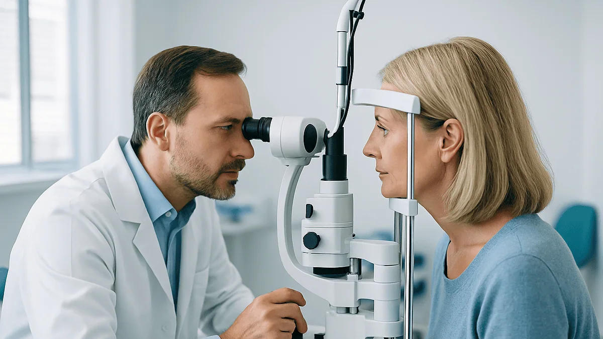
Key Takeaways
- Contact lenses temporarily mold the cornea through pressure, reduced oxygen and altered tear film, and that borrowed shape can distort corneal topography, thickness readings and the prescription programmed into the laser.
- Commonly cited lens-free intervals run around two weeks for soft lenses and three to four weeks or longer for rigid gas-permeable lenses, with orthokeratology requiring the longest break, but the surgeon sets the final figure.
- The break must cover the pre-operative measurement visit, not just the surgery date, because the entire laser plan is built from those scans.
- Surgeons confirm the cornea has settled by repeating topography and refraction and checking that consecutive measurements agree, which is why some people need a longer interval than first quoted.
- Lens warpage can mimic early keratoconus on imaging or hide genuine irregularity, so an adequate break protects against both wrongly excluding and wrongly operating on a candidate.
- The so-called 3:1:1 rule for contacts does not appear in mainstream guidance from major medical sources on laser eye surgery preparation; written instructions from the surgical team are the only standard that applies.
Contact lenses rest on the cornea and gently flatten or mold it, so measurements taken while the eye still carries that borrowed shape can be inaccurate. Surgeons ask patients to stop wearing contacts before LASIK, commonly about two weeks for soft lenses and several weeks for rigid lenses, so the cornea returns to its natural curve and the laser plan rests on true measurements rather than lens-shaped ones.
The reminder usually arrives as a short line in a pre-assessment email: please stop wearing contacts before LASIK, and bring your glasses. Then comes the quiet scramble. The glasses are in a drawer somewhere, the prescription is a few years old, and the frames pinch. People who have worn lenses since their teens often describe the first morning in spectacles as oddly disorienting, like watching the world through a window they had forgotten was there.
It feels like an inconvenience tacked onto the paperwork. In fact, it is one of the more consequential instructions in the whole process, because the surgery is planned to a fraction of a hair’s width, and a lens-shaped cornea quietly skews every number the surgeon relies on.
This explainer walks through what those weeks in glasses are actually doing to the eye, which measurements change, why the interval differs by lens type, and what to ask the team if the schedule feels impossible to keep.
What actually happens when you stop wearing contacts before LASIK
LASIK, short for laser-assisted in situ keratomileusis, reshapes the cornea, the clear dome at the front of the eye, so that light focuses on the retina without glasses. The cornea does roughly two thirds of the eye’s focusing work, and it is a little over half a millimeter thick at its center, according to Cleveland Clinic. That makes it both powerful and sensitive: a tiny change in its curvature produces a noticeable change in vision.
A contact lens sits directly on this surface for most waking hours. Soft lenses drape over it; rigid lenses perch on a thin layer of tears above it. Either way, the cornea is a living tissue that responds to sustained pressure, reduced oxygen and altered tear flow. Over months and years it adapts to the lens, in the same way a foot adapts to a shoe that is worn every day.
Take the lens away and the tissue begins to rebound toward its native shape. The surface cells that were slightly flattened or thickened recover, the swelling that comes with lower oxygen settles, and the curvature drifts back to where genetics and age have placed it. The point of the waiting period is simply to let that rebound finish before anyone measures anything.
Why not measure on the day and correct for the lens? Because the degree of molding varies from person to person and cannot be predicted from the lens box. The only reliable way to know the cornea’s true shape is to let it show you. Mayo Clinic’s patient guidance describes stopping lenses and wearing only glasses for at least a few weeks before the evaluation and the surgery, with the exact interval set by the surgeon based on lens type and years of wear.
Corneal warpage from contacts: how a lens molds living tissue
Eye specialists use the phrase corneal warpage for a reversible change in corneal shape caused by contact lens wear. It is not a disease and it is not permanent in the vast majority of cases, but it is real, and it shows up on imaging long before a person notices anything.

Three mechanisms are at work. The first is mechanical. A lens presses on the cornea with every blink, and a lens that sits slightly off-center presses more on one side than the other, so the cornea can develop a subtle tilt or asymmetry. The second is oxygen. The cornea has no blood vessels and draws oxygen directly from the air through the tear film; a lens reduces that supply, and the tissue swells slightly in response, which alters its thickness and curvature. The third is the tear film itself. Lenses change how tears spread and evaporate, and an unstable tear film makes the corneal surface optically uneven.
Rigid gas-permeable lenses, the small hard lenses some people wear for astigmatism or keratoconus, produce the most pronounced molding because they do not flex with the eye. Orthokeratology lenses, which are worn overnight specifically to flatten the cornea and provide lens-free daytime vision, are the extreme case: they are designed to warp the cornea on purpose, and that effect can persist for weeks or months after stopping.
Soft lenses mold less, but they are not innocent. Toric soft lenses, which are weighted to stay oriented for astigmatism, press unevenly by design. Even standard daily disposables leave a measurable imprint in long-term wearers. The reassuring part is that warpage from contacts almost always reverses with time out of lenses. The practical part is that nobody can be certain how much time without checking.
Which measurements change, and why fractions of a millimeter matter
A LASIK assessment is less a single eye test than a stack of overlapping maps. Each one can be distorted by a cornea that has not yet returned to shape.
Corneal topography is a color-coded map of the surface curvature, and it is the test most obviously affected. A warped cornea can look irregular or steep in places, which can mimic early keratoconus, a condition in which the cornea thins and bulges. Because keratoconus is a reason to decline LASIK, a false appearance of it can wrongly exclude someone, or a lens that happens to flatten a genuinely suspicious area can hide it.
Pachymetry measures corneal thickness. Surgeons need enough tissue to create the flap and remove the planned amount beneath it while leaving a safe residual bed; Mayo Clinic lists a thin cornea among the reasons a person may not be a good candidate. Lens-related swelling can add micrometers of thickness that are not really there to be spent.
The refraction, the glasses prescription itself, also shifts. If the cornea is temporarily flatter or more astigmatic, the measured prescription is off, and the laser treats the wrong number. Wavefront analysis, which maps subtle optical imperfections called higher-order aberrations, is similarly thrown by an uneven surface.
| Measurement | What it tells the surgeon | How contact lens molding can distort it |
|---|---|---|
| Corneal topography | Surface curvature and regularity | False irregularity resembling keratoconus, or masking of true irregularity |
| Pachymetry | Corneal thickness | Lens-related swelling overstates available tissue |
| Refraction | Prescription to program into the laser | Temporary flattening or astigmatism shifts the target |
| Wavefront analysis | Fine optical imperfections | Uneven tear film and surface exaggerate aberrations |
The surgical plan is only as good as the weakest map. That is the whole argument for patience.
How long to stop wearing contacts before LASIK: typical ranges by lens type
There is no single number, and anyone who quotes one with certainty is simplifying. The interval depends on the type of lens, how many years it has been worn, how many hours a day it is worn, and what the first set of scans shows.

Patient-facing guidance from Mayo Clinic frames it as stopping lenses completely and wearing only glasses for at least a few weeks before both the evaluation and the surgery. Guidance from Cleveland Clinic and similar centers typically describes roughly two weeks for standard soft lenses, somewhat longer for toric soft lenses, and about three to four weeks or more for rigid gas-permeable lenses, with orthokeratology lenses requiring the longest break, often measured in months. Treat these as commonly cited starting points rather than fixed rules; the surgeon may extend any of them.
| Lens type | Commonly cited minimum break | Why |
|---|---|---|
| Soft spherical (daily or monthly) | Around 2 weeks | Flexible material molds the cornea least |
| Soft toric (for astigmatism) | Often 2–3 weeks | Weighted design presses unevenly |
| Rigid gas-permeable | Around 3–4 weeks, sometimes longer | Rigid material produces more pronounced warpage |
| Orthokeratology (overnight reshaping) | Weeks to months, surgeon-directed | Lens is designed to reshape the cornea |
Two details often surprise people. First, the break applies to the pre-operative measurement visit, not just the surgery date, because the plan is built from those scans. Second, decades of wear generally mean a longer break than a few years of wear, because the tissue has had longer to adapt. The honest summary is that the calendar is set by the cornea, confirmed by repeat imaging, and signed off by the treating team.
Contact lenses before a LASIK consultation: what the first visit checks
Many people assume the first appointment is a quick suitability chat and that the serious measuring happens later. Usually it is the reverse. The initial consultation is often the most data-heavy visit of the whole process, which is exactly why teams ask for contact lenses to be out of the picture before it.
The visit typically includes a detailed refraction, corneal topography, pachymetry, a check of pupil size in dim light, a tear film assessment for dry eye, and an examination of the retina and the front of the eye with the pupils dilated. MedlinePlus notes that LASIK is generally considered for adults whose glasses or contact lens prescription has been stable, so the team will also want to see previous prescriptions if available.
If someone arrives having worn lenses that morning, the team has a choice: take measurements that may be wrong, or reschedule. Most will measure anyway for a provisional picture, then insist on a repeat set after the appropriate break. That means two visits instead of one, which is the hidden cost of ignoring the instruction.
Some practical points help the appointment go smoothly. Bring current glasses, even if they are an old prescription, so the refraction can start from a known point. Expect the eye drops used for dilation to blur near vision and increase light sensitivity for several hours, so arranging a lift home is sensible; Mayo Clinic advises against driving after a dilated examination. Mention any history of dry eye, previous eye infections, autoimmune conditions, or pregnancy, since each can influence timing. None of this is a test to pass. It is the team gathering the information they need to tell you honestly whether LASIK is a sound option, and a lens-free cornea is what lets them answer.
Who this applies to, and who is usually asked to wait longer
Every contact lens wearer considering corneal laser surgery is asked to take a break before measurement. The same logic applies to related procedures such as PRK, in which the surface layer is removed rather than a flap created, and lens-based options, because candidacy for all of them starts with an accurate corneal map.
Some groups are typically asked to stay out of lenses longer, or to have their scans repeated more than once before the plan is finalized:
- Long-term rigid gas-permeable wearers, especially those with more than a decade of daily use
- Anyone who has used orthokeratology lenses, even if they stopped some time ago
- People whose first topography shows irregularity that could be either warpage or early keratoconus
- Wearers of toric soft lenses with higher degrees of astigmatism
- People with significant dry eye, because tear film instability can make scans noisy
- Those whose prescription has changed between visits, which may signal that the cornea has not yet settled
The decision to extend is not a sign that something is wrong. It reflects a principle the surgeon follows throughout: operate on a stable eye, not a changing one. Mayo Clinic lists an unstable prescription, thin or irregular corneas, severe dry eye and certain autoimmune conditions among the factors that may make a person a poor candidate, and the lens break is partly a way of distinguishing a temporary lens effect from a true underlying issue.
A small number of people are told, after an adequate break, that their corneas are genuinely irregular and that laser surgery is not advisable. That is disappointing to hear, but it is the system working. Discovering irregularity on a scan is considerably better than discovering it after a laser has thinned the tissue further. The treating team weighs these findings and makes the final call; the patient’s job is to give them a true picture to work from.
What the weeks in glasses usually look like
The first few days are mostly about adjustment. Glasses sit farther from the eye than a lens does, so the image is slightly smaller for nearsighted people and slightly larger for farsighted people, and peripheral vision has frames at its edges. Side vision when driving, judging steps, and reading in bed all feel subtly different. Most people stop noticing within a week.
If the glasses prescription is out of date, the blur can be more than a nuisance. It is reasonable to ask the surgical team whether an updated pair of glasses is worthwhile for the interval; some teams can provide a temporary refraction once the cornea has started to settle, while others prefer to wait. Do not fill the gap by slipping lenses back in for a day. Even a few hours can partially reset the molding and prolong the wait.
Around the midpoint of the break, many long-term wearers notice their eyes feel less dry and less red than they expected. The cornea is receiving full oxygen for the first time in years, and the tear film is spreading over a bare surface. Others notice the opposite for a few days as the eye readjusts its blink and tear habits. Both patterns are common and usually brief.
Toward the end of the interval the team repeats the scans. If two sets of topography taken a week or more apart match closely, the cornea is considered stable and the plan can be finalized. If they differ, the break is extended and the scans repeated. Mayo Clinic’s guidance frames the whole pre-operative period as preparation for an eye that will be measured and treated in a single, stable state; the glasses weeks are simply how that state is reached.
How surgeons know the cornea has settled
The waiting period is not run on faith. The surgeon is looking for a specific signal: measurements that stop moving.
Corneal topography is the main tool. On the first visit, a long-term lens wearer’s map may show a central flattening with a steeper ring around it, an off-center tilt, or a mild bow-tie pattern that exaggerates astigmatism. A second map a week or two later is overlaid on the first. Modern topographers can produce a difference map that subtracts one from the other, so any remaining drift shows up as color where there should be none. When the difference map is essentially flat, and the keratometry readings, which are the numerical curvature values in the steepest and flattest meridians, agree within the instrument’s normal variation, the cornea is judged stable.
The refraction is the second check. A prescription that matches between two visits supports stability; a measurable shift, particularly in the astigmatism component or its axis, suggests the tissue is still relaxing. Pachymetry readings that have stopped falling complete the picture, since lens-related swelling resolves as oxygen supply normalizes.
This is why the question “how long to stop wearing contacts before LASIK” has no universal answer. The interval is a hypothesis; the repeat scan is the test. Some people are stable at the first repeat. Others, especially rigid lens wearers, need a third or fourth set. Cleveland Clinic and Mayo Clinic both describe the surgeon individualizing this period rather than applying a fixed rule, and the willingness of a team to repeat imaging before proceeding is a reasonable marker of careful practice.
There is one more benefit. Scans taken once the cornea has settled serve as the true baseline for life, the reference point against which any future change, from age or disease, can be judged.
Why can't you wear contacts during surgery?
It sounds like a question with an obvious answer, yet it is genuinely searched, so it deserves a plain explanation.
LASIK works directly on the cornea. During the procedure, after numbing drops, the surgeon creates a thin hinged flap in the surface of the cornea, either with a precise blade or a femtosecond laser, folds it back, and uses an excimer laser to remove a microscopic, pre-programmed amount of tissue from the exposed layer beneath. The flap is then laid back into place, where it adheres on its own. MedlinePlus describes the whole process as typically taking around ten to fifteen minutes per eye.
A contact lens would sit exactly where the surgeon needs to work. It would physically block both the flap creation and the laser, and it would prevent the eye-tracking system, which follows tiny eye movements and adjusts the laser hundreds of times per second, from locking onto the natural landmarks of the iris and pupil. Suction devices used to stabilize the eye during flap creation also need direct contact with the eye’s surface.
Infection is the other concern. The cornea is briefly opened during surgery, and a contact lens carries bacteria, biofilm and debris even when well cared for. The CDC notes that improper lens wear is a leading risk factor for serious eye infections in otherwise healthy eyes. Nobody wants a lens anywhere near an eye whose surface barrier is temporarily interrupted.
The question may also stem from confusion with a different, legitimate practice. After PRK, and occasionally after LASIK if the flap needs support, the surgeon may place a sterile bandage contact lens on the eye for a few days to protect the healing surface and reduce discomfort. That lens is medical-grade, placed and removed by the clinician, and has no optical power. It is not the patient’s own lens, and it is never worn into surgery.
Can you wear contacts after LASIK, even 20 years later?
Yes, in principle. LASIK does not make the eye allergic to lenses, and plenty of people who had surgery decades ago wear contacts today, usually for one of a few reasons.
The most common is presbyopia. The eye’s natural lens stiffens with age, and from the mid-forties onward, reading without help becomes harder regardless of any previous surgery. LASIK corrects distance vision; it does not stop this process. Some people then choose reading glasses, others try multifocal contact lenses, and some consider further surgery, a conversation for an eye specialist.
The second reason is regression or new change. A proportion of people experience a gradual drift in prescription over the years, and a small residual correction may be wanted for driving at night or fine work. The third is simply preference for lenses on certain occasions, for sport or events, even when glasses do the job day to day.
The practical complication is fit. A cornea treated for nearsightedness is flatter in the center than untreated corneas, which is a shape that standard soft lenses were not designed for. A lens that fits a normal dome may be loose, decenter, or move excessively on a post-LASIK eye. Specialty designs exist, including reverse-geometry lenses whose center is flatter than their edge, and scleral lenses that vault over the cornea entirely and rest on the white of the eye. These require a fitting by an eye care professional experienced with post-surgical corneas, not an online reorder of a pre-surgery prescription.
Timing matters too. Mayo Clinic describes healing and visual stabilization after LASIK as taking weeks to months, and the flap interface remains a consideration for the eye’s surface health. Any decision to wear lenses after surgery, whether at twenty months or twenty years, belongs with the eye care team that can examine the cornea and confirm the surface is healthy enough.
What people often get wrong
Misunderstandings about this instruction are common, and several are worth correcting directly.
“Dailies don’t count.” Daily disposables are softer and thinner than many lenses, but they still sit on the cornea for most waking hours and still alter oxygen supply and tear film. Guidance from Mayo Clinic and Cleveland Clinic makes no exception for them; the soft-lens interval applies.
“One lens-free day before the scan is enough.” A day lets the tear film recover. It does not let the corneal shape rebound, which happens over days to weeks depending on lens type and years of wear.
“I can wear lenses between the consultation and the surgery.” Returning to lenses after the measurement visit can partially re-mold the cornea, so the laser plan no longer matches the eye on the day. The break covers the whole period from before the first measurements through to surgery.
“The 3:1:1 rule tells me what to do.” This phrase circulates online, but it does not correspond to any rule in mainstream guidance from Mayo Clinic, Cleveland Clinic, MedlinePlus or the NHS on preparing for laser eye surgery. Versions of it describe ratios of weeks per lens type or years of wear, none consistently, and it appears to be an informal mnemonic rather than a clinical standard. The surgeon’s written instructions, confirmed by repeat imaging, are the only rule that matters.
“If my scan looks irregular, I have keratoconus.” Not necessarily. Lens warpage can mimic keratoconus on topography, which is one reason the break exists. Only a stable scan after an adequate lens-free period can distinguish the two, and that interpretation sits with the treating team.
“Glasses for a few weeks will weaken my eyes.” No. Glasses neither strengthen nor weaken the eye. They simply focus light; the eye underneath is unchanged, and if anything is healthier for the extra oxygen.
What can go wrong if the instruction is ignored
The consequences of skipping the break fall on a spectrum, from inconvenient to genuinely harmful.
At the mild end is wasted time. A scan taken on a lens-molded cornea is usually recognized as unreliable, and the team asks for a repeat after a proper break. The surgery date moves. Nobody is hurt, but the schedule everyone arranged around unravels.
In the middle is a flawed plan that is caught late. If the refraction was taken on a temporarily altered cornea and the error is small, it may only be noticed when the pre-surgery check on the day disagrees with the consultation. Careful teams measure again before treating and will postpone if the numbers do not match. Again, inconvenient rather than dangerous.
At the serious end is an inaccurate treatment. Programming the laser with a prescription or astigmatism axis that was distorted by lens molding means removing tissue in the wrong pattern. The likely results are residual nearsightedness, farsightedness/">farsightedness or astigmatism, and sometimes irregular astigmatism that glasses cannot fully correct. Mayo Clinic lists undercorrection, overcorrection and astigmatism from uneven tissue removal among the known risks of LASIK, and an unsettled cornea raises the odds of all three. Enhancement surgery may be possible, but it spends more corneal thickness, and some people do not have it to spare.
There is also the diagnostic risk described earlier: a lens that flattens a genuinely abnormal area can hide early keratoconus. Performing LASIK on a cornea with undetected keratoconus can accelerate thinning and bulging, a complication known as ectasia. It is uncommon, and screening exists precisely to prevent it, but screening only works on an unmolded cornea.
None of this is intended to alarm. The break is a low-effort safeguard against a high-consequence error, and the overwhelming majority of people who follow it have an uneventful assessment. It is one of the rare situations in medicine where patience alone removes a real risk.
Questions to ask your care team
The pre-assessment is a two-way conversation, and the lens break is a good place to start it. A short list, tailored to your own situation, tends to produce clearer answers than a general “anything I should know?”
- Given the type of lenses I wear and how long I have worn them, how many weeks do you want me out of lenses before my measurement visit, and before surgery?
- Will you repeat my corneal topography to confirm the shape has stabilized, and how will I know the result?
- If my current glasses are out of date, is it worth updating them for the waiting period, or would you prefer to wait until the cornea settles?
- Does my first scan show anything that could be either lens warpage or an underlying corneal condition, and how will you tell the difference?
- Is my corneal thickness comfortably sufficient for the planned correction, with room to spare?
- How will you measure and manage dry eye before and after surgery, since long-term lens wear often affects the tear film?
- If I accidentally wear lenses during the break, what should I do, and does the clock restart?
- After surgery, when would it be safe to consider contact lenses again if I ever wanted them, and what kind of fitting would that involve?
- What alternatives would you suggest if the scans show I am not a good candidate for LASIK?
Write down the answers, particularly the lens-free interval, because it is easy to misremember weeks later. If two members of the team give different numbers, ask which applies. The NHS advises patients considering laser eye surgery to ask about the surgeon’s experience, the specific risks for their eyes and what happens if the result is not as expected, and the same openness should extend to the preparation.
Every one of these questions has a reasonable answer. A team that welcomes them is a team that takes the measurements seriously, and that is exactly what you want from the people planning to reshape your cornea.
When to call your doctor
The lens-free period is usually uneventful, and the surgery itself has a predictable recovery. Even so, certain symptoms warrant a prompt call to the surgical team or eye specialist rather than waiting for the next scheduled visit.
During the weeks before surgery, contact the team if you develop a red, painful eye, sudden light sensitivity, discharge, or a feeling that something is in the eye that does not clear with blinking. Long-term lens wearers can carry a low-grade surface problem that only becomes obvious once the lens is off, and an active infection or inflammation would need to be treated and fully settled before any surgery is scheduled. The CDC describes eye pain, redness, blurred vision, discharge and light sensitivity as signs of a possible lens-related infection that need same-day professional assessment.
After surgery, Mayo Clinic and MedlinePlus both describe some grittiness, mild discomfort, watering, light sensitivity and fluctuating vision in the first days as expected. The following are not expected and should prompt an urgent call, or an emergency department visit if the team cannot be reached:
- Severe or worsening eye pain, particularly if it increases rather than eases after the first day
- A sudden drop in vision, or vision that was improving and then deteriorates
- Increasing redness, thick discharge, or a white or gray spot visible on the cornea
- A direct knock, rub or poke to the eye in the early healing period, because the flap can be displaced
- New flashes of light, a shower of floaters, or a curtain-like shadow across part of the vision, which can indicate a retinal problem needing immediate care
Do not put a contact lens into a painful or recently operated eye to “see if it helps.” Do not use leftover drops from a previous prescription without checking. Call, describe what you see and feel, and let the team decide whether you need to be seen today. They would far rather hear from you about a false alarm than miss a treatable problem.
Frequently asked questions
How long to stop wearing contacts before LASIK?
Commonly cited ranges are about two weeks for soft lenses and roughly three to four weeks or more for rigid gas-permeable lenses, with orthokeratology lenses requiring the longest break. Mayo Clinic frames it as at least a few weeks in glasses before both the evaluation and the surgery. The surgeon confirms the interval with repeat scans, so treat these numbers as starting points rather than fixed rules.
What is the 3:1:1 rule for contacts?
It is not a recognized clinical rule. The phrase circulates online with inconsistent meanings, sometimes as weeks per lens type and sometimes as a ratio tied to years of wear, but it does not appear in guidance from Mayo Clinic, Cleveland Clinic, MedlinePlus or the NHS on preparing for laser eye surgery. Follow the written instructions from your surgical team instead.
Why can't you wear contacts during surgery?
A lens would sit exactly where the surgeon works, blocking flap creation and the laser, and preventing the eye-tracking system from locking onto the iris and pupil. Lenses also carry bacteria, and the corneal surface is briefly opened during surgery. A sterile bandage lens is sometimes placed by the clinician afterward, but that is a medical device, not the patient’s own lens.
Can you wear contacts 20 years after LASIK?
Often yes, after assessment by an eye care professional. A LASIK-treated cornea is flatter centrally, so standard soft lenses may fit poorly, and specialty designs such as reverse-geometry or scleral lenses are sometimes needed. Many people return to lenses for presbyopia or minor prescription drift. The decision depends on the health of the corneal surface and should be made with the eye care team.
Can I buy contact lenses without a current prescription?
Contact lenses are regulated medical devices, and in the United States a valid prescription from an eye care professional is required. The CDC notes that lenses obtained or worn without proper fitting and follow-up carry a higher risk of serious eye infection. If you need lenses while waiting for surgery or afterward, ask your eye care team rather than reusing an old prescription.
What is corneal warpage from contacts, and is it permanent?
Corneal warpage is a reversible change in corneal shape caused by lens wear, through pressure, reduced oxygen and altered tear flow. It shows up on topography as flattening, tilt or exaggerated astigmatism. In the great majority of people it resolves after an adequate lens-free period, with rigid and orthokeratology lenses taking longest. Repeat imaging confirms when the shape has stabilized.
Do I need to stop wearing contacts before the LASIK consultation or only before surgery?
Before the consultation, because that visit usually includes the topography, thickness and refraction measurements the plan is built from. Lenses should then stay out through to surgery, since returning to them can partially re-mold the cornea. If you arrive in lenses, teams often take provisional measurements and insist on a repeat set after the appropriate break.
Do daily disposable lenses count, or is the break only for hard lenses?
They count. Daily disposables are thin and flexible, but they still cover the cornea for most waking hours and alter oxygen supply and tear film. Guidance from Mayo Clinic and Cleveland Clinic applies the soft-lens interval to all soft lenses, including dailies. Rigid lenses simply require a longer break because they mold the cornea more.
What happens if I accidentally wear my contacts during the break?
Tell the surgical team. A short, single wear may have little effect, especially with soft lenses, but the team may extend the interval or repeat the scans to be sure the cornea has settled again. Honesty matters more than the slip itself, because an undetected change in corneal shape is what leads to inaccurate measurements.
Can a lens-molded cornea be mistaken for keratoconus on a LASIK scan?
Yes. Warpage from contact lenses can produce topography patterns that resemble early keratoconus, a condition in which the cornea thins and bulges and which is a reason to decline LASIK. The opposite can also happen, with a lens masking true irregularity. Only a stable scan after an adequate lens-free period lets the surgeon tell the two apart.
References
This article is for general information only and is not a substitute for professional medical advice. Please consult a qualified doctor about your individual situation.

More from the Blog

How Long Lip Filler Lasts, and How Long It Takes to Settle
Most hyaluronic acid lip fillers last roughly six to twelve months, and some people see results for up to eighteen months, according to the…

Why Surgeons Ask You to Stop Smoking Before Disease Reconstruction Surgery
Surgeons ask patients to stop smoking before reconstructive surgery because nicotine narrows small blood vessels and carbon monoxide lowers the oxygen reaching tissue, so…

RF Microneedling Results Timeline: Why Collagen Remodeling Takes Months, Not Days
Most people see little change in the first two weeks after RF microneedling because the treatment works by triggering a wound-healing response, and new…

How Cryoablation (CRYO) Works: Image-Guided Probes, the Freeze Cycles and What You Feel
Cryoablation destroys a targeted patch of tissue by freezing it. A doctor guides one or more thin probes into the target using ultrasound, CT…

Dermal Fillers vs Botulinum Toxin: Volume vs Movement and Why Many Plans Use Both
Dermal fillers and botulinum toxin (widely known by the brand name Botox) treat different problems. Fillers are injected gels that restore lost volume and…

Collagen-Stimulating Treatments vs Fillers: Building Your Own Collagen or Adding Volume
Dermal fillers add volume right away by placing a soft gel, most often hyaluronic acid, beneath the skin. Collagen-stimulating treatments work differently: they prompt…