Flatfoot (Pes Planus)
Learn about flatfoot (pes planus): common symptoms, causes, how doctors confirm the diagnosis, and treatment options from supportive footwear to surgery.
Quick answer
Flatfoot (pes planus) is a condition in which the inner arch of the foot is low or absent, so the sole rests flat on the ground. It is often painless and common in children, but adults may develop it when the tendon supporting the arch weakens. Treatment ranges from supportive shoes and exercises to surgery.
What is flatfoot (pes planus)?
Flatfoot, also called pes planus (the Latin medical term for a flat foot), is a condition in which the arch on the inside of the foot is lower than usual or absent, so that most or all of the sole touches the ground when a person stands. The arch normally acts like a spring that spreads body weight across the foot and helps absorb shock during walking and running.
Flatfoot is very common. Almost all babies and toddlers have flat feet, because the arch has not yet formed and the foot still contains a pad of soft fat. In most children the arch develops gradually during early childhood. Some people simply never develop a full arch and keep flexible flat feet for life without ever having a problem. Other people develop flatfoot later in life, often because the tendons and ligaments that support the arch weaken or become injured. Doctors call this adult-acquired flatfoot.
Doctors usually describe flatfoot in two broad ways:
- Flexible flatfoot: the arch is visible when the person sits or stands on tiptoe but disappears when the foot bears weight. This is the most common type and is often painless.
- Rigid flatfoot: the arch is absent whether or not the foot is bearing weight, and the foot is stiff. This type is less common and is more often linked to a bone or joint problem.
Flatfoot symptoms
Many people with flatfoot have no symptoms at all and only notice the shape of their foot, a worn-down inner edge on their shoes, or a comment from a family member or shoe fitter. When flatfoot symptoms do occur, they tend to relate to strain on the structures that would normally be supported by the arch. Common symptoms include:
- Aching or tiredness in the arch or heel, especially after standing or walking for a long time
- Pain along the inside of the ankle, where the main supporting tendon runs
- Swelling on the inside of the ankle or along the arch
- Pain in the outer ankle in more advanced cases, where bones may begin to press against each other
- Heels that tilt outward (doctors call this heel valgus) when viewed from behind
- Difficulty standing on tiptoe on the affected foot
- Shoes that wear down unevenly, mainly on the inner edge
- Pain in the knees, hips, or lower back that some people attribute to altered walking patterns
Symptoms often differ by type and stage. Children with flexible flatfoot usually have no pain, although some report tired legs or cramps at night, or may be reluctant to walk long distances. Adults with acquired flatfoot often describe a gradual onset of pain and swelling on the inner ankle that worsens with activity. As the condition progresses, the arch may flatten visibly, the heel may drift outward, and the foot may become stiffer. In rigid flatfoot, stiffness and pain in the middle of the foot are more typical, and symptoms may appear during adolescence when the bones finish growing.
Causes and risk factors
Flatfoot causes vary depending on when the condition appears. In children, flexible flatfoot is usually a normal variation in foot shape that runs in families. Loose ligaments (the bands of tissue that connect bones to each other) allow the arch to sag under body weight. Rigid flatfoot in children and teenagers is often caused by a tarsal coalition, an abnormal connection between two bones in the back of the foot that is present from birth and limits movement. Rarely, flatfoot in children is linked to a neurological or muscular condition such as cerebral palsy.
In adults, the most common cause of newly acquired flatfoot is a problem with the posterior tibial tendon. This tendon runs from a muscle in the calf, behind the inner ankle bone, and attaches under the arch. It is the main dynamic support for the arch. When it becomes inflamed, stretched, or torn, a condition doctors call posterior tibial tendon dysfunction, the arch gradually collapses. Other flatfoot causes in adults include:
- Injury, such as a fracture or dislocation of the bones in the middle of the foot
- Arthritis, including rheumatoid arthritis, which can damage the joints that hold the arch together
- Nerve damage (neuropathy), for example from long-standing diabetes, which can lead to a severe form of joint collapse called Charcot foot
- Stretching of ligaments over many years, sometimes linked to very tight calf muscles
Several factors increase the likelihood of developing flatfoot or of flatfoot becoming painful:
- A family history of flat feet
- Obesity, which increases the load on the tendons and ligaments of the foot
- Increasing age, particularly for adult-acquired flatfoot
- Diabetes and high blood pressure, which may affect tendon health and blood supply
- Rheumatoid arthritis and other inflammatory joint diseases
- Previous foot or ankle injury
- Pregnancy, because of hormonal changes that loosen ligaments and extra body weight
- Occupations or sports that involve prolonged standing or repetitive impact
Flatfoot diagnosis
Flatfoot diagnosis usually begins with a conversation about your symptoms, your general health, any injuries, and when you first noticed the change in your foot. The doctor will then examine your feet while you sit, stand, and walk. Several parts of this examination are particularly informative:
- Observation of the arch and heel: the doctor looks at the height of the arch when you are standing and checks whether the heel tilts outward. Viewed from behind, several toes may be visible on the outer side of a flat foot, a finding sometimes called the too-many-toes sign.
- Tiptoe test: you are asked to stand on tiptoe. In flexible flatfoot the arch reappears and the heel turns inward. Difficulty rising onto the toes of one foot may suggest weakness of the posterior tibial tendon.
- Single-leg heel rise: a more demanding version of the tiptoe test done on one leg at a time.
- Flexibility and strength testing: the doctor moves the foot and ankle to check for stiffness and asks you to push against resistance to assess tendon strength.
- Gait analysis: watching how you walk and how your shoes have worn.
Imaging is not always necessary, especially for painless flexible flatfoot in children. When it is needed, weight-bearing X-rays (taken while you stand) are the usual first step because they show the alignment of the bones under load and can reveal arthritis or a tarsal coalition. A CT scan (computed tomography, a detailed X-ray that produces cross-sectional images) may be used to look more closely at bone connections. MRI (magnetic resonance imaging, which uses magnets and radio waves to image soft tissue) or ultrasound may be ordered when the doctor suspects a tendon tear or inflammation. At Acibadem, flatfoot is assessed and managed within the Orthopedics & Joint Center, where foot and ankle specialists coordinate examination, imaging, and follow-up.
Flatfoot treatment options
Flatfoot treatment depends on whether the condition causes symptoms, its type, its stage, and your age and activity level. Painless flexible flatfoot generally does not require treatment at all. When treatment is recommended, doctors usually start with non-surgical measures and reserve surgery for people whose symptoms persist despite these efforts.
Observation. In children with flexible flatfoot and no pain, doctors most often recommend simply watching the feet over time. Special shoes and inserts have not been shown to change how the arch develops, so they are usually suggested only if the child has discomfort.
Activity changes and weight management. Reducing high-impact activities during a painful period, and maintaining a healthy body weight, can lower the load on the tendons and ligaments of the foot.
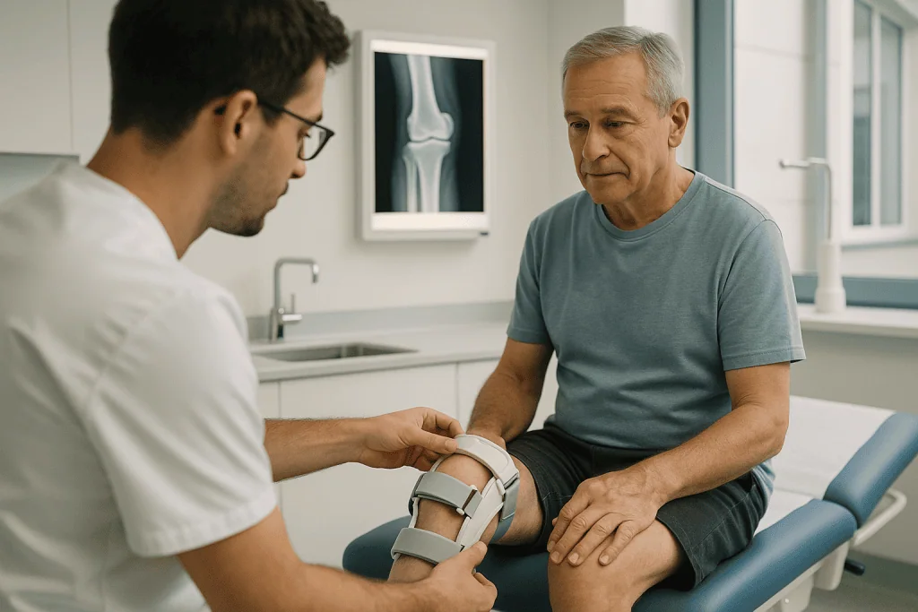
Supportive footwear and orthotics. Shoes with a firm heel counter and good arch support may reduce discomfort. Orthotics (shoe inserts designed to support the arch) may be off-the-shelf or custom-made. For more advanced adult-acquired flatfoot, a doctor may recommend an ankle brace or a custom device that supports both the foot and ankle.
Physical therapy. A therapist can guide stretching of tight calf muscles and strengthening exercises for the posterior tibial tendon and other muscles that support the arch. Exercises are often a central part of treatment for adult-acquired flatfoot in its earlier stages.
Medication. Over-the-counter pain relievers such as acetaminophen or non-steroidal anti-inflammatory drugs (NSAIDs, for example ibuprofen) may help with pain and swelling. These are generally used for short periods, and your doctor or pharmacist can advise whether they are suitable for you. Steroid injections into the posterior tibial tendon are usually avoided because they may weaken the tendon.
Immobilization. When the tendon is acutely inflamed, a short period in a walking boot or cast may be recommended to allow it to rest.
Surgery. Surgery is considered when pain and disability continue despite several months of non-surgical treatment, or when the foot has become rigid or severely deformed. The procedure depends on the cause and stage. Options a surgeon may discuss include repairing or replacing a damaged tendon using another tendon from the foot, lengthening a tight calf muscle or Achilles tendon, cutting and repositioning bones to rebuild the arch (osteotomy), removing a tarsal coalition, or fusing arthritic joints so they no longer move (arthrodesis). Recovery often involves weeks in a cast or boot without bearing weight, followed by months of rehabilitation. As with any operation, there are risks, including infection, nerve irritation, stiffness, and incomplete relief of symptoms, which your surgeon will explain in detail.
Living with flatfoot (pes planus) and outlook
For most people, flatfoot is a lifelong foot shape rather than a disease, and it does not stop them from living an active life. Children with flexible flatfoot usually grow into adults with no pain or limitation, whether or not an arch eventually develops. Many adults with flat feet participate in sports and demanding jobs without difficulty, sometimes with the help of supportive shoes or inserts.
When flatfoot does cause symptoms, the outlook depends largely on the cause and how early it is addressed. Adult-acquired flatfoot from posterior tibial tendon dysfunction tends to progress if it is ignored, so early attention to pain on the inner ankle may help preserve the arch and avoid more complex treatment later. Non-surgical care relieves symptoms for many people, though it does not restore a collapsed arch. Surgery can reduce pain and improve alignment in many cases, but results vary, recovery takes time, and some stiffness may remain, particularly after joint fusion.
Practical steps that many people find helpful include choosing well-fitting shoes with good support, replacing worn footwear, keeping calf muscles flexible, staying at a healthy weight, and paying attention to new or worsening pain rather than pushing through it. People with diabetes or reduced sensation in their feet should check their feet regularly, because changes in foot shape can lead to pressure points and skin problems.
Frequently asked questions
Is flatfoot a serious condition?
In most cases, no. Flexible flatfoot without pain is a common variation of normal anatomy and rarely leads to problems. Flatfoot becomes a medical concern mainly when it is painful, stiff, rapidly changing, or associated with another condition such as arthritis, tendon damage, or nerve disease. A doctor can help determine which situation applies.
What are the first flatfoot symptoms in adults?
Adult-acquired flatfoot often begins with aching, tenderness, or swelling along the inside of the ankle and arch, particularly after activity or prolonged standing. Some people notice that one foot looks flatter than the other or that they can no longer rise comfortably onto their toes on that side. These early flatfoot symptoms are worth discussing with a doctor because the condition may progress if untreated.
Can flatfoot be corrected without surgery?
Non-surgical flatfoot treatment such as supportive footwear, orthotics, physical therapy, and weight management often reduces pain and improves function. However, these measures generally do not rebuild an arch that has already collapsed; they support the foot rather than reshape it. Surgery is the only treatment that changes the structure of the foot, and it is reserved for people whose symptoms do not respond to other approaches.
Do children with flatfoot need special shoes or insoles?
Usually not. Research to date has not shown that special shoes or insoles change the natural development of the arch in children with flexible flatfoot. Doctors generally recommend comfortable, well-fitting footwear and suggest inserts only if a child has pain or tires easily. Rigid or painful flatfoot in a child is different and should be evaluated by a doctor.
How is flatfoot diagnosis made if I do not have pain?
Flatfoot diagnosis is largely based on physical examination, so a doctor can identify flat feet by looking at your arch while standing, checking how your heel is positioned, and asking you to stand on tiptoe. If there is no pain, no stiffness, and no other concerning finding, imaging is often unnecessary. X-rays or other scans are typically reserved for painful, rigid, or progressive cases.
Are flatfoot causes different in children and adults?
Yes. In children, flatfoot is most often inherited and related to loose ligaments, or less commonly caused by abnormal bone connections present from birth. In adults, the most frequent cause is wear or injury of the posterior tibial tendon, which supports the arch, along with arthritis, injury, or nerve damage. Because the causes differ, the approach to evaluation and treatment also differs by age.
Can flatfoot cause knee, hip, or back pain?
Flat feet can change the way the leg rotates during walking, and some people with flatfoot report pain in the knees, hips, or lower back. The connection is not fully understood, and many people with flat feet never have these problems. If you have both flat feet and pain elsewhere in the leg or back, a doctor can help decide whether the two are related.
When to see a doctor
Painless flat feet do not usually need medical attention. It is reasonable to arrange a routine appointment if you or your child have flat feet together with foot or ankle pain, tiredness in the legs that limits activity, a change in the shape of one or both feet, shoes that wear out very unevenly, or difficulty finding comfortable footwear. A child whose flat foot is stiff, painful, or affects only one side should also be evaluated.
Seek prompt medical care if you notice any of the following warning signs:
- Sudden collapse of the arch, or a foot that changes shape quickly over days or weeks
- Severe pain, swelling, or inability to bear weight after an injury
- A foot that is red, hot, and swollen, especially if you have diabetes or reduced sensation, since this may indicate infection or Charcot foot
- Numbness, tingling, or weakness in the foot or leg
- An open sore, blister, or skin breakdown on the foot that is not healing
- Fever together with foot pain and swelling
- Persistent pain that does not improve with rest and supportive footwear over several weeks
A doctor can confirm whether flatfoot is the cause of your symptoms, rule out other conditions, and discuss which treatment options are appropriate for your situation.
Medically reviewed by the Acıbadem International Medical Board September 13, 2026
See our medical review board →
Update history
- PublishedSeptember 13, 2026
- Medical review approvedSeptember 13, 2026
- Last content updateSeptember 13, 2026