Microtia
Microtia is a congenital ear difference. Learn about microtia symptoms, hearing effects, diagnosis, reconstruction and when to seek care.

Quick answer
Microtia is a congenital condition in which the external ear is underdeveloped or absent, sometimes affecting hearing on the same side. Treatment depends on the severity and may include hearing assessment, hearing support, and reconstructive surgery to improve ear shape and function, with care planned by ear, hearing, and plastic surgery specialists at Acibadem in Turkey.
Microtia is a congenital condition in which the outer ear is smaller than usual, underdeveloped, or absent at birth. It may occur with a narrow or missing ear canal, so evaluation by specialists is important to assess hearing, speech development, and reconstructive options.
Overview
Microtia is a congenital ear difference in which the external ear, also called the pinna or auricle, is smaller than usual, incompletely formed, or rarely absent. It develops before birth and is visible at delivery. Microtia can range from a mildly small ear with recognizable structures to a very small ear remnant with little typical ear shape.
Microtia most commonly affects one ear, often the right ear, but it can affect both ears. In many children, the condition is limited to the outer ear. In others, it occurs with aural atresia, meaning the ear canal is closed or absent, or with a very narrow ear canal. When the ear canal or middle ear is affected, sound may not travel normally to the inner ear, causing conductive hearing loss.
Microtia is not usually painful, and it is not caused by anything a child or parent did after birth. The main medical priorities are to evaluate hearing, support speech and language development, and consider safe reconstructive options when the child and family are ready. Care often involves a team that may include a plastic and reconstructive surgeon, ear, nose and throat specialist, audiologist, pediatrician, speech-language therapist, and sometimes genetics or craniofacial specialists.
Symptoms
The most obvious sign of microtia is an underdeveloped outer ear. The ear may be smaller, folded, positioned differently, or missing many of the normal curves and contours. Some children have a small skin-and-cartilage remnant, while others have a partially formed ear that still has some recognizable features.
Microtia is often described in grades according to severity. In milder cases, the ear is small but many structures are present. In more severe cases, only a small tissue remnant is visible, and in the rarest form, called anotia, the external ear is absent. These descriptions help doctors communicate about anatomy, but each child’s ear is assessed individually.
Possible features associated with microtia include:
- A small, misshapen, or absent external ear
- A narrow, closed, or absent ear canal
- Reduced hearing on the affected side, especially when aural atresia is present
- Skin tags or small tissue bumps near the ear or cheek
- Differences in jaw, cheek, or facial growth in some children
- Speech or listening difficulties if hearing loss is not identified and supported early
Microtia itself usually does not cause ear pain or infection. However, if there is a narrow ear canal, wax buildup or infections may occur more easily. Families may also notice practical challenges such as difficulty positioning glasses, hearing devices, or masks, depending on the ear shape.
Causes & Risk Factors
Microtia occurs when the outer ear does not form typically during early pregnancy. In most cases, the exact cause is not known. Ear development is complex and involves genetic signals, blood supply, and environmental influences during a very early stage of fetal growth. For many families, microtia happens unexpectedly and is not linked to a clear inherited pattern.
Some cases occur as part of a broader craniofacial or genetic syndrome, especially when there are differences involving the jaw, cheek, eye, spine, heart, kidneys, or both ears. Conditions that may include microtia can vary widely in severity, so doctors look for additional signs during the newborn and pediatric assessment.
Risk factors reported in medical literature include a family history of ear differences, certain maternal health conditions, and some exposures during pregnancy. However, having a risk factor does not mean a child will develop microtia, and many children with microtia have no identifiable risk factor. Parents should not assume blame; the condition is typically related to early developmental processes outside their control.
If microtia appears along with other physical findings, a pediatrician or specialist may recommend genetic counseling or additional evaluations. This is not because every child with microtia has a syndrome, but because identifying associated conditions early can guide safe, complete care.
Diagnosis
Microtia is usually diagnosed by physical examination soon after birth. A doctor examines the shape of the ear, the position of the ear, the ear canal opening, facial symmetry, and any skin tags or other visible findings. The child’s general health and development are also reviewed, because microtia can occasionally be associated with other congenital differences.
Hearing assessment is an essential part of diagnosis. Newborn hearing screening may suggest whether the inner ear is receiving sound normally, but children with a closed ear canal may need additional tests by an audiologist. These tests help determine whether hearing loss is present, whether it affects one or both ears, and what type of hearing support may be helpful.
Imaging, such as a CT scan of the temporal bone, may be considered later in childhood if ear canal surgery or detailed middle ear evaluation is being discussed. It is not always needed in infancy, and timing is chosen carefully to balance the value of information with the child’s age and clinical needs. Imaging can show the ear canal, middle ear bones, and nearby structures important for surgical planning.
Depending on the examination, doctors may recommend checks for related conditions, such as kidney ultrasound, heart evaluation, eye examination, or spine assessment. These tests are not required for every child, but they may be useful when there are additional signs or when microtia is bilateral or syndromic.
Treatment Options
Microtia treatment is individualized. The right approach depends on the degree of ear difference, whether aural atresia is present, hearing test results, the child’s age and growth, skin and cartilage availability, family preferences, and the experience of the treating team. A specialist should assess the child before any treatment plan is chosen.
Hearing care is often the first priority, especially when the ear canal is absent or both ears are affected. Options may include regular hearing monitoring, bone-conduction hearing devices, classroom listening support, and speech-language support when needed. Early hearing management helps children develop communication skills and reduces listening strain at home and school.
Ear reconstruction can be considered for cosmetic and functional reasons, such as improving ear shape or helping with glasses and hearing device placement. General reconstructive categories include autologous reconstruction using the child’s own rib cartilage, implant-based reconstruction using a biocompatible framework covered by tissue, and a removable prosthetic ear designed to match the other side. Each option has advantages, limitations, timing considerations, and possible risks, so families should discuss them carefully with a reconstructive surgeon.
In selected children with aural atresia, ear canal reconstruction may be considered by an ENT surgeon with expertise in this area. This surgery is not suitable for every child, because success depends on middle ear anatomy, hearing potential, and long-term canal care. When both hearing and outer ear reconstruction are planned, coordination between ENT and plastic surgery teams is important so one procedure does not compromise another.
Living With / Prognosis
Many children with microtia grow, learn, and participate in daily life very well, particularly when hearing needs are identified and supported early. If only one ear is affected and the other ear hears normally, speech development is often good, but some children may still have difficulty locating sounds or hearing in noisy classrooms. Regular audiology follow-up helps families and teachers understand the child’s listening needs.
Emotional and social support is also important. Some children become aware of visible differences as they start school or social activities. Age-appropriate explanations, supportive teachers, and access to peer or family counseling can help build confidence. Decisions about reconstruction should include the child’s voice when developmentally appropriate, as treatment often occurs over several years.
Long-term follow-up may include monitoring facial growth, hearing, speech, ear canal health if present, and the condition of any reconstruction or prosthesis. Children with bilateral microtia or associated syndromes may require more coordinated care, but a planned multidisciplinary approach can address medical, developmental, and reconstructive needs step by step.
For international families seeking coordinated evaluation, Acibadem International’s multidisciplinary specialists and JCI-accredited hospitals diagnose and treat microtia, including hearing assessment and reconstructive planning. As with any medical condition, expected results and treatment choices should be discussed directly with qualified specialists after a full assessment.
When to See a Doctor
A baby with microtia should be evaluated by a pediatrician and referred for hearing assessment as early as possible. Even when the child appears to respond to sounds, formal testing is important because hearing loss in one ear can still affect sound localization and listening in noisy environments. Early audiology care is especially urgent if both ears are affected.
Families should seek specialist advice if there is no visible ear canal, if hearing screening is not passed, if speech or language milestones seem delayed, or if the child has repeated ear infections or wax problems in a narrow canal. Medical review is also important if there are other findings, such as facial asymmetry, jaw differences, eye concerns, kidney problems, heart concerns, or spine differences.
A consultation with a plastic and reconstructive surgeon or craniofacial team can help families understand future options, even if reconstruction is not planned immediately. Early counseling can clarify timing, treatment pathways, and how hearing devices or future surgeries may interact.
Urgent medical care is rarely needed for microtia itself. However, prompt evaluation is appropriate for fever with ear pain, drainage from an ear canal, sudden change in hearing, dizziness, or any concern about the child’s general health. Parents should always consult a qualified doctor rather than delaying care or relying only on online information.
Frequently asked questions
What is microtia?
Microtia is a congenital condition in which the outer ear is small, underdeveloped, or absent at birth. It may affect one ear or both ears. Some children also have a closed or missing ear canal, which can affect hearing.
Does microtia always cause hearing loss?
Microtia does not always cause significant hearing loss, but hearing loss is common when the ear canal is narrow, closed, or absent. The inner ear may still work normally, while sound cannot pass through the outer or middle ear effectively. A formal hearing test is needed to understand the child’s hearing status.
Can microtia be corrected?
Microtia can often be improved with reconstructive treatment, but the best option depends on the child’s anatomy, age, hearing needs, and family preferences. Options may include reconstruction using rib cartilage, implant-based reconstruction, or a prosthetic ear. A specialist can explain realistic goals, timing, benefits, and risks.
When is ear reconstruction usually considered for microtia?
Timing varies by technique and by the child’s growth and medical situation. Some methods are usually considered when the child is older and has enough tissue or cartilage, while other approaches may be possible earlier in selected cases. The timing should be decided with an experienced reconstructive surgeon and coordinated with hearing care.
Is microtia inherited?
Most cases of microtia occur without a clear inherited cause. A family history can increase the chance in some cases, and microtia can sometimes be part of a genetic or craniofacial syndrome. Genetic counseling may be recommended if both ears are affected, if there are other physical findings, or if there is a family history.
Will a child with microtia have normal speech development?
Many children with microtia develop normal speech, especially when one ear hears normally and hearing needs are monitored. However, hearing loss in one or both ears can affect listening, speech, and learning if it is not supported. Early audiology evaluation and appropriate hearing support help protect communication development.
Which doctors treat microtia?
Microtia is often managed by a multidisciplinary team. This may include a pediatrician, plastic and reconstructive surgeon, ENT specialist, audiologist, speech-language therapist, and sometimes a geneticist or craniofacial specialist. Team-based care helps coordinate hearing, development, and reconstruction safely.
This article is for general information only and is not a substitute for professional medical advice. Please consult a qualified doctor about your individual situation.
Medically reviewed by the Acıbadem International Medical Board June 20, 2026
See our medical review board →
Update history
- PublishedJune 8, 2026
- Medical review approvedJune 20, 2026
- Last content updateJune 12, 2026
Treatments for This Condition
Care at Acibadem
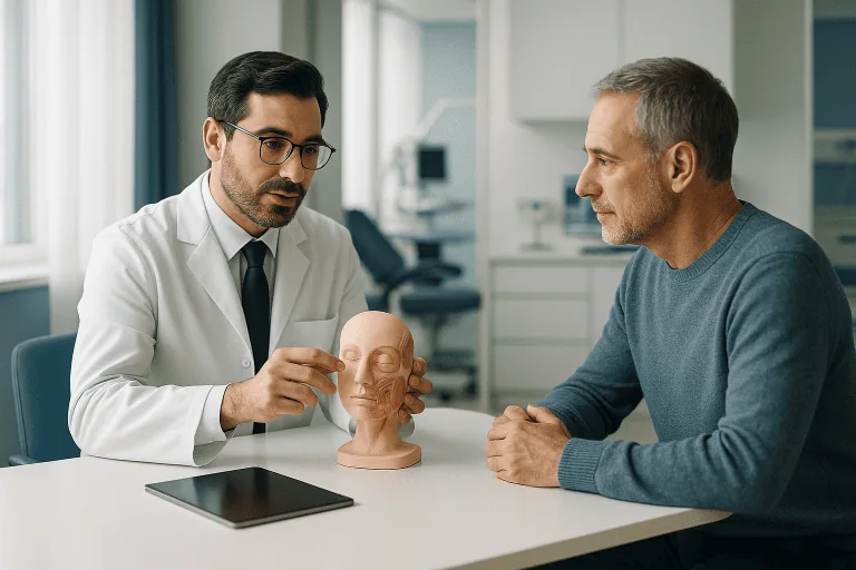
Doctors Who Treat This Condition



Aesthetic Plastic & Reconstructive Surgery
Prof. Mehmet Veli Karaaltın, MD

Aesthetic Plastic & Reconstructive Surgery
Assoc. Prof. Ahmet Küçükçelebi, MD

Aesthetic Plastic & Reconstructive Surgery
Assoc. Prof. Erdem Güven, MD
Aesthetic Plastic & Reconstructive Surgery
Assoc. Prof. Mehmet Altıparmak, MD

Aesthetic Plastic & Reconstructive Surgery