Thymoma
Thymoma is a rare tumor of the thymus. Learn about thymoma symptoms, diagnosis, treatment options and when to see a specialist.

Quick answer
Thymoma is a tumor that develops in the thymus gland in the chest and may be found incidentally or cause symptoms such as cough, chest pain, or signs linked to myasthenia gravis. At Acibadem in Turkey, evaluation typically includes imaging and tissue diagnosis, and treatment depends on the stage and type, often combining surgery with radiotherapy, chemotherapy, or other therapies…
Thymoma is a rare tumor that starts in the epithelial cells of the thymus, a small immune-system organ in the upper chest behind the breastbone. It often grows slowly, but it can spread locally or recur, so specialist diagnosis, staging and long-term follow-up are important.
Overview
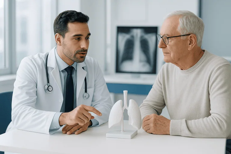
Thymoma is a rare tumor of the thymus gland, an organ located in the front of the chest, behind the breastbone and between the lungs. The thymus is most active in childhood, when it helps the immune system develop T lymphocytes, a type of white blood cell. In adulthood, the thymus usually becomes smaller and less active, but thymic cells can still give rise to tumors.
Most thymomas begin in the epithelial cells that form the structure of the thymus. They are different from thymic carcinoma, which is usually more aggressive, and from lymphomas or germ cell tumors that can also occur in the same chest area. Thymoma is often described as slow growing, but it is still considered a tumor with malignant potential because it can invade nearby tissues, spread within the chest or recur after treatment.
Many thymomas are found in the anterior mediastinum, the front compartment of the central chest. Some are discovered by chance during imaging performed for another reason. Others are found because they cause chest symptoms or are associated with immune-related conditions, especially myasthenia gravis. A careful evaluation by specialists helps determine whether the tumor is localized, whether it can be removed safely, and which treatment approach is most appropriate.
Symptoms
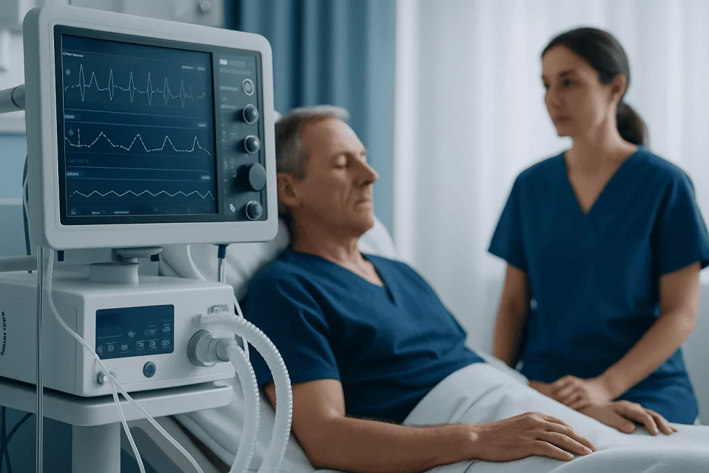
Thymoma symptoms vary widely. Some people have no symptoms, and the tumor is detected incidentally on a chest X-ray, CT scan or other imaging test. When symptoms occur, they often happen because the tumor presses on nearby structures in the chest, such as the lungs, airways, blood vessels or nerves.
Possible symptoms of thymoma include persistent cough, chest pain or pressure, shortness of breath, wheezing, hoarseness or difficulty swallowing. Less commonly, swelling of the face, neck or upper chest may occur if a large tumor affects blood flow through major veins. These symptoms can also be caused by many non-cancerous conditions, so they require medical evaluation rather than self-diagnosis.
Thymoma is notable for its association with autoimmune and immune-system disorders. The most common is myasthenia gravis, which can cause muscle weakness that worsens with activity and improves with rest. Symptoms of myasthenia gravis may include drooping eyelids, double vision, difficulty chewing or swallowing, slurred speech, weakness in the arms or legs, and in severe cases breathing difficulty. Other less common associated conditions include pure red cell aplasia, which can cause anemia-related fatigue, and hypogammaglobulinemia, which may increase susceptibility to infections.
Causes & Risk Factors
The exact cause of thymoma is not known. In most people, it develops without a clear inherited or lifestyle-related trigger. Unlike some cancers, thymoma is not strongly linked to smoking, diet or a specific environmental exposure. It is also not contagious and cannot be passed from one person to another.
Thymoma is more often diagnosed in adults than in children, and it is usually found in middle-aged or older adults. It can occur in both men and women. Because the tumor is rare, many people have never heard of it before diagnosis, and it is commonly evaluated in centers with experience in thoracic tumors and mediastinal masses.
The most important clinical association is with immune dysregulation. A person with myasthenia gravis may be evaluated for thymic disease, including thymoma, because the thymus can play a role in abnormal immune signaling. Conversely, a person diagnosed with thymoma may be assessed for symptoms or laboratory signs of autoimmune conditions. Having one of these immune conditions does not mean thymoma is always present, but it can guide the medical team toward appropriate testing.
- Known risk factors are limited and not fully understood.
- Age and thymus-related immune abnormalities may be relevant in some cases.
- Family history is not usually a major factor.
- There is no proven screening test for the general population.
Diagnosis
Diagnosis of thymoma usually begins with imaging of the chest. A chest X-ray may show an abnormal shadow, but a CT scan with contrast is commonly used to define the size, location and relationship of the tumor to nearby structures. MRI may be helpful in selected cases, particularly when more detail is needed about invasion into surrounding tissues. PET imaging may be considered when doctors need more information about metabolic activity or possible spread, although its role depends on the individual situation.
Doctors also assess symptoms, medical history and physical examination findings, especially signs of myasthenia gravis or other immune-related conditions. Blood tests may be ordered to evaluate anemia, immune function or antibodies associated with myasthenia gravis. Pulmonary function tests or cardiac assessment may be recommended before surgery or other treatments, especially if the tumor is near important chest structures or the person has other health conditions.
A tissue diagnosis is sometimes obtained with a needle biopsy or surgical biopsy, particularly if the tumor cannot be removed immediately, if another type of mediastinal tumor is possible, or if systemic treatment is being considered first. In some cases where imaging strongly suggests a resectable thymoma, the medical team may proceed directly to surgical removal and confirm the diagnosis by pathology afterward. The decision about biopsy must be individualized, because planning the safest and most effective procedure is important.
Pathologists examine the tumor under a microscope and classify it using recognized systems, such as the World Health Organization histologic classification. The tumor is also staged, commonly using systems such as Masaoka-Koga or TNM staging, which describe whether it is confined to the thymus, invades nearby tissues or has spread. Stage and completeness of removal are key factors in planning treatment and follow-up.
Treatment Options
Thymoma treatment depends on the stage of the tumor, whether it can be completely removed, its microscopic features, associated conditions such as myasthenia gravis, and the person’s overall health. The right approach is decided by a specialist team after assessment. This team may include thoracic surgeons, medical oncologists, radiation oncologists, neurologists, radiologists, pathologists and anesthesiology or intensive care specialists when needed.
Surgery is a central treatment for many thymomas, especially when the tumor appears localized and can be removed safely. The operation often involves removal of the thymus and the tumor, and sometimes nearby tissue if there is local invasion. Surgical planning may use open, minimally invasive or robotic-assisted approaches depending on tumor size, location, extent and the expertise of the surgical team. If myasthenia gravis is present, neurologic stabilization before and after surgery is especially important.
Radiotherapy may be recommended after surgery in some cases, particularly if there is concern about microscopic residual disease, invasion into surrounding tissues or a higher risk of local recurrence. It may also be used when surgery is not possible or as part of combined treatment for more advanced disease. Systemic therapy, such as chemotherapy or other drug-based treatment, may be considered for tumors that are advanced, unresectable, recurrent or need to be reduced in size before surgery. The choice of systemic treatment is made by oncology specialists based on tumor behavior and patient-specific factors.
Some very small, stable or uncertain thymic abnormalities may be monitored with planned imaging when the medical team judges that immediate intervention is not needed. However, confirmed or suspected thymoma should not be ignored, because careful staging and follow-up are essential. Supportive care may include treatment for myasthenia gravis, rehabilitation after surgery, nutrition support, breathing exercises, pain control and management of treatment side effects.
Living With / Prognosis
Living with thymoma often involves a period of testing, treatment planning and long-term surveillance. Many thymomas can be treated effectively, particularly when found at an early stage and removed completely. Prognosis varies from person to person and depends on stage, tumor type, whether complete surgical removal was possible, response to therapy, and the presence of associated autoimmune conditions.
Follow-up is important because thymoma can recur, sometimes years after treatment. Follow-up care may include periodic chest imaging, physical examinations and review of symptoms. The schedule is individualized by the treating team, and it may be longer than for some other tumors because late recurrence is recognized in thymoma care.
People with thymoma and myasthenia gravis may need coordinated follow-up with both oncology or thoracic surgery specialists and a neurologist. Muscle weakness, swallowing difficulty or breathing changes should be communicated promptly, because these symptoms may need specific neurologic management. Emotional support is also important; a rare diagnosis can feel isolating, and clear explanations from the care team can help patients and families make informed decisions.
Healthy daily habits cannot replace medical treatment, but they can support recovery and general wellbeing. These include staying physically active as advised, following breathing or rehabilitation exercises if prescribed, eating a balanced diet, avoiding tobacco smoke, keeping vaccinations and infection-prevention advice up to date when appropriate, and attending all follow-up visits. Acibadem International’s multidisciplinary specialists and JCI-accredited hospitals diagnose and treat thymoma for international patients, with care plans tailored after specialist assessment.
When to See a Doctor
A doctor should evaluate persistent or unexplained chest symptoms, especially cough, chest discomfort, shortness of breath, hoarseness or difficulty swallowing. These symptoms do not necessarily mean thymoma, but they should be assessed if they do not resolve, worsen or occur with abnormal imaging findings. A person who has been told there is a mediastinal mass or thymic lesion on imaging should be referred to an appropriate specialist for further evaluation.
Medical attention is also important for symptoms suggestive of myasthenia gravis, such as drooping eyelids, double vision, weakness that worsens during the day, difficulty chewing, swallowing problems or unusual fatigue of the arms and legs. Breathing difficulty, rapidly worsening weakness, severe chest pain or swelling of the face and neck requires urgent medical assessment. These situations need prompt evaluation to identify the cause and provide safe treatment.
Anyone previously treated for thymoma should contact their care team if new chest symptoms, unexplained weight loss, persistent fatigue, neurologic symptoms or recurrent infections develop. Regular follow-up appointments should be kept even when a person feels well, because imaging and clinical assessment can detect changes before symptoms appear. Decisions about testing, treatment and follow-up should always be made with a qualified healthcare professional familiar with thymic tumors.
Frequently asked questions
What is thymoma?
Thymoma is a rare tumor that begins in the epithelial cells of the thymus gland, located in the front part of the chest. It often grows slowly, but it can invade nearby tissues or recur, so it requires specialist evaluation and follow-up.
Is thymoma cancer?
Thymoma is generally considered a tumor with malignant potential because it can spread locally, recur or, less commonly, spread beyond the chest. It is different from thymic carcinoma, which is typically more aggressive. The exact risk depends on the stage, tumor features and completeness of treatment.
What are the most common thymoma symptoms?
Some people have no symptoms and the tumor is found incidentally on imaging. When symptoms occur, they may include cough, chest pressure, shortness of breath, hoarseness or difficulty swallowing. Thymoma may also be associated with myasthenia gravis, which can cause drooping eyelids, double vision and muscle weakness.
How is thymoma diagnosed?
Diagnosis usually involves chest imaging such as CT, and sometimes MRI or PET imaging. Doctors may also order blood tests and neurologic evaluation if myasthenia gravis is suspected. Tissue confirmation may come from biopsy or from pathology after surgical removal, depending on the case.
What is the treatment for thymoma?
Treatment may include surgery, radiotherapy, systemic therapy or observation in selected situations. Surgery is commonly used when the tumor can be removed safely, while radiotherapy or drug-based treatments may be considered for higher-risk, advanced or recurrent disease. The treatment plan should be decided by a specialist team after full assessment.
Can thymoma come back after treatment?
Yes, thymoma can recur, including years after treatment. This is why long-term follow-up with periodic imaging and clinical review is important. The follow-up schedule depends on the original stage, treatment received and the patient’s overall condition.
Is thymoma related to myasthenia gravis?
Thymoma is associated with myasthenia gravis in some patients, although not everyone with thymoma has this condition and not everyone with myasthenia gravis has thymoma. Myasthenia gravis affects communication between nerves and muscles, causing fluctuating weakness. If symptoms suggest it, a neurologist can help confirm the diagnosis and guide management.
This article is for general information only and is not a substitute for professional medical advice. Please consult a qualified doctor about your individual situation.
Medically reviewed by the Acıbadem International Medical Board June 20, 2026
See our medical review board →
Update history
- PublishedJune 9, 2026
- Medical review approvedJune 20, 2026
- Last content updateJune 9, 2026