3D Mammography: Breast Tomosynthesis for Screening and Diagnosis

3D mammography takes multiple low-dose X-ray images from different angles to create a layered view of the breast. It is used for both screening and diagnostic breast imaging, often together with 2D mammography or a synthetic 2D image.
Key Takeaways
- 3D mammography takes multiple low-dose X-ray images from different angles to create a layered view of the breast.
- It is used for both screening and diagnostic breast imaging, often together with 2D mammography or a synthetic 2D image.
- Breast tomosynthesis may reduce callbacks and can be especially helpful for people with dense breast tissue.
- The radiation dose is kept within accepted safety standards, and the benefit of early detection is considered important for eligible patients.
- A suspicious mammogram does not mean cancer; additional imaging or biopsy may be needed to confirm a diagnosis.
3D mammography, also called digital breast tomosynthesis, is an advanced breast imaging technique that creates thin, layered images of breast tissue. It can support routine breast cancer screening and help doctors evaluate symptoms or unclear findings more accurately.
Overview
3D mammography is a modern form of breast imaging used to look for early signs of breast cancer and to investigate breast symptoms. Its medical name is digital breast tomosynthesis. Unlike a standard 2D mammogram, which produces flat images, 3D mammography captures multiple images from different angles and reconstructs them into thin layers that a radiologist can review one by one.
This layered view can make it easier to see small cancers that may be hidden by overlapping breast tissue. It can also help distinguish normal tissue patterns from findings that need further assessment. For many patients, this means clearer information and, in some situations, fewer repeat imaging appointments after a screening mammogram.
3D mammography is not a separate test for a different disease; it is an advanced way of performing mammographic breast imaging. It may be used as part of routine screening for people with no symptoms, or as part of diagnostic evaluation when there is a breast lump, nipple change, pain in one area, or an abnormal result on another imaging test.
How Breast Tomosynthesis Works
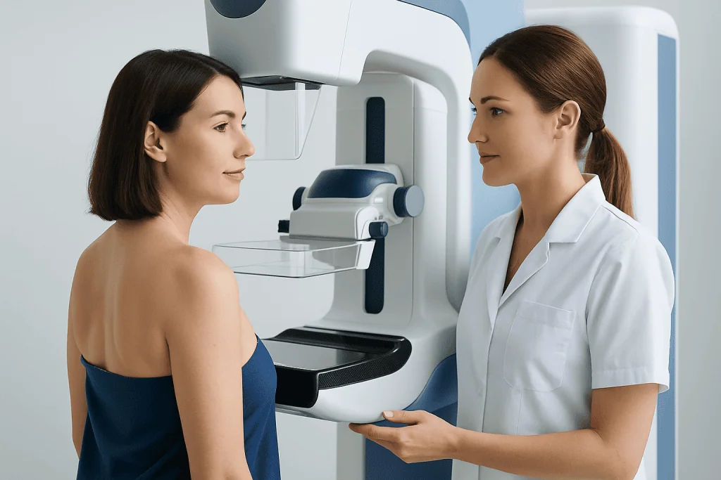
During 3D mammography, the breast is gently compressed between two plates, similar to a conventional mammogram. The X-ray tube moves in an arc over the breast, taking a series of low-dose images from several angles. Computer software then reconstructs these images into thin slices, usually about 1 millimeter thick, allowing the radiologist to examine the breast tissue layer by layer.
The process helps reduce the visual effect of tissue overlap. In a 2D mammogram, structures at different depths can appear stacked on top of each other, which may obscure a small abnormality or create the appearance of a false suspicious area. With tomosynthesis, radiologists can scroll through the tissue and better assess the shape, location, and borders of a finding.
In many centers, 3D mammography is performed together with a 2D mammogram. Some systems can create a synthetic 2D image from the 3D data, which may reduce the need for separate 2D exposures. The exact technique depends on the equipment, clinical question, national guidelines, and the radiologist’s protocol.
Screening and Diagnostic Uses
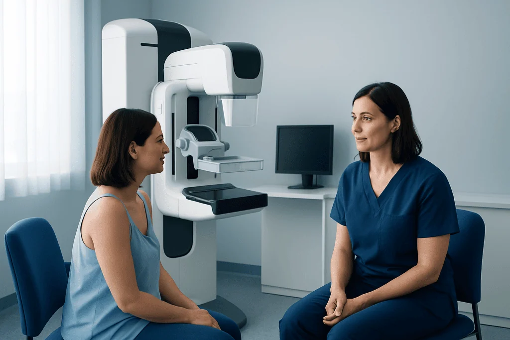
Screening mammography is performed when a person has no breast symptoms, with the goal of detecting cancer at an early and more treatable stage. 3D mammography may be offered as a screening option for many women, depending on age, risk factors, breast density, local availability, and clinical guidance. Screening schedules vary between countries and professional organizations, so individuals should discuss the best timing with their doctor.
Diagnostic mammography is different. It is used to evaluate a specific concern, such as a breast lump, nipple discharge, skin thickening, focal pain, or an abnormal screening result. In this setting, breast tomosynthesis may be combined with additional mammographic views, targeted breast ultrasound, or other imaging to clarify whether a finding is benign, probably benign, or suspicious.
3D mammography does not replace all other breast imaging tests. Ultrasound can be useful for evaluating cysts, solid lumps, and findings in dense breasts. Breast MRI may be recommended for certain high-risk patients or for specific diagnostic and treatment-planning situations. If imaging remains suspicious, a biopsy is the standard method for confirming whether cancer or another condition is present.
Benefits and Limitations
A key benefit of 3D mammography is improved visualization of breast tissue, particularly when the tissue is dense. Dense breast tissue and many breast cancers both appear white on mammograms, which can make interpretation more challenging. By separating tissue into layers, tomosynthesis can help radiologists detect abnormalities that may be less visible on 2D images.
Breast tomosynthesis may also reduce the number of false-positive callbacks, which occur when a screening mammogram suggests a possible abnormality that later proves to be benign. Fewer callbacks can reduce anxiety, additional imaging, and time away from daily activities. However, callbacks still happen, and they are an important part of careful screening when something needs a closer look.
Like all screening tests, 3D mammography has limitations. It cannot detect every breast cancer, and some findings may still be uncertain. It can identify very slow-growing cancers that may never have caused harm, a concept known as overdiagnosis. It may also lead to additional testing, including biopsy, for findings that turn out not to be cancer.
Patients should understand that an abnormal mammogram is not a diagnosis of cancer. Many mammographic findings are benign, such as cysts, fibroadenomas, calcifications, or overlapping normal tissue. The purpose of follow-up imaging is to reach a clearer answer and guide the next appropriate step.
Who May Benefit and Safety Considerations
3D mammography may be particularly helpful for people with dense breasts, those who have had previous callbacks due to overlapping tissue, and patients undergoing diagnostic evaluation of a subtle mammographic finding. It can also be used in routine screening for average-risk patients when available and appropriate. The decision should consider personal risk factors, previous imaging history, and local screening recommendations.
Breast density is common and is usually not something a person can determine by touch. It is assessed on mammography and reported by the radiologist. Dense breast tissue can slightly increase breast cancer risk and can make cancers harder to see on mammograms. Some patients with dense breasts may be advised to consider supplemental imaging, such as ultrasound or MRI, depending on their overall risk profile.
3D mammography uses ionizing radiation, but the dose is low and regulated. When 3D images are combined with synthetic 2D images, the dose can be similar to or only modestly different from standard mammography, depending on the system. For eligible patients, the potential benefit of early breast cancer detection generally outweighs the small radiation risk.
Pregnancy is an important safety consideration. Routine screening mammography is usually postponed during pregnancy unless there is a specific medical reason. If a pregnant or possibly pregnant patient has a breast symptom, they should tell the imaging team and doctor before the exam so the safest and most appropriate diagnostic pathway can be chosen.
Preparing for the Exam and What to Expect
Preparation for 3D mammography is simple. Patients are usually advised not to use deodorant, powder, lotion, or perfume on the underarms or breast area on the day of the exam, because these products can sometimes appear as artifacts on images. If previous mammograms were performed at another facility, bringing or transferring those images can help the radiologist compare changes over time.
During the exam, a mammography technologist positions the breast on the imaging platform and applies compression for a short time. Compression may feel uncomfortable, but it should not be severely painful. It is necessary because it spreads the tissue, reduces motion, improves image quality, and helps use the lowest reasonable radiation dose.
Most breast tomosynthesis appointments are brief. Each compression lasts only seconds, although the full visit may take longer for registration, positioning, and image review. For screening, images are typically taken of both breasts from standard angles. For diagnostic imaging, additional views may be taken, and the radiologist may request an ultrasound during the same visit.
Patients who have breast implants can still have mammography, but they should inform the scheduling team and technologist. Special implant-displacement views may be used to see as much breast tissue as possible. People with limited shoulder movement, prior breast surgery, or significant tenderness should also tell the technologist so positioning can be adapted when possible.
Results, Follow-Up, and When to See a Doctor
Mammography results are usually reported using a standardized system called BI-RADS, which helps describe whether findings are negative, benign, probably benign, suspicious, or highly suggestive of malignancy. A screening result may be normal, or it may recommend additional imaging. A diagnostic result may lead to short-term follow-up, biopsy, or routine screening, depending on the level of concern.
Patients should contact a qualified doctor promptly if they notice a new breast lump, persistent focal breast pain, nipple discharge that is bloody or occurs without squeezing, nipple inversion, skin dimpling, redness, thickening, or a change in breast size or shape. These symptoms do not necessarily mean cancer, but they should be evaluated. A normal recent mammogram should not delay assessment of a new or persistent symptom.
Follow-up is an important part of breast health. If additional imaging or biopsy is recommended, it is best to complete it within the timeframe advised by the doctor or radiologist. Keeping copies of reports, understanding personal breast density, and sharing family history can help clinicians tailor screening and diagnostic decisions.
For international patients seeking coordinated breast imaging and care, Acibadem International’s multidisciplinary specialists and JCI-accredited hospitals diagnose and treat breast conditions, including the use of advanced imaging such as breast tomosynthesis when appropriate. Patients should always discuss their individual screening plan and results with a qualified healthcare professional.
Frequently asked questions
Is 3D mammography better than regular mammography?
3D mammography can provide a clearer layered view of breast tissue than standard 2D mammography. It may improve detection of some breast cancers and reduce false-positive callbacks, especially in dense breasts. However, the best test depends on the patient’s age, breast density, risk factors, symptoms, and local medical guidance.
Does 3D mammography hurt?
The exam involves breast compression, which can feel tight or uncomfortable for a short time. Compression usually lasts only seconds for each image and is important for image quality and safety. Patients should tell the technologist if they feel significant pain so positioning can be adjusted when possible.
Is the radiation from breast tomosynthesis safe?
3D mammography uses low-dose X-rays, and modern systems are designed to keep exposure within accepted safety standards. In many cases, synthetic 2D images can be created from the 3D data to help manage dose. Patients who are pregnant or may be pregnant should inform their doctor and the imaging team before the exam.
Who should consider 3D mammography?
It may be considered for routine screening in many eligible women and can be especially useful for those with dense breast tissue or prior callbacks from overlapping tissue. It is also used in diagnostic evaluation when a symptom or unclear mammogram finding needs more detail. The decision should be made with a healthcare professional based on individual risk and local recommendations.
Can 3D mammography detect all breast cancers?
No imaging test can detect every breast cancer. 3D mammography improves tissue visualization, but some cancers may still be difficult to see, particularly in very dense breasts or certain tumor types. New breast symptoms should be evaluated even if a recent mammogram was normal.
What happens if my 3D mammogram is abnormal?
An abnormal result often means that the radiologist needs more information, not that cancer has been diagnosed. Additional mammographic views, ultrasound, MRI, or comparison with prior images may be recommended. If a finding remains suspicious, a biopsy may be performed to provide a definitive diagnosis.
References
- American College of Radiology
- Radiological Society of North America
- National Cancer Institute
- World Health Organization
- European Society of Breast Imaging
This article is for general information only and is not a substitute for professional medical advice. Please consult a qualified doctor about your individual situation.
Oncology care in Turkey — second opinion and treatment plan
JCI-accredited · board-certified surgeons · reply within 24h
Add us as a Preferred Source to see more of our trusted health content across Google Search, AI Overviews and Discover.