Actinic Keratosis: Diagnosis, Outlook, and Modern Treatment Approaches

Actinic keratosis usually appears on sun-exposed skin such as the face, scalp, ears, hands, and forearms. These lesions are caused by cumulative ultraviolet damage and are more common with age and fair skin, but they can affect anyone.
Key Takeaways
- Actinic keratosis usually appears on sun-exposed skin such as the face, scalp, ears, hands, and forearms.
- These lesions are caused by cumulative ultraviolet damage and are more common with age and fair skin, but they can affect anyone.
- A dermatologist often diagnoses actinic keratosis by examination, and sometimes a skin biopsy is needed to confirm it.
- Treatment may include freezing, topical medicines, light-based therapy, or removal of suspicious lesions.
- Daily sun protection and regular skin checks help prevent new lesions and support early detection.
Actinic keratosis is a common rough, scaly skin patch caused by long-term sun exposure. It is considered a precancerous lesion, which means prompt evaluation and appropriate treatment can reduce the chance of progression to skin cancer.
Overview: what actinic keratosis means
Actinic keratosis is a rough, scaly patch or spot on the skin caused by years of ultraviolet (UV) exposure from sunlight or tanning devices. It is not always dangerous in the short term, but it matters because it is considered a precancerous lesion. That means some lesions can develop into a type of skin cancer called squamous cell carcinoma if they are not monitored or treated.
These spots often develop slowly and may be easier to feel than to see at first. Many people notice a sandpaper-like area on the face, scalp, ears, lips, forearms, or backs of the hands. The skin may look red, pink, tan, brown, or skin-colored, depending on the person’s natural skin tone and the lesion itself.
Actinic keratosis is very common, especially in adults with a long history of sun exposure. Having one lesion also suggests that the surrounding skin has been affected by sun damage, sometimes called “field damage” or “field cancerization.” This is one reason a dermatologist may recommend treating not only the visible spot but also the nearby sun-damaged area.
Because actinic keratosis can resemble other skin conditions, expert assessment is useful. Some lesions stay stable or even go away on their own, but others return or change over time. Regular follow-up helps identify lesions that need closer attention.
Symptoms and how it may look or feel
Actinic keratosis often begins as a small, dry, crusty, or scaly patch. It may feel rough, tender, itchy, or mildly irritated. In some people, the main symptom is texture rather than color, making the lesion easier to notice by touch while washing the face or applying skin care products.
Appearance can vary. Some lesions are flat; others are slightly raised. A few become thick and wart-like. On the lips, a related sun-damage change called actinic cheilitis can cause persistent dryness, cracking, or a rough pale patch. This area deserves careful review because chronic lip lesions can sometimes behave differently from lesions on other parts of the skin.
Common features include:
- Rough, scaly, or crusted skin patch
- Pink, red, tan, brown, gray, or skin-colored spot
- Burning, stinging, or tenderness
- Lesion on sun-exposed areas such as the scalp, face, ears, neck, forearms, and hands
- Persistent spot that does not fully heal
Although actinic keratosis can be subtle, certain changes deserve prompt attention. A lesion that becomes thicker, more painful, bleeds easily, ulcerates, or grows quickly may need urgent assessment to rule out skin cancer, including skin cancer.
Causes and risk factors

The main cause of actinic keratosis is cumulative UV radiation damage to skin cells. This damage builds up over time, so lesions often appear later in adulthood, even though the harmful sun exposure started years earlier. Both natural sunlight and artificial UV sources, such as tanning beds, can contribute.
Risk is influenced by a combination of skin type, lifestyle, environment, and immune health. People with fair skin, light eyes, blond or red hair, or a tendency to burn rather than tan have a higher risk, but darker skin tones do not eliminate the possibility. Living in sunny climates, spending many hours outdoors for work or recreation, and having a history of severe sunburns also increase the chance of developing these lesions.
Other factors that raise risk include:
- Older age
- Male sex, partly due to cumulative outdoor exposure in some populations
- Weakened immune system, including after organ transplantation or during certain medical treatments
- Previous actinic keratoses or previous nonmelanoma skin cancers
- Use of tanning beds
- Balding scalp or thinning hair that leaves skin less protected
Actinic keratosis is part of a broader pattern of chronic sun damage. A person who develops these lesions may also have other sun-related skin changes or conditions, such as basal cell carcinoma, so a full skin examination can be helpful.
How diagnosis is made
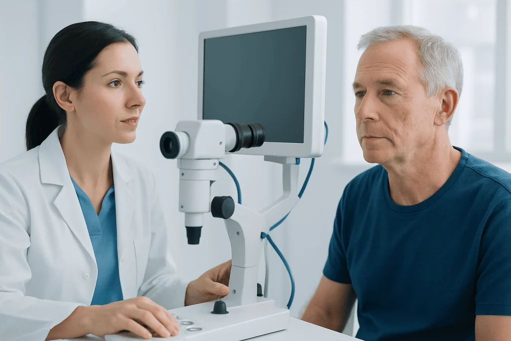
Diagnosis usually begins with a clinical skin examination by a dermatologist or other qualified doctor. The clinician looks at the lesion’s texture, borders, thickness, color, and location, while also checking the surrounding skin for signs of additional sun damage. In many cases, an experienced specialist can identify actinic keratosis based on appearance and feel.
If the spot is unusually thick, inflamed, tender, rapidly changing, or not responding to standard treatment, a biopsy may be recommended. A biopsy involves removing part or all of the lesion so it can be examined under a microscope. This helps confirm the diagnosis and exclude squamous cell carcinoma or another skin condition.
Diagnosis is not only about naming a single lesion. It also includes evaluating how widespread the sun damage is, whether there are multiple lesions, and whether treatment should target one spot or a broader area of affected skin. This is important because visible lesions may represent only part of the total sun injury.
In specialist settings, diagnostic planning may involve dermoscopy, a noninvasive tool that helps visualize surface and subsurface skin structures. If a suspicious lesion needs removal or tissue sampling, this may be arranged through dermatology evaluation and treatment or, when tissue confirmation is necessary, skin biopsy.
Modern treatment approaches and outlook
Treatment aims to remove visible actinic keratoses, reduce the risk of progression to skin cancer, relieve symptoms, and treat the wider area of sun-damaged skin when needed. The right approach depends on the number of lesions, their thickness, location, the patient’s medical history, and cosmetic considerations. Many people need more than one type of treatment over time.
For isolated lesions, common options include cryotherapy, which freezes the lesion with liquid nitrogen; curettage or minor surgical removal; and sometimes laser-based approaches. These methods are often chosen for thicker or more clearly defined spots. If a lesion is especially suspicious, complete removal may also provide tissue for pathologic examination.
When there are multiple lesions or broader field damage, doctors may recommend topical medicines or light-based therapy. Topical treatments can help treat visible and subclinical lesions across a wider area, though they may temporarily cause redness, crusting, or irritation as damaged cells are targeted. Photodynamic therapy is another option for selected patients and can be useful for field treatment on the face or scalp.
The outlook is generally good when actinic keratosis is recognized and managed early. Still, recurrence is common because the underlying sun damage remains. Long-term follow-up is important, especially in people with repeated lesions, a weakened immune system, or a history of squamous cell carcinoma. In advanced or uncertain cases, care may be coordinated with specialists in oncology if cancer is diagnosed.
Prevention and self-care after diagnosis
Prevention focuses on reducing additional UV damage. Even after treatment, new actinic keratoses can develop if the skin continues to receive high sun exposure. Consistent sun protection helps lower this risk and supports the success of treatment.
Helpful daily measures include using a broad-spectrum sunscreen with adequate SPF, wearing wide-brimmed hats and protective clothing, seeking shade during peak sunlight hours, and avoiding tanning beds. Sunscreen should be applied generously and reapplied as directed, especially after sweating or swimming.
Self-care also includes becoming familiar with the skin. People who have had actinic keratosis benefit from checking for new rough patches, persistent red or scaly spots, nonhealing areas, or lesions that bleed. Taking photos over time can help track changes, though photos do not replace medical evaluation.
Skin care should be gentle while lesions are healing from treatment. Following the clinician’s instructions for cleansing, moisturizing, wound care, and sun avoidance can improve comfort and recovery. Near the end of the treatment journey, some international patients may choose assessment at Acibadem International, where multidisciplinary specialists in JCI-accredited hospitals diagnose and treat skin conditions with individualized follow-up plans.
When to seek medical care
Medical review is important for any persistent rough, scaly, or crusted patch on sun-exposed skin, especially if it does not improve or repeatedly returns. A person should also seek care if they have many lesions, significant sun damage, or a personal history of skin cancer. Early evaluation makes treatment simpler and helps identify lesions that need biopsy.
More urgent assessment is recommended if a spot grows quickly, becomes hard or raised, bleeds easily, develops an open sore, becomes painful, or changes in color or shape. These features do not always mean cancer, but they should not be ignored. People with a weakened immune system should be especially careful, as their risk of progression can be higher.
Regular dermatology follow-up may be advised after treatment, particularly for those with recurrent lesions or extensive sun-damaged skin. The exact schedule varies, but ongoing surveillance supports early detection and timely management of both actinic keratosis and other sun-related skin problems.
Frequently asked questions
Is actinic keratosis cancer?
Actinic keratosis is not the same as skin cancer, but it is considered precancerous. This means some lesions can develop into squamous cell carcinoma over time, which is why medical evaluation and follow-up are important.
Can actinic keratosis go away on its own?
Some lesions may fade or seem to disappear, especially if sun exposure is reduced. However, they can return, and it is not possible to predict which lesions may progress, so a doctor should assess persistent or recurring spots.
What does actinic keratosis feel like?
Many people describe it as a rough, dry, or sandpaper-like patch. It can also itch, sting, feel tender, or catch on clothing or during shaving, depending on its location.
How is actinic keratosis different from seborrheic keratosis?
Actinic keratosis is linked to sun damage and has precancerous potential, while seborrheic keratosis is a benign skin growth that is not caused by UV damage in the same way. Because they can sometimes look similar, a clinician may need to examine the lesion closely.
Will treatment leave a scar?
Some treatments can cause temporary redness, crusting, pigment changes, or mild scarring, depending on the method and the depth of the lesion. The doctor usually chooses the approach that balances effectiveness, cosmetic outcome, and the need for tissue diagnosis.
Can younger adults get actinic keratosis?
Yes, although it is more common with increasing age because UV damage builds up over time. Younger adults may develop it if they have significant sun exposure, tanning bed use, fair skin, or other risk factors.
References
- American Academy of Dermatology
- National Cancer Institute
- American Cancer Society
- British Association of Dermatologists
- National Health Service
This article is for general information only and is not a substitute for professional medical advice. Please consult a qualified doctor about your individual situation.
Oncology care in Turkey — second opinion and treatment plan
JCI-accredited · board-certified surgeons · reply within 24h
Add us as a Preferred Source to see more of our trusted health content across Google Search, AI Overviews and Discover.