Barnacles on Humans — Explained by Medical Evidence, Not Myths
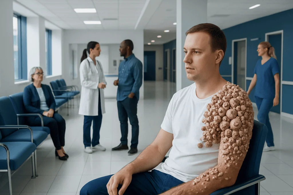
True marine barnacles do not grow on living human skin. The phrase usually refers to skin lesions that look crusted, stuck on, or clustered.
Key Takeaways
- True marine barnacles do not grow on living human skin.
- The phrase usually refers to skin lesions that look crusted, stuck on, or clustered.
- Common explanations include seborrheic keratoses, warts, callused areas, or other dermatologic conditions.
- A doctor may need to examine the skin closely or perform a biopsy to confirm the diagnosis.
- Any lesion that changes quickly, bleeds, ulcerates, or looks unlike the person’s other spots should be checked promptly.
Barnacles on humans is not a medical diagnosis. In most cases, people use this phrase to describe rough, attached-looking skin growths or clustered bumps, which may have several different causes and should be assessed by a qualified clinician if they are new, changing, painful, or bleeding.
Overview: What people mean by 'barnacles on humans'
Barnacles on humans are not actual sea barnacles attaching to and growing from human skin. Medically, this phrase is usually a nonmedical description for rough, raised, crusted, or clustered lesions that appear to be “stuck on” the skin surface. In other words, the term describes how something looks, not a specific disease.
Many different skin conditions can create this appearance. Some are benign and common, such as age-related or sun-related growths. Others may be infectious, inflammatory, or occasionally cancerous. Because photographs online can be misleading, it is safest to avoid self-diagnosing based on shape alone.
A useful clinical approach is to ask what the lesions feel like, how quickly they appeared, whether they itch or bleed, and whether they are isolated or widespread. This helps narrow the possibilities more reliably than the word “barnacles,” which has no formal role in dermatology.
Why true barnacles do not grow on human skin
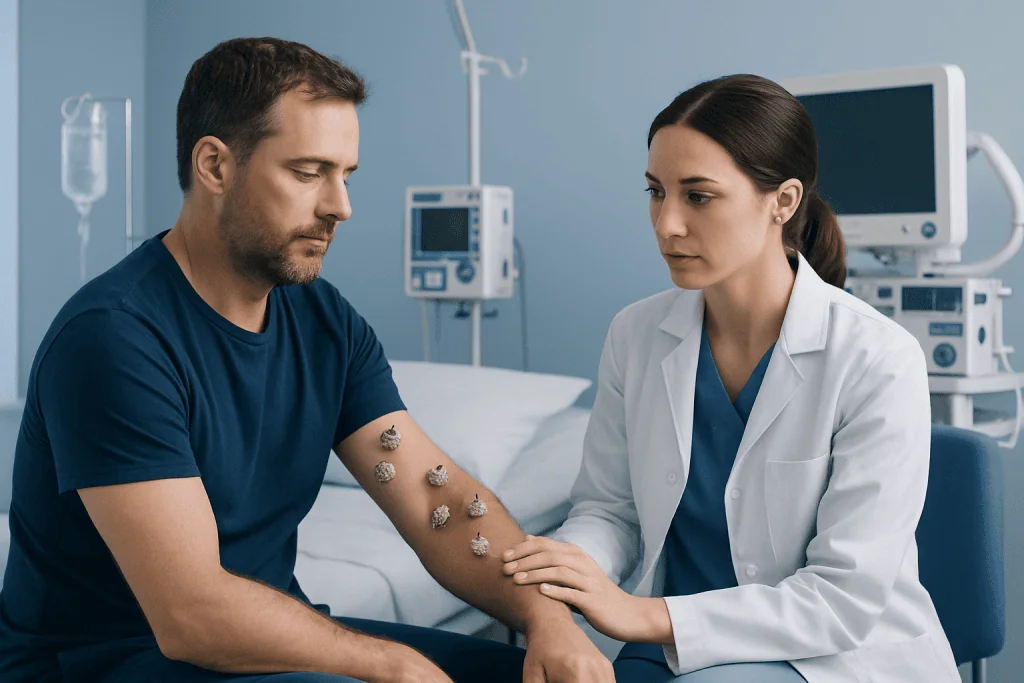
Barnacles are marine crustaceans that live in saltwater and attach to hard surfaces such as rocks, shells, piers, and boat hulls. Their biology is not suited to living human tissue. They require environmental conditions that human skin does not provide, so true barnacle colonization of intact living skin does not occur in normal medical practice.
Confusion may arise after ocean exposure, when a person develops cuts, stings, rashes, or infected abrasions. In these situations, marine material or debris can become embedded in a wound, or seawater exposure can lead to irritation or infection. That is very different from barnacles growing on the body.
This distinction matters because myths can delay proper care. A lesion that resembles a shell, plaque, wart, or cluster may have a routine dermatologic explanation and can often be diagnosed with a simple skin examination. If there has been a sea injury with retained material, cleaning the wound and checking for infection become the main concerns rather than removing “barnacles.”
Medical conditions that can look like barnacles

One of the most common explanations is seborrheic keratosis, a benign skin growth that often appears waxy, brown, black, tan, or rough with a stuck-on look. These lesions can occur singly or in groups and are especially common with age. They are harmless in many cases, but a new or unusual lesion still deserves professional review because some skin cancers can imitate benign growths.
Warts are another possibility. Caused by certain strains of human papillomavirus, they may be rough, raised, and clustered, especially on hands, feet, or areas exposed to friction. Thickened calluses or corns can also resemble attached plaques, particularly on pressure points. In some people, chronic scratching or eczema can lead to lichenified, thickened patches that feel crusted or uneven.
Less commonly, scaly precancerous lesions such as actinic keratoses may appear rough and persistent on sun-exposed skin. Some skin cancers, including forms of skin cancer, can present as scaly, bleeding, ulcerated, or nonhealing growths. For this reason, the appearance alone is not enough to separate harmless from concerning lesions.
- Seborrheic keratoses: waxy, stuck-on, benign growths
- Warts: rough viral lesions, sometimes clustered
- Calluses or corns: thickened skin from friction or pressure
- Actinic keratoses: sun-related rough spots that may need treatment
- Skin cancer: changing, bleeding, or nonhealing lesions that require assessment
Symptoms and warning signs that help doctors distinguish causes
Doctors look at more than texture. Important details include color, border, size, symmetry, depth, tenderness, itch, and whether the lesion is flat, raised, crusted, or ulcerated. They also ask when it first appeared and whether it has changed over weeks or months.
Features that often support a benign process include long-standing stability, a typical stuck-on surface, and the presence of several similar spots elsewhere on the body. However, even common lesions can become irritated by clothing, shaving, or scratching, which may make them red or tender and more difficult to judge without an exam.
Warning signs include rapid growth, spontaneous bleeding, dark or uneven pigmentation, persistent pain, drainage, ulceration, or a sore that does not heal. A lesion that looks clearly different from a person’s other spots should also be assessed. If there are associated symptoms such as fever, expanding redness, or swelling after an injury, infection becomes an important consideration.
How diagnosis is made
Diagnosis begins with a skin history and physical examination. A clinician will review the lesion’s onset, changes over time, history of sun exposure, previous skin problems, recent travel or water exposure, personal or family history of skin cancer, and any medications that affect the immune system.
The appearance may be clear enough for a doctor to identify the lesion in the clinic. In dermatology, magnified examination with a dermatoscope can reveal surface and pigment patterns that are not visible to the naked eye. If a lesion is suspicious or uncertain, a skin biopsy may be recommended to confirm the diagnosis under a microscope.
Tests are selected based on what the doctor suspects. A biopsy is not needed for every rough skin growth, but it is an important tool when there is concern for precancer or cancer. If a person has a sea-related wound, the doctor may also check for retained debris, signs of infection, and the need for wound care or tetanus review.
Treatment options depend on the actual cause
Because “barnacles on humans” is not one condition, treatment varies. Benign lesions such as seborrheic keratoses may not need treatment unless they are irritated, repeatedly traumatized, or cosmetically bothersome. In appropriate cases, removal may be performed with office-based methods such as cryotherapy, curettage, or other dermatology treatments chosen by the physician.
Warts may be treated with topical medicines, cryotherapy, or other procedures depending on location and persistence. Thickened calluses improve with pressure reduction, protective footwear, and careful skin care. Eczema-like or inflammatory lesions are managed according to the underlying diagnosis, often with moisturization and prescription anti-inflammatory treatment when needed.
If the lesion is precancerous or malignant, management becomes more targeted. This may include local destruction, excision, or structured cancer care depending on the pathology result. When there is concern for more serious disease, patients may be referred for skin cancer treatment or, if a suspicious lesion involves a larger surface area or complex reconstruction is needed, oncology care. Any home treatment should be discussed with a doctor first, because acids, scraping, or picking can worsen irritation and may delay diagnosis.
Self-care, prevention, and safe next steps
It is generally best not to pick, cut, or scrape rough lesions at home. This can lead to bleeding, infection, scarring, and confusion about the original appearance. Keeping the skin clean, moisturized, and protected from friction may reduce irritation while waiting for an appointment.
Sun protection is one of the most practical preventive steps for many rough or precancerous lesions. Broad-spectrum sunscreen, protective clothing, hats, and limiting intense sun exposure can help reduce cumulative damage. Regular skin self-checks are also helpful, especially for people with many moles, significant sun exposure, fair skin, or a personal or family history of skin cancer.
People who notice repeated skin changes may benefit from periodic dermatology review. Near the end of the care pathway, if specialist evaluation or treatment is needed, Acibadem International’s multidisciplinary specialists and JCI-accredited hospitals diagnose and treat skin conditions for international patients in a coordinated setting.
When to seek medical care
Medical care should be sought if a lesion is new and persistent, changing in size or color, bleeding without clear injury, becoming painful, or failing to heal. Prompt evaluation is also important if there are signs of infection such as increasing redness, warmth, swelling, pus, or fever, especially after a cut or marine exposure.
An urgent visit is advisable for rapidly enlarging growths, darkly pigmented lesions with irregular borders, or sores that ulcerate. Immunocompromised individuals and people with a past history of skin cancer should have a lower threshold for getting checked.
Even when the cause turns out to be benign, a professional diagnosis can provide reassurance and prevent unnecessary myths or unsafe self-treatment. If there is uncertainty, a qualified doctor or dermatologist is the right source of guidance.
Frequently asked questions
Can barnacles really grow on human skin?
No. True marine barnacles do not grow on living human skin. When people use this phrase, they are usually describing skin growths or rough lesions that only resemble attached barnacles.
What is the most common medical explanation for a barnacle-like skin growth?
A common explanation is seborrheic keratosis, which is a benign growth with a waxy or stuck-on appearance. Warts, calluses, and some sun-related lesions can also look similar, so a medical exam may still be needed.
Are barnacle-like lesions always dangerous?
No, many are harmless. However, some concerning conditions, including precancerous lesions or skin cancer, can mimic benign growths, especially if they bleed, ulcerate, or change quickly.
Should a person try to remove these lesions at home?
Home removal is not recommended without a diagnosis. Picking, cutting, or applying strong products can cause infection, scarring, and make it harder for a doctor to identify the lesion correctly.
How do doctors tell the difference between a harmless lesion and skin cancer?
Doctors assess the lesion's history, appearance, texture, color, and any change over time. If there is uncertainty, a biopsy may be done to examine the tissue under a microscope.
Can ocean exposure cause skin problems that people mistake for barnacles?
Yes. Sea injuries, embedded debris, stings, and wound infections after saltwater exposure can create unusual-looking skin changes. These are different from true barnacles and may need wound care or infection treatment.
This article is for general information only and is not a substitute for professional medical advice. Please consult a qualified doctor about your individual situation.
Explore treatments in Turkey — costs, top hospitals & a free quote
JCI-accredited · board-certified surgeons · reply within 24h
Add us as a Preferred Source to see more of our trusted health content across Google Search, AI Overviews and Discover.

More from the Health Library

Chipped Tooth: A Complete Medical Overview

Pectineus: An Evidence-Based Guide for Patients

Deltoid: A Complete Medical Overview

Ophthalmologist: An Evidence-Based Guide for Patients

Magnesium Citrate for Constipation: An Evidence-Based Guide for Patients