

Bone Densitometry (DEXA) Scan: What It Measures and Who Should Get One
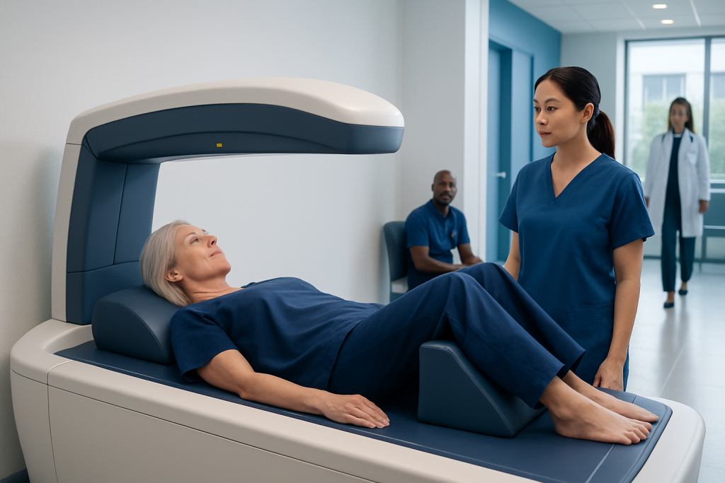
A DEXA scan measures bone mineral density, usually at the hip and spine. It is the standard test for diagnosing osteoporosis and osteopenia.
Key Takeaways
- A DEXA scan measures bone mineral density, usually at the hip and spine.
- It is the standard test for diagnosing osteoporosis and osteopenia.
- The scan is painless, fast, and uses very low levels of radiation.
- Postmenopausal women, older adults, and people with certain risk factors may benefit from testing.
- Results are interpreted using T-scores and sometimes Z-scores to guide follow-up and treatment.
A bone densitometry (DEXA) scan is a quick, low-radiation test that measures bone mineral density and helps estimate fracture risk. It is commonly used to screen for osteoporosis, monitor bone health over time, and guide treatment decisions.
Overview: What a Bone Densitometry (DEXA) Scan Measures
Bone densitometry, often called a DEXA or DXA scan, is an imaging test used to measure bone mineral density. In simple terms, it helps show how strong or fragile a person’s bones may be. The test is most often performed on the lower spine and hip because these areas are especially important for predicting the risk of fractures.
DEXA stands for dual-energy X-ray absorptiometry. The scan uses two low-dose X-ray beams to estimate how much mineral, mainly calcium, is present in bone. Because bone mineral density is closely linked to bone strength, the test is a key tool in identifying low bone mass, osteopenia, and osteoporosis.
Doctors use bone densitometry both for screening and for follow-up. It can help detect bone loss before a fracture happens, estimate future fracture risk, and monitor whether lifestyle changes or medications are helping. For many people, it is one of the most useful tests in long-term bone health planning.
Why the Test Is Important
Bone loss often develops gradually and does not usually cause symptoms at first. Many people do not realize their bones have become weaker until they experience a fracture after a minor fall or even everyday movement. A DEXA scan can identify reduced bone density early, when there may be a better chance to reduce future risk.
The test is especially valuable in osteoporosis screening. Osteoporosis is a condition in which bones become less dense and more likely to break. Common fracture sites include the hip, wrist, and spine. Spinal fractures may sometimes happen silently, causing height loss or stooped posture over time rather than sudden pain.
Bone densitometry is also useful for treatment monitoring. If a person is taking medication for osteoporosis or has known risk factors for bone loss, repeat scans may show whether bone density is stable, improving, or decreasing. This helps doctors make more informed decisions about care.
In some situations, a DEXA scan may be part of a broader evaluation that includes blood tests, fracture history, and assessment of related conditions such as osteoporosis. The scan result is important, but it is only one part of the full picture of bone health.
Who Should Get a DEXA Scan
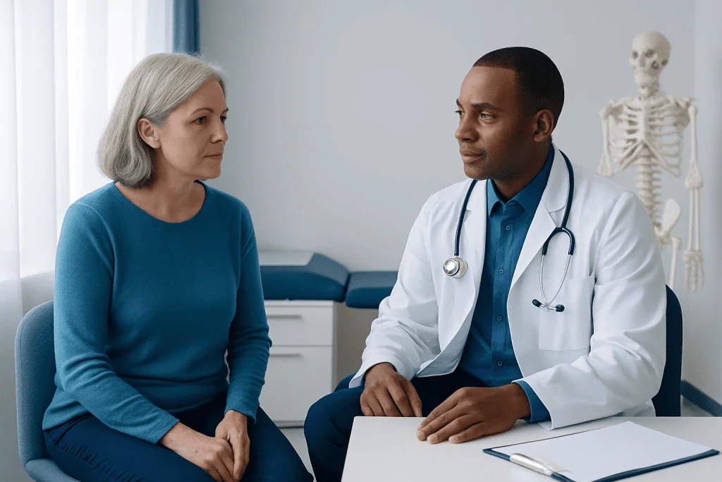
A doctor may recommend a bone densitometry (DEXA) scan based on age, sex, medical history, and individual risk factors. It is commonly advised for women aged 65 and older and for men aged 70 and older, even if they do not have symptoms. Younger postmenopausal women and younger men may also need testing if they have higher fracture risk.
Several factors can increase the need for earlier screening. These include a previous fracture from a minor injury, long-term corticosteroid use, low body weight, smoking, heavy alcohol use, a family history of hip fracture, early menopause, certain hormone-related conditions, and diseases that affect nutrient absorption or bone metabolism.
People with chronic medical conditions may also benefit from assessment. Examples include inflammatory disorders, some endocrine conditions, kidney disease, and conditions linked with poor nutrition or reduced mobility. A doctor may also suggest testing if there has been noticeable height loss, back pain related to possible vertebral fracture, or X-ray evidence of thinning bones.
For patients who are being evaluated for bone health more broadly, a physician may combine bone densitometry with other diagnostic tools such as MRI scan or CT scan when symptoms suggest additional structural problems. These tests do not replace DEXA for bone density measurement, but they may help answer different clinical questions.
What Happens During the Scan
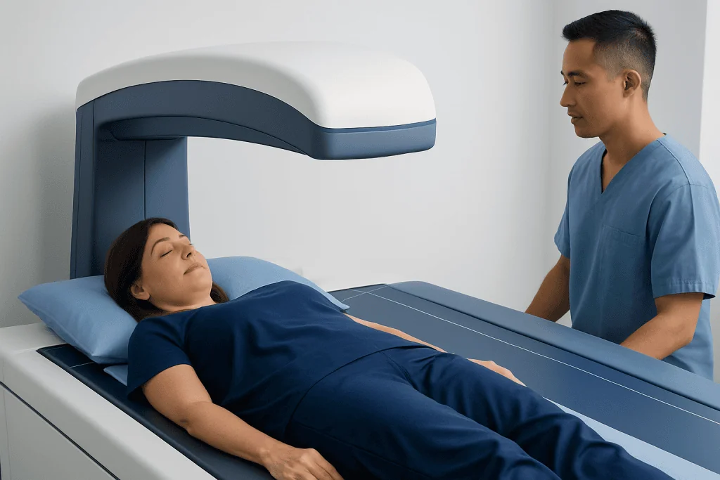
A DEXA scan is simple, painless, and usually completed within a short appointment. In most cases, the patient lies on a padded table while the scanner passes over the body. The person typically remains awake and does not need sedation. The test itself often takes only a few minutes, although timing can vary depending on the area being scanned.
The most common sites measured are the hip and lower spine. In some cases, the forearm may be tested instead, especially if hip or spine measurements are not possible. Whole-body DEXA scans can also be used in selected situations, including body composition analysis, but standard osteoporosis screening usually focuses on the hip and spine.
Preparation is usually minimal. Patients may be asked to avoid calcium supplements for a short period before the scan and to inform the care team if they are pregnant or may be pregnant. It is also helpful to mention any recent contrast imaging studies or nuclear medicine tests, since these can sometimes affect timing.
The amount of radiation used in DEXA is very low compared with many other imaging tests. That makes it a reassuring option for routine bone density measurement. If a person has questions about imaging safety, a healthcare professional can explain how the benefits and risks apply to their situation.
Understanding the Results
DEXA results are usually reported as T-scores and sometimes Z-scores. A T-score compares a person’s bone density with that of a healthy young adult at peak bone mass. This score helps doctors classify bone density as normal, low bone mass, or osteoporosis.
In general, a T-score of -1.0 or above is considered within the expected range for bone density. A T-score between -1.0 and -2.5 suggests low bone mass, often called osteopenia. A T-score of -2.5 or lower is consistent with osteoporosis. These numbers are important, but they are interpreted alongside age, fracture history, medications, and other health factors.
A Z-score compares bone density with what is typical for someone of the same age and sex. It may be especially helpful in younger adults, children, or people in whom secondary causes of bone loss are being considered. A low Z-score may prompt a doctor to look more closely for an underlying condition.
Doctors may also use bone density results together with fracture risk tools to estimate the likelihood of future fractures. If findings suggest significant bone weakness or past compression fractures, further assessment might include X-ray imaging of the spine or other affected areas.
Causes of Low Bone Density and Related Risk Factors
Low bone density can happen for many reasons. Aging is one of the most common, because bones naturally lose some strength over time. Hormonal changes, especially the drop in estrogen after menopause, can accelerate bone loss. Lower testosterone levels in men may also play a role.
Lifestyle factors matter as well. Not getting enough calcium or vitamin D, limited physical activity, smoking, and high alcohol intake can all contribute to weaker bones. Long periods of immobility may also reduce bone strength, since bones stay healthiest when they are regularly used and loaded through movement.
Some medications and medical conditions increase risk. Long-term use of corticosteroids is a well-known example. Other causes may include overactive thyroid disease, parathyroid disorders, chronic digestive diseases that impair absorption, kidney problems, and low body weight or eating disorders.
Because bone health is influenced by many factors, the best interpretation of a DEXA scan comes from a full medical review. This is why doctors often ask about diet, exercise, medications, menstrual history, prior fractures, and family history when discussing results and next steps.
Treatment Options and Follow-Up After a DEXA Scan
Treatment depends on the scan findings and the person’s overall fracture risk. If bone density is only mildly reduced, a doctor may focus first on nutrition, exercise, fall prevention, and monitoring. If osteoporosis is present or fracture risk is high, medication may be recommended to help slow bone loss or improve bone strength.
Self-care measures are an important part of any plan. These often include getting enough calcium and vitamin D, doing weight-bearing and muscle-strengthening exercise, avoiding smoking, and limiting alcohol. Fall prevention strategies, such as improving home safety, checking vision, and reviewing medications that cause dizziness, can also lower fracture risk.
Repeat DEXA scanning may be advised, but the timing varies. Some people need follow-up after a few years, while others being actively treated for osteoporosis may need monitoring sooner. A doctor considers the starting result, age, treatment use, and rate of expected bone change when planning repeat testing.
When care is complex, patients may be evaluated by specialists in endocrinology, rheumatology, geriatrics, or orthopedics. Near the end of the care pathway, it may be reassuring to know that Acibadem International’s multidisciplinary specialists in JCI-accredited hospitals diagnose and treat bone health conditions for international patients.
When to Speak With a Doctor
It is sensible to speak with a doctor about bone densitometry if there has been a fracture after a minor fall, unexplained loss of height, new stooped posture, or persistent back pain that could suggest a vertebral fracture. A consultation is also helpful for anyone with strong risk factors, even if there are no symptoms.
People who take long-term steroid medication or who have conditions that affect hormones, kidneys, digestion, or nutrition should ask whether bone density testing is appropriate. Postmenopausal women and older men who have never been screened may also benefit from discussing timing with a healthcare professional.
A DEXA scan is not usually an emergency test, but it can play an important role in preventing future fractures and preserving mobility and independence. The most useful next step is an individualized discussion with a qualified doctor who can interpret risk in context and recommend the right screening plan.
Frequently asked questions
Is a bone densitometry (DEXA) scan painful?
No. A DEXA scan is painless and noninvasive. The patient usually lies still on a table for a short time while the scanner measures bone density.
How long does a DEXA scan take?
The scan itself is usually quick and often takes only several minutes. The full appointment may be a little longer to allow for registration, positioning, and instructions.
What is the difference between osteopenia and osteoporosis?
Osteopenia means bone density is lower than normal but not low enough to meet the definition of osteoporosis. Osteoporosis means the bones are significantly weaker and the risk of fracture is higher.
How often should someone have a DEXA scan?
There is no single schedule that fits everyone. The timing depends on age, risk factors, previous results, and whether the person is receiving treatment for bone loss.
Does a DEXA scan expose the patient to a lot of radiation?
No. DEXA uses a very low dose of radiation compared with many other imaging tests. For most people, this makes it a suitable option for routine bone density assessment.
Can men need a DEXA scan too?
Yes. Men can develop low bone density and osteoporosis, especially with aging, low body weight, steroid use, hormone problems, or a history of fractures. Doctors may recommend testing based on risk rather than symptoms alone.
References
- World Health Organization
- National Institute of Arthritis and Musculoskeletal and Skin Diseases
- International Osteoporosis Foundation
- National Osteoporosis Foundation
- Radiological Society of North America
This article is for general information only and is not a substitute for professional medical advice. Please consult a qualified doctor about your individual situation.
Joint and spine care in Turkey — expert assessment & treatment
JCI-accredited · board-certified surgeons · reply within 24h
Add us as a Preferred Source to see more of our trusted health content across Google Search, AI Overviews and Discover.

More from the Health Library


Related Specialists

Prof. Dr. Halit Çavuşoğlu
Neurosurgery
Dr. Ayça Özgen
Cardiovascular Surgery
Prof. Dr. Muzaffer Olcay Çizmeli
Interventional Neuroradiology
Dr. Erdem Kaplan
Emergency Service