

Bones of the Cranium: What Patients Need to Know
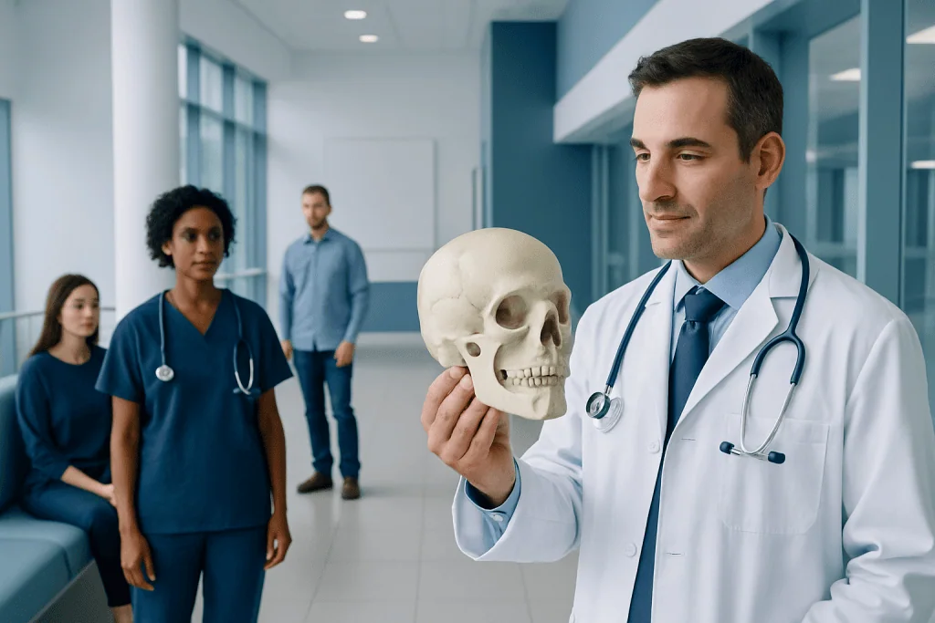
The cranium is made of eight bones: one frontal, two parietal, two temporal, one occipital, one sphenoid, and one ethmoid bone. Cranial bones protect the brain, house sensory structures, and provide attachment points for muscles and connective tissues.
Key Takeaways
- The cranium is made of eight bones: one frontal, two parietal, two temporal, one occipital, one sphenoid, and one ethmoid bone.
- Cranial bones protect the brain, house sensory structures, and provide attachment points for muscles and connective tissues.
- Most cranial bones meet at sutures, which are flexible during infancy and gradually become more fixed with age.
- Head injuries, persistent headaches, neurological symptoms, or visible skull changes should be assessed by a qualified clinician.
- Imaging tests such as CT or MRI may be used when a skull or brain condition is suspected.
The bones of the cranium are eight bones that surround and protect the brain. Together, they form the upper and back portion of the skull, connect at immovable joints called sutures, and support important structures of the head.
Overview: What Are the Bones of the Cranium?
The bones of the cranium are the eight bones that create the protective bony case around the brain. They form the upper, side, and rear portions of the skull. The cranium is distinct from the facial skeleton, which includes bones such as the jaw, cheekbones, and nasal bones.
Although the skull may seem like one solid structure, it is made of separate bones joined by strong bands of fibrous tissue called sutures. These joints allow the skull to grow during childhood and create a stable protective enclosure in adulthood. The cranial cavity inside these bones contains the brain, its protective membranes, cerebrospinal fluid, and major blood vessels.
The cranium has several roles beyond brain protection. Its bones help form the eye sockets, ear structures, nasal cavity, and the base of the skull. Openings in the skull base allow nerves, blood vessels, and the spinal cord to pass between the brain and the rest of the body.
The Eight Cranial Bones and Where They Are Located
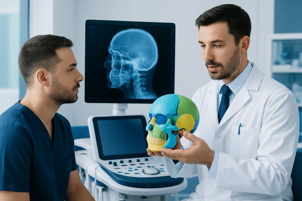
There are eight cranial bones. Four are single bones located in the midline, while two pairs are located on both sides of the head. Their shapes are complex because they must fit together closely while accommodating the brain, sensory organs, nerves, and blood vessels.
- Frontal bone: forms the forehead, the upper part of the eye sockets, and part of the front of the cranial cavity.
- Parietal bones: a pair of large bones that make up much of the roof and upper sides of the skull.
- Temporal bones: a pair of bones at the lower sides of the skull, near the ears. They contain structures involved in hearing and balance.
- Occipital bone: forms the back and lower part of the skull. It contains the foramen magnum, a large opening through which the spinal cord connects with the brain.
- Sphenoid bone: a central bone at the skull base that helps form the eye sockets and connects with many other skull bones.
- Ethmoid bone: a small bone between the eye sockets that contributes to the nasal cavity, nasal septum, and part of the eye sockets.
These bones are often described in anatomy diagrams individually, but in daily life they function as an integrated protective framework. Their position and structure are important when clinicians assess head injury, sinus disease, neurological symptoms, hearing problems, or congenital skull differences.
Cranial Sutures and Skull Development
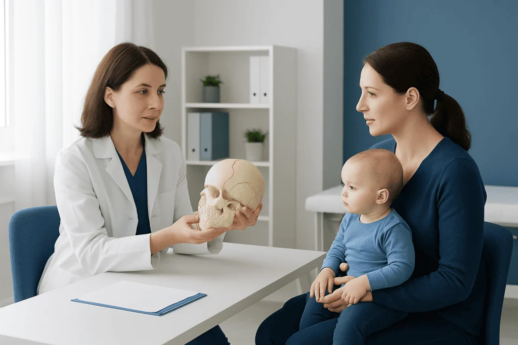
Sutures are specialized fibrous joints between cranial bones. Major sutures include the coronal suture between the frontal and parietal bones, the sagittal suture between the two parietal bones, the lambdoid suture between the parietal and occipital bones, and squamous sutures near the temporal bones.
In babies, the sutures are not fully fused. This allows the head to change shape slightly during birth and gives the skull room to expand as the brain grows rapidly in early life. Wider membrane-covered spaces where several sutures meet are called fontanelles, sometimes known as “soft spots.” These are a normal part of infant skull development.
Over time, sutures become more rigid. Early fusion of one or more sutures can affect skull shape and may sometimes affect brain growth. This condition is called craniosynostosis and requires medical assessment. In contrast, most adult skull sutures are stable and are not designed to move like joints in the arms or legs.
How the Cranium Protects the Brain
The cranial bones provide a firm outer barrier for the brain. Their curved shape helps distribute force over a wider area, and the skull base supports the lower regions of the brain. Inside the cranium, the brain is additionally protected by three membranes called meninges and by cerebrospinal fluid, which provides cushioning.
Protection is important, but the skull cannot prevent every injury. A forceful blow, rapid acceleration and deceleration, or penetrating trauma can still cause concussion, bleeding, swelling, or a skull fracture. Symptoms may occur even when there is no visible cut, bruise, or fracture.
The bones at the base of the skull are especially intricate. They contain channels and openings for cranial nerves and arteries. For this reason, injuries or conditions affecting this region need careful clinical evaluation, often with detailed imaging. Head trauma can also be associated with concussion, which may affect thinking, balance, vision, sleep, or mood even if routine scans are normal.
Common Conditions That Can Affect Cranial Bones
Many people never have a medical problem involving the cranial bones. When concerns do arise, they may relate to injury, development, infection, tumors, or disorders that influence bone strength. The appropriate evaluation depends on a person’s age, symptoms, medical history, and the nature of the concern.
Skull fractures can occur after significant head trauma. A fracture may be simple and stable, or it may be associated with damage to the brain or nearby tissues. Some fractures are not obvious from appearance alone. A clinician may recommend a CT scan when there is concern for fracture or bleeding after an injury.
Developmental conditions, including craniosynostosis, can alter skull shape in infants and young children. Bone disorders, severe infections, and benign or malignant growths can also involve the skull, although these are less common. A noticeable new lump, a progressively changing skull contour, or unexplained localized tenderness should be evaluated rather than self-diagnosed.
Some symptoms attributed to “skull problems,” such as headache or facial pressure, are more often related to other causes. For example, sinus inflammation may create pressure around the forehead or eyes without affecting the strength of the cranial bones. A clinician can help distinguish among possible causes.
How Doctors Assess the Cranium and Skull Symptoms
Assessment usually begins with a medical history and physical examination. A clinician may ask about a recent injury, loss of consciousness, vomiting, headache pattern, vision changes, hearing symptoms, seizure activity, fever, or changes in behavior and alertness. They may examine the scalp, face, eyes, ears, balance, strength, sensation, and coordination.
Imaging is not necessary for every headache or minor bump to the head. However, it can be important when symptoms suggest a possible fracture, bleeding, infection, structural abnormality, or neurological problem. CT is often used in urgent head trauma because it provides detailed images of bone. MRI may be more useful for evaluating the brain, nerves, soft tissues, and certain longer-term concerns.
For children with an unusual head shape, assessment may include measurements of head growth and specialist review. For suspected bone disease, doctors may also consider blood tests or other studies. The goal is to identify whether the cranial bones themselves are involved and to address any underlying cause safely.
Caring for Skull Health and Reducing Injury Risk
Healthy cranial bones depend on the same foundations that support bone health throughout the body: adequate nutrition, regular physical activity when appropriate, avoidance of tobacco, and management of medical conditions that affect bones. Calcium and vitamin D needs vary by age, diet, health status, and location, so individuals should seek personalized advice before taking supplements.
Preventing head injury is also important. Seat belts, age-appropriate child restraints, helmets for cycling and relevant sports, safe work practices, and fall-prevention measures can reduce risk. Helmets lower the risk of some head injuries but do not make high-impact activities completely risk-free.
After a suspected concussion or head injury, returning to sport, driving, school, or work should be guided by a healthcare professional when symptoms persist or the injury was significant. Rest and a gradual, individualized return to normal activities are commonly advised, depending on the injury and the person’s symptoms.
Acibadem International’s multidisciplinary specialists and JCI-accredited hospitals assess head, skull, and neurological concerns for international patients when specialist evaluation is needed.
When to Seek Medical Care
Urgent medical assessment is needed after a head injury if there is loss of consciousness, worsening or severe headache, repeated vomiting, seizure, confusion, unusual sleepiness, weakness, numbness, trouble speaking, unequal pupils, difficulty walking, or fluid or blood coming from the nose or ears. Emergency care is also important after a high-impact injury, even if symptoms seem mild at first.
Parents and caregivers should seek prompt advice for an infant with a bulging fontanelle when calm and upright, persistent vomiting, unusual drowsiness, seizures, feeding difficulties, or a rapidly changing head shape. A healthcare professional should also assess a baby or child with concerns about head growth or a persistent asymmetrical skull shape.
Non-urgent medical review is appropriate for recurring headaches, a new or enlarging skull lump, persistent pain after an injury, changes in hearing or vision, or concerns about bone health. A qualified doctor can determine whether observation, imaging, referral, or treatment is appropriate.
Frequently asked questions
How many bones are in the cranium?
The cranium is made of eight bones: the frontal, occipital, sphenoid, and ethmoid bones, plus two parietal and two temporal bones. These bones join together to form the protective case around the brain.
What is the difference between the cranium and the skull?
The cranium is the part of the skull that encloses the brain. The full skull includes the cranium as well as the facial bones, such as the upper jaw, cheekbones, nasal bones, and lower jaw.
Do cranial bones move?
In adults, cranial bones are connected by sutures that are largely immovable. In infants and young children, the sutures are more flexible to allow brain and skull growth.
Can a cranial bone fracture heal?
Many uncomplicated skull fractures can heal with observation and supportive care, but treatment depends on the type and location of the fracture and whether there is brain injury. Any suspected skull fracture should be assessed promptly by a healthcare professional.
Can headaches come from the cranial bones?
Most headaches do not come directly from the cranial bones. They are more commonly related to nerves, blood vessels, muscles, tension, migraine, infection, or other conditions. New, severe, or changing headaches should be discussed with a clinician.
Why does a baby have soft spots on the head?
Soft spots, or fontanelles, are normal spaces between cranial bones in infants. They allow the skull to adapt during birth and expand as the brain grows, and they gradually close as the child develops.
References
- National Institute of Neurological Disorders and Stroke
- American Academy of Orthopaedic Surgeons
- MedlinePlus, U.S. National Library of Medicine
- Mayo Clinic
- World Health Organization
This article is for general information only and is not a substitute for professional medical advice. Please consult a qualified doctor about your individual situation.
Joint and spine care in Turkey — expert assessment & treatment
JCI-accredited · board-certified surgeons · reply within 24h
Add us as a Preferred Source to see more of our trusted health content across Google Search, AI Overviews and Discover.

More from the Health Library

Related Specialists
Assoc. Prof. Dr. Murat Baştopcu
Cardiovascular Surgery
Prof. Dr. Lütfi Tunç
Urology
Dr. Mukhtar Shahgaldıyev
Physical Medicine & Rehabilitation
Prof. Dr. İsmail Hamzaoğlu
General Surgery