Brain Aneurysm Flow Diversion: Candidates, Procedure Steps, and Follow-Up

Flow diversion is most often considered for unruptured or previously treated brain aneurysms with specific shapes, sizes, or locations. The procedure is performed through the blood vessels, usually from the wrist or groin, without open brain surgery.
Key Takeaways
- Flow diversion is most often considered for unruptured or previously treated brain aneurysms with specific shapes, sizes, or locations.
- The procedure is performed through the blood vessels, usually from the wrist or groin, without open brain surgery.
- Patients typically need antiplatelet medicines before and after treatment to reduce the risk of clot formation on the device.
- Aneurysm closure happens gradually, so scheduled imaging follow-up is an important part of care.
- The decision to use a flow diverter depends on aneurysm features, patient health, bleeding risk, and specialist assessment.
Brain aneurysm flow diversion is a minimally invasive endovascular treatment used for selected intracranial aneurysms, especially those that are wide-necked, large, or difficult to treat with standard coiling. It works by placing a specialized stent-like device inside the parent blood vessel to redirect blood flow and support gradual healing.
Overview
Brain Aneurysm Flow Diversion is an endovascular treatment for certain intracranial aneurysms. A brain aneurysm is a weakened, bulging area in the wall of a blood vessel in the brain. Some aneurysms are small and monitored over time, while others may need treatment to reduce the risk of growth, rupture, or symptoms caused by pressure on nearby structures.
A flow diverter is a fine, flexible, mesh-like tube placed inside the main artery across the neck of the aneurysm. Instead of filling the aneurysm sac directly, it changes the pattern of blood flow so that less blood enters the bulge. Over time, blood inside the aneurysm clots, and the artery lining grows over the device, helping reconstruct the vessel wall.
This approach is different from traditional coiling, in which tiny coils are placed inside the aneurysm sac. Flow diversion can be especially useful when an aneurysm has a wide neck, a large or giant size, a fusiform shape, or has recurred after prior treatment. It is not suitable for every aneurysm, so careful evaluation by a neurointerventional specialist is essential.
Who May Be a Candidate
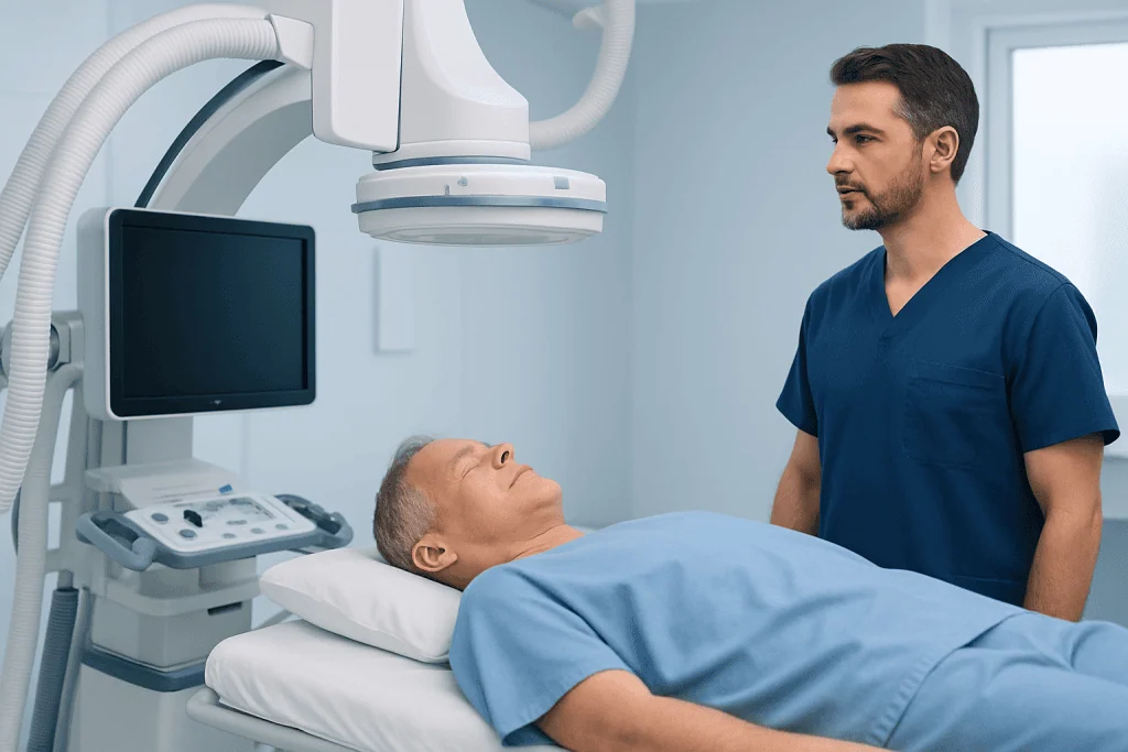
Candidacy for Brain Aneurysm Flow Diversion depends on the aneurysm’s size, shape, location, rupture status, and relationship to nearby branch arteries. It is commonly considered for certain unruptured aneurysms of the internal carotid artery and other large vessels at the base of the brain. It may also be considered for aneurysms that have widened again after previous coiling or clipping.
Doctors evaluate whether the benefits of treatment outweigh the risks of observation or other treatment options. Important patient factors include age, general health, kidney function, allergy history, bleeding tendency, pregnancy status, and the ability to take antiplatelet medicines safely. People who cannot take these medicines may need another approach, because flow diverters require clot-prevention treatment.
Flow diversion may not be the first choice for some recently ruptured aneurysms, very small aneurysms, or aneurysms located where the device could block important small branches. In those cases, coiling, stent-assisted coiling, microsurgical clipping, bypass surgery, or monitoring may be more appropriate. The final recommendation is individualized after reviewing diagnostic images and overall medical risk.
Symptoms and How Aneurysms Are Found

Many unruptured brain aneurysms cause no symptoms and are found incidentally during imaging for headaches, dizziness, trauma, or another medical concern. When symptoms occur, they may be related to the aneurysm pressing on nearby nerves or brain structures. Possible symptoms include double vision, drooping eyelid, facial pain, changes in vision, or a persistent localized headache.
A ruptured aneurysm is a medical emergency and usually presents very differently. A sudden, severe headache, loss of consciousness, vomiting, neck stiffness, weakness, confusion, or seizure requires urgent emergency care. Flow diversion is generally planned after careful assessment, but in selected situations it may be used as part of treatment for complex ruptured aneurysms in experienced centers.
Because symptoms can overlap with other neurological conditions, diagnosis cannot be made by symptoms alone. Imaging tests are needed to understand whether an aneurysm is present and whether it has features that make treatment advisable. A calm, structured evaluation helps patients understand their options without assuming that every aneurysm requires immediate intervention.
Diagnosis and Pre-Procedure Planning
Planning begins with detailed imaging of the brain arteries. Common tests include computed tomography angiography, magnetic resonance angiography, and catheter-based digital subtraction angiography. Digital subtraction angiography is often considered the most detailed test because it shows blood vessel anatomy, aneurysm neck shape, branch vessels, and flow patterns in real time.
Before a flow diverter procedure, the care team reviews medical history and current medicines. Blood tests may check kidney function, blood counts, and clotting status. Because the device is a foreign surface inside the artery, patients are usually started on antiplatelet medicines before treatment, and some centers perform tests to assess response to these medicines.
Pre-procedure planning also includes discussing alternatives and expected follow-up. The specialist explains why flow diversion is being recommended, what the likely benefits are, and what risks need to be considered. Patients should tell their doctor about previous strokes, bleeding problems, stomach ulcers, planned surgeries, allergies to contrast dye, and all prescription or non-prescription medicines.
Procedure Steps
Brain Aneurysm Flow Diversion is performed in an angiography suite or hybrid operating environment by a neurointerventional team. The patient usually receives general anesthesia, although the exact approach depends on the hospital and clinical situation. A thin catheter is inserted into an artery, commonly through the groin or wrist, and guided through the blood vessels to the brain using X-ray imaging and contrast dye.
Once the catheter reaches the target vessel, the flow diverter stent is positioned across the opening of the aneurysm. The device is then carefully released so it opens against the artery wall. The team confirms placement, checks blood flow through the parent artery and nearby branches, and ensures the device covers the aneurysm neck as intended.
Some aneurysms are treated with a flow diverter alone, while others may also need coils or additional devices depending on size, shape, and flow characteristics. After the device is placed, the catheter is removed and the access site is closed. Patients are monitored closely in a recovery area or specialized unit to watch neurological status, blood pressure, and the puncture site.
The procedure does not require opening the skull, but it is still a specialized treatment involving delicate brain blood vessels. Potential risks include bleeding at the access site, clot formation, stroke, vessel injury, contrast reaction, kidney strain from contrast dye, and, rarely, aneurysm rupture. The treating team explains these risks in relation to the patient’s specific aneurysm and health profile.
Recovery and Follow-Up
Hospital stay varies depending on the complexity of the aneurysm, the patient’s health, and local practice. Many patients treated electively are observed for at least one night. During this period, nurses and doctors check for headache changes, weakness, speech difficulty, vision changes, and access-site bleeding or swelling.
Recovery instructions usually include taking antiplatelet medicines exactly as prescribed. Stopping these medicines too early can increase the risk of clot formation on the device, while taking extra doses may increase bleeding risk. Patients should ask their doctor before using additional anti-inflammatory pain medicines, supplements, or any new medication that may affect bleeding.
Follow-up imaging is essential because aneurysm healing after flow diversion is gradual. The aneurysm may shrink and close over months rather than immediately. Doctors may schedule magnetic resonance angiography, computed tomography angiography, or digital subtraction angiography at intervals such as several months after treatment and later as needed.
Patients can support recovery by following activity restrictions, caring for the catheter access site, attending all imaging appointments, and reporting new neurological symptoms promptly. Acibadem International’s multidisciplinary specialists and JCI-accredited hospitals provide diagnosis, endovascular treatment, and follow-up planning for international patients with complex brain aneurysms.
Benefits, Limitations, and Alternatives
The main advantage of flow diversion is that it treats the diseased segment of the parent artery rather than only filling the aneurysm sac. This can be helpful for aneurysms that are difficult to secure with coils because of a wide neck or unusual shape. It can also reduce the need to place material inside fragile or complex aneurysm sacs.
However, flow diversion has limitations. It usually requires dual antiplatelet therapy for a period determined by the treating physician, and aneurysm occlusion may take time. It may not be suitable when important branch vessels arise from the aneurysm neck or when the risk of antiplatelet therapy is high.
Alternative treatments include observation with periodic imaging, endovascular coiling, balloon-assisted coiling, stent-assisted coiling, intrasaccular flow disruption devices, microsurgical clipping, or bypass procedures. Each option has different advantages and risks. A patient-centered decision considers aneurysm rupture risk, treatment risk, life expectancy, symptoms, and personal preferences.
When to See a Doctor
A person diagnosed with a brain aneurysm should see a neurosurgeon or neurointerventional specialist for individualized assessment, even if the aneurysm was found by chance. Specialist review helps determine whether monitoring or treatment is the safer strategy. Patients should bring prior imaging studies, reports, medication lists, and details of family history if available.
Urgent medical care is needed for sudden severe headache, fainting, seizure, new weakness, facial drooping, speech difficulty, confusion, severe neck stiffness, or sudden vision changes. These symptoms do not always mean an aneurysm has ruptured, but they require immediate evaluation. After a flow diverter procedure, any new neurological symptom, significant access-site swelling, chest pain, shortness of breath, or uncontrolled bleeding should also be reported promptly.
Patients should contact their treating team before dental procedures, surgery, or stopping antiplatelet medicines. Good communication between the neurointerventional team, primary doctor, and other specialists helps reduce medication-related risks and keeps follow-up on schedule.
Frequently asked questions
What is a flow diverter for a brain aneurysm?
A flow diverter is a fine mesh stent-like device placed inside a brain artery across the opening of an aneurysm. It redirects blood flow away from the aneurysm so clotting can occur inside the bulge. Over time, the vessel lining may grow over the device and help reconstruct the artery.
Is flow diversion the same as coiling?
No. Coiling places small coils inside the aneurysm sac to promote clotting. Flow diversion places a device in the parent artery across the aneurysm neck, which changes blood flow and supports gradual vessel healing.
How long does it take for an aneurysm to close after flow diversion?
Closure is usually gradual and may take months. Some aneurysms close earlier, while others require longer observation with repeat imaging. The treating specialist will recommend a follow-up schedule based on the aneurysm and procedure result.
Will the flow diverter stay in the artery permanently?
Yes, the device is designed to remain in place. Over time, the inner lining of the artery can grow over it, incorporating it into the vessel wall. Patients generally do not feel the device once it is implanted.
Why are antiplatelet medicines needed after the procedure?
Antiplatelet medicines reduce the chance of blood clots forming on the flow diverter while the artery heals. They must be taken exactly as prescribed unless the treating doctor advises otherwise. Patients should not stop them on their own, even if they feel well.
Can all brain aneurysms be treated with flow diversion?
No. Suitability depends on aneurysm size, shape, location, rupture status, and nearby branch vessels, as well as the patient’s overall health. Some aneurysms are better treated with coiling, clipping, other endovascular devices, or careful monitoring.
References
- American Heart Association
- American Stroke Association
- Society of NeuroInterventional Surgery
- European Stroke Organisation
- National Institute of Neurological Disorders and Stroke
This article is for general information only and is not a substitute for professional medical advice. Please consult a qualified doctor about your individual situation.
Heart care in Turkey — expert evaluation and treatment
JCI-accredited · board-certified surgeons · reply within 24h
Add us as a Preferred Source to see more of our trusted health content across Google Search, AI Overviews and Discover.