Brain MRI vs CT for Neurological Symptoms: How Doctors Choose Imaging
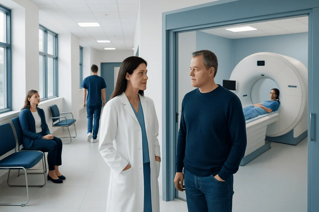
CT is often preferred first in emergencies because it is fast and excellent for detecting bleeding, major injury, and some urgent complications. MRI provides more detailed images of brain tissue and is often better for subtle stroke, tumors, inflammation, infections, and many chronic neurological conditions.
Key Takeaways
- CT is often preferred first in emergencies because it is fast and excellent for detecting bleeding, major injury, and some urgent complications.
- MRI provides more detailed images of brain tissue and is often better for subtle stroke, tumors, inflammation, infections, and many chronic neurological conditions.
- The best scan depends on the clinical question, not on which test is generally considered more advanced.
- Contrast material may be used with either CT or MRI when doctors need to see blood vessels, tumors, inflammation, or infection more clearly.
- People with sudden weakness, severe headache, confusion, seizures, or head injury should seek urgent medical care rather than choosing an imaging test themselves.
Brain MRI and CT are both valuable imaging tests, but they answer different clinical questions. Doctors choose between them by considering the symptom pattern, urgency, safety factors, and what condition is most likely.
Overview: Why Brain Imaging Is Used
Neurological symptoms can arise from many different causes, including migraine, stroke, infection, inflammation, seizures, trauma, tumors, nerve disorders, and medication effects. Because the brain and nervous system are complex, doctors often combine a detailed history, neurological examination, blood tests, and imaging to narrow the diagnosis. Brain MRI and CT are two of the most commonly used imaging methods.
A CT scan, or computed tomography, uses X-rays processed by a computer to create cross-sectional images. It is quick, widely available, and especially useful when doctors need to identify bleeding, skull fractures, swelling, or major structural changes quickly. In many emergency situations, CT is the first scan because speed can be important for decision-making.
MRI, or magnetic resonance imaging, uses a strong magnetic field and radio waves to create detailed images of soft tissues. It can show small or subtle changes in the brain that may not be visible on CT, such as early ischemic stroke, multiple sclerosis lesions, certain tumors, inflammation, and abnormalities in the brainstem or posterior fossa. The choice is therefore not simply “MRI is better” or “CT is better”; it depends on the medical question being asked.
Brain MRI vs CT: Key Differences
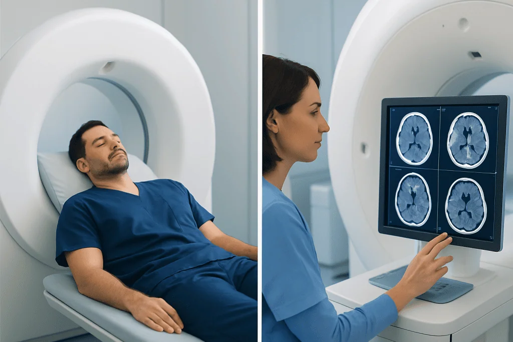
The main difference between brain MRI vs CT is how the images are created and what each method shows best. CT is fast and highly effective for evaluating fresh bleeding, major head injury, bone injury, and some urgent complications such as significant brain swelling. MRI takes longer but provides higher detail for brain tissue, nerves, small blood vessel changes, and many non-emergency neurological conditions.
Radiation exposure is another difference. CT uses ionizing radiation, while MRI does not. For a single medically needed CT, the benefit usually outweighs the radiation risk, especially in emergencies. However, doctors are thoughtful about repeated CT scans, particularly in children and younger patients, and may use MRI or ultrasound when appropriate.
The experience of the scan also differs. A CT scan often takes only a few minutes and is usually easier for people who are very unwell, in pain, confused, or unable to stay still for long. MRI usually requires the person to lie still in a narrower scanner for a longer time, and it can be noisy. Some patients with claustrophobia, implanted devices, or difficulty lying flat may need special planning before MRI.
- CT is often chosen for: trauma, suspected bleeding, sudden severe headache, emergency stroke assessment, and rapid evaluation.
- MRI is often chosen for: subtle stroke, seizures, multiple sclerosis, tumors, unexplained neurological symptoms, inflammation, and follow-up of known brain conditions.
When CT Is Usually Chosen First

In emergency medicine, head CT is frequently the first imaging test for sudden or severe neurological symptoms. It is particularly useful when doctors need to quickly check for bleeding in or around the brain, a skull fracture, significant swelling, or a mass effect that may require urgent treatment. For example, a patient with a head injury, loss of consciousness, worsening confusion, or a sudden “worst headache” may need a CT scan without delay.
CT is also commonly used in the early evaluation of suspected stroke. A non-contrast CT can rapidly distinguish bleeding in the brain from many cases of ischemic stroke, where a blood vessel is blocked. This distinction matters because treatments for ischemic and hemorrhagic stroke are different. In some cases, CT angiography or CT perfusion may be added to evaluate blood vessels and blood flow.
Doctors may choose CT when MRI is not immediately available, would take too long, or is not safe for a specific patient. For people with certain pacemakers, older implanted devices, metal fragments near the eye, or severe instability requiring life-support equipment, CT may be the more practical and safer option. CT can also be helpful when evaluating bone structures, sinus disease affecting the skull base, or calcifications that may be relevant to diagnosis.
When MRI Is Usually Preferred
Brain MRI is often preferred when doctors need a more detailed view of the brain tissue itself. It can detect many abnormalities that may be very small or subtle on CT. This is especially important for symptoms that are persistent, recurrent, unexplained, or not clearly related to an emergency injury or bleeding event.
MRI is commonly used for evaluating seizures, suspected multiple sclerosis, brain tumors, infections, inflammation, pituitary and brainstem problems, dizziness related to central nervous system causes, and certain memory or cognitive symptoms. It can also be very helpful after a CT scan if the CT result is normal but symptoms remain concerning. Different MRI sequences can highlight different tissues, fluid, blood products, diffusion changes, or old injuries.
For ischemic stroke, MRI with diffusion-weighted imaging can detect early changes that may not yet be obvious on CT, especially in smaller strokes or strokes in the posterior circulation. However, MRI is not always the first test if rapid emergency treatment decisions are needed. In practice, a patient may have CT first for speed, followed by MRI for detail once the person is stable or when the diagnosis remains uncertain.
How Doctors Decide Which Scan Is Best
Doctors choose imaging by matching the test to the patient’s symptoms, examination findings, medical history, and urgency. The first question is usually whether the situation is time-sensitive. Sudden weakness on one side, facial drooping, speech difficulty, severe headache, seizure, significant head injury, or a rapidly changing level of consciousness may require emergency imaging, often starting with CT.
The second question is what condition is most likely. If bleeding or fracture is a major concern, CT is often the fastest and most useful first step. If the concern is inflammation, demyelinating disease, a small tumor, subtle stroke, infection, or a condition affecting the brainstem, MRI may provide more useful detail. Sometimes the best plan is not either-or: CT may answer the urgent question, and MRI may later clarify the underlying cause.
Patient-specific safety factors also matter. Doctors ask about pregnancy, kidney function, allergies to contrast materials, implanted medical devices, previous surgery, metal injuries, claustrophobia, and the ability to lie still. Children, older adults, and people with chronic illnesses may need individualized planning. The goal is to obtain the clearest and safest information with the least unnecessary testing.
Availability and timing can also influence the decision. A high-quality scan is most useful when it can be performed promptly and interpreted by experienced radiologists in the context of the patient’s symptoms. For this reason, imaging decisions are usually made by the treating physician together with radiology specialists rather than by the patient alone.
Contrast, Safety, and What to Expect
Both CT and MRI can be performed with or without contrast material. CT contrast is usually iodine-based and is often used to evaluate blood vessels, tumors, inflammation, infection, or blood flow. MRI contrast is usually gadolinium-based and can help show tumors, active inflammation, infection, scarring, and blood-brain barrier changes. Not every scan needs contrast; many urgent head CT scans, for example, are done without it.
Before contrast is given, the healthcare team may ask about kidney disease, previous contrast reactions, asthma or allergies, pregnancy, breastfeeding, and current medications. Most people tolerate contrast well, but mild reactions can occur, and rare serious reactions are possible. People with reduced kidney function may need special assessment before contrast, especially if the scan is not urgent.
For MRI, safety screening is essential because the magnet is very strong. Patients should tell the team about pacemakers, cochlear implants, aneurysm clips, metal fragments, implanted pumps, neurostimulators, and any prior metal injury. Many modern implants are MRI-conditional, meaning MRI may be possible under specific conditions, but this must be verified before the scan.
During CT, the patient lies on a table that moves through a short ring-shaped scanner. During MRI, the patient lies on a table that moves into a tunnel-like scanner, and ear protection is used because the machine makes loud tapping sounds. Staying still helps image quality. If anxiety or movement is a concern, the medical team can discuss comfort measures or, in selected cases, sedation planning.
When to Seek Medical Care
People should seek urgent medical care for sudden neurological symptoms rather than trying to decide which imaging test they need. Warning symptoms include sudden weakness or numbness on one side of the body, difficulty speaking, facial drooping, new confusion, loss of consciousness, seizure, sudden vision loss, severe or unusual headache, fever with stiff neck, or symptoms after a significant head injury. These symptoms do not always mean a serious condition is present, but they should be assessed promptly.
Non-urgent symptoms also deserve medical evaluation if they are persistent, worsening, or unexplained. Examples include recurrent headaches with a new pattern, dizziness with neurological signs, progressive memory changes, repeated fainting-like episodes, new tremor, balance problems, or changes in sensation or coordination. A doctor can decide whether imaging is needed and which test is most appropriate.
Patients can help by bringing a clear timeline of symptoms, a medication list, previous imaging reports, implant cards, and information about allergies or kidney disease. This helps the care team choose the safest and most informative scan. If a scan is performed, the result should be interpreted together with symptoms and examination findings, because imaging can sometimes show incidental findings that are not the cause of the problem.
Acibadem International’s multidisciplinary specialists and JCI-accredited hospitals diagnose and treat neurological conditions for international patients, including situations where advanced imaging is part of the evaluation. Patients should always follow the advice of their treating physician or emergency team for urgent symptoms.
Frequently asked questions
Is MRI always better than CT for brain symptoms?
No. MRI provides more detail for many brain tissue problems, but CT is often better as the first test in emergencies because it is fast and excellent for detecting bleeding and major injury. The best scan depends on the suspected condition, urgency, and patient safety factors.
Why do doctors often order a CT scan first for stroke symptoms?
A CT scan can be performed quickly and can help identify bleeding in the brain, which changes treatment decisions. In many stroke evaluations, speed is essential. MRI may be used later or in selected cases when more detail is needed.
Does a normal CT scan mean nothing is wrong?
Not always. CT can be normal in some early or subtle conditions, including small strokes, inflammation, certain infections, or seizure-related changes. If symptoms continue or the examination suggests a neurological problem, doctors may recommend MRI or other tests.
Is brain MRI safe if a person has a pacemaker or implant?
It depends on the device. Some modern devices are MRI-conditional and can be scanned under specific safety rules, while others may not be safe for MRI. Patients should tell the imaging team about all implants and bring device information whenever possible.
Does CT radiation make the test dangerous?
A medically necessary CT scan is generally considered safe when the expected benefit outweighs the risk. Doctors avoid unnecessary radiation and choose the most appropriate test for the situation. Patients who have had multiple scans should inform their doctor so prior imaging can be reviewed.
When is contrast needed for brain MRI or CT?
Contrast may be used when doctors need to see blood vessels, tumors, infection, inflammation, or abnormal enhancement more clearly. It is not required for every scan, and many emergency CT scans are done without contrast. Kidney function, allergy history, pregnancy status, and the clinical question are considered before contrast is given.
This article is for general information only and is not a substitute for professional medical advice. Please consult a qualified doctor about your individual situation.
Explore treatments in Turkey — costs, top hospitals & a free quote
JCI-accredited · board-certified surgeons · reply within 24h
Add us as a Preferred Source to see more of our trusted health content across Google Search, AI Overviews and Discover.

More from the Health Library
How to Read Common Blood Test Results Without Panicking
Neuropsychological Testing: Memory, Attention, and Brain Function Assessment

How to Prepare for a Medical Imaging Test: Fasting, Contrast, and Medications
EEG Test: What Brain Wave Monitoring Can Help Diagnose
Cerebral Angiography: What to Expect During Brain Vessel Imaging