Nerve Ultrasound in Neuromuscular Disease: When Imaging Adds Clarity

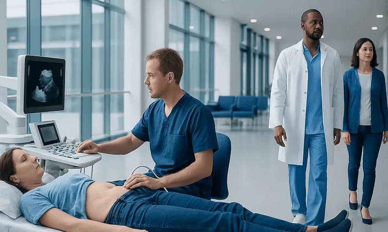
Nerve ultrasound uses high-frequency sound waves to evaluate peripheral nerves in real time. It can help identify nerve enlargement, compression, injury, inflammation, tumors or structural changes around the nerve.
Key Takeaways
- Nerve ultrasound uses high-frequency sound waves to evaluate peripheral nerves in real time.
- It can help identify nerve enlargement, compression, injury, inflammation, tumors or structural changes around the nerve.
- The test is often used alongside electromyography and nerve conduction studies, not as a replacement for them.
- It is noninvasive, does not use radiation and can be repeated when follow-up imaging is helpful.
- Results are most useful when interpreted by clinicians experienced in neuromuscular disorders and ultrasound imaging.
Nerve ultrasound is a painless imaging method that can show changes in peripheral nerves and nearby tissues. In selected neuromuscular conditions, it adds useful anatomical information to the clinical examination and nerve conduction studies.
Overview
Nerve ultrasound is an imaging technique that uses high-frequency sound waves to create detailed pictures of peripheral nerves. Peripheral nerves are the nerves outside the brain and spinal cord; they carry signals for movement, sensation and automatic body functions. In neuromuscular disease, these nerves or the muscles they control may be affected, leading to symptoms such as numbness, weakness, pain, tingling or muscle wasting.
Traditionally, doctors evaluate neuromuscular problems with a neurological examination, blood tests, magnetic resonance imaging when needed, and electrodiagnostic tests such as nerve conduction studies and electromyography. Nerve ultrasound adds a different kind of information: it can show the nerve’s shape, size, continuity and relationship to surrounding structures. This can be especially helpful when a doctor needs to understand where a nerve is being compressed, whether a nerve is swollen, or whether a focal lesion is present.
The procedure is typically performed with the patient sitting or lying comfortably. A small amount of gel is placed on the skin, and a handheld ultrasound probe is moved over the path of the nerve. The test does not use ionizing radiation, does not require injections in most cases, and is generally painless. Because it can be performed dynamically, the clinician may also ask the patient to move a joint to see how the nerve behaves during motion.
When Nerve Ultrasound Adds Clarity
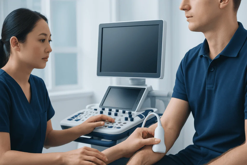
Nerve ultrasound is most helpful when the clinical question is anatomical. For example, a nerve conduction study may show that a nerve signal slows across the wrist, suggesting carpal tunnel syndrome. Ultrasound can then show whether the median nerve is enlarged, flattened or compressed, and whether a nearby structure such as a cyst, tendon abnormality or anatomical variation may be contributing.
It can also be valuable when symptoms and electrodiagnostic findings do not completely match. Some patients have pain or sensory symptoms in a specific nerve distribution, but the electrical test is borderline or technically difficult. Ultrasound may show focal swelling, scarring, traumatic disruption or entrapment at a site that helps explain the symptoms. In other cases, ultrasound helps distinguish a localized nerve problem from a more generalized neuropathy.
Common situations in which nerve ultrasound may be considered include:
- Suspected entrapment neuropathies, such as carpal tunnel syndrome or ulnar neuropathy at the elbow.
- Traumatic nerve injury after cuts, fractures, dislocations or surgery.
- Evaluation of nerve tumors or cyst-like lesions near a nerve.
- Inflammatory neuropathies, where nerves may appear enlarged in characteristic patterns.
- Guidance for selected procedures, such as targeted injections or biopsy planning, when clinically appropriate.
Symptoms That May Lead to Testing

Nerve ultrasound is not usually ordered simply because a symptom is present. It is considered when the doctor suspects that imaging of a peripheral nerve may change diagnosis, treatment planning or follow-up. Symptoms that may prompt a neuromuscular evaluation include numbness, tingling, burning pain, electric-shock sensations, weakness, clumsiness, muscle cramps or visible muscle thinning.
The pattern of symptoms is important. Tingling in the thumb, index and middle fingers may suggest median nerve involvement at the wrist, while numbness in the ring and little fingers may point toward the ulnar nerve. Foot drop may raise concern for peroneal nerve injury, a nerve root problem in the spine, or another neurological condition. A careful examination helps decide whether ultrasound should focus on a specific nerve or whether broader testing is needed.
Ultrasound may also be considered when symptoms change over time. For example, a person with known neuropathy who develops a new focal weakness may need evaluation for a superimposed nerve entrapment. Similarly, persistent symptoms after a fracture, laceration or surgical procedure may require imaging to assess whether the nerve is continuous, tethered by scar tissue or compressed by surrounding structures.
What the Test Can Show
During a nerve ultrasound, the examiner looks at the nerve in short-axis and long-axis views. A healthy peripheral nerve often has a fascicular pattern, sometimes described as a honeycomb appearance on cross-section. The examiner may measure the cross-sectional area of the nerve, compare it with the opposite side, and assess whether the nerve is swollen, flattened, displaced or interrupted.
In entrapment neuropathies, ultrasound may show nerve enlargement before the site of compression and flattening at the compressed segment. In traumatic injury, it may help identify partial or complete nerve discontinuity, neuroma formation, foreign bodies, scar tissue or a nearby fracture callus. In inflammatory neuropathies, multiple nerves may appear enlarged, although ultrasound findings must be interpreted in the context of symptoms, examination and electrodiagnostic results.
Nerve ultrasound can also evaluate the tissues around the nerve. Tendons, muscles, blood vessels, joints and soft-tissue masses may all contribute to nerve irritation or compression. This is one reason ultrasound can be helpful when symptoms appear mechanical, change with movement, or occur after an injury. However, ultrasound has limits: deep nerves, nerves behind bone and very small branches may be difficult to visualize, and image quality depends on equipment and operator expertise.
Nerve Ultrasound, EMG and Other Tests
Nerve ultrasound and electrodiagnostic tests answer different questions. Nerve conduction studies and electromyography evaluate how nerves and muscles function electrically. They can show whether signals are slowed, blocked or reduced, and whether muscles show evidence of denervation or chronic nerve injury. Ultrasound, by contrast, shows the structure of the nerve and surrounding anatomy.
For many patients, the two methods are complementary. Electrodiagnostic testing may identify the type and severity of nerve dysfunction, while ultrasound may localize a compressive lesion or reveal a structural cause. In some cases, ultrasound can guide where the electrodiagnostic examination should focus, especially when symptoms are complex or more than one nerve could be involved.
Other imaging tests may still be needed. Magnetic resonance imaging can be better for evaluating deep structures, the spine, nerve roots, muscles or larger areas of soft tissue. Blood tests, genetic tests or cerebrospinal fluid studies may be considered when a generalized neuropathy, immune-mediated disease or inherited neuromuscular condition is suspected. The choice of tests depends on the patient’s history, examination and the diagnostic question.
How to Prepare and What to Expect
Preparation for nerve ultrasound is usually simple. Patients may be asked to wear comfortable clothing that allows access to the area being examined, such as the wrist, elbow, shoulder, knee or ankle. Jewelry, watches or tight sleeves may need to be removed. In most cases, patients can eat, drink and take regular medications as usual unless their doctor gives different instructions.
During the examination, the clinician applies ultrasound gel to the skin and moves the probe along the course of the nerve. Mild pressure may be felt, especially over tender areas, but the test should not be painful. The examiner may compare both sides of the body, take measurements, and ask the patient to change position or move a joint so the nerve can be seen dynamically.
The length of the examination varies depending on the number of nerves being assessed and the complexity of the symptoms. After the test, patients can usually return to normal activities immediately. Results may be discussed at the time of examination or provided later in a report, depending on the clinic workflow. The most useful interpretation combines ultrasound findings with the medical history, neurological examination and any previous test results.
Treatment Planning, Follow-up and When to See a Doctor
Nerve ultrasound does not treat neuromuscular disease by itself, but it can support treatment decisions. If it confirms a localized entrapment, treatment may include activity modification, splinting, physical or occupational therapy, medication for symptoms, image-guided procedures or surgical consultation when appropriate. If it suggests inflammatory or generalized nerve involvement, further neurological evaluation may be needed to guide disease-specific care.
Ultrasound can also be useful in follow-up. In selected cases, clinicians may monitor nerve size or structural changes after treatment, injury or surgery. However, improvement in symptoms and function remains very important; imaging findings are only one part of the overall assessment. A nerve may remain enlarged for some time even if symptoms improve, so results should be interpreted carefully.
A person should seek medical advice if numbness, tingling or weakness is persistent, worsening, one-sided, associated with loss of hand or foot function, or follows an injury. Urgent care is needed for sudden weakness, new problems with speech or facial drooping, loss of bladder or bowel control, or rapidly progressive neurological symptoms, as these may require immediate evaluation for conditions beyond peripheral nerve disease. Acibadem International’s multidisciplinary specialists and JCI-accredited hospitals diagnose and treat neuromuscular conditions for international patients, using imaging and neurophysiology when clinically indicated.
Frequently asked questions
Is nerve ultrasound painful?
Nerve ultrasound is generally not painful. The examiner places gel on the skin and moves a probe over the area being studied. Some people may feel mild pressure, especially if the area is already tender.
Does nerve ultrasound replace EMG or nerve conduction studies?
Usually, it does not replace them. EMG and nerve conduction studies show how nerves and muscles are functioning electrically, while ultrasound shows the nerve’s structure and surrounding tissues. Many neuromuscular evaluations use these tests together.
What conditions can nerve ultrasound help diagnose?
It can help evaluate nerve entrapments, traumatic nerve injuries, nerve swelling, some inflammatory neuropathies and masses near nerves. It may also help explain symptoms when a nerve is compressed by a cyst, scar tissue or other nearby structure. The final diagnosis depends on the full clinical assessment.
Is nerve ultrasound safe?
Yes, diagnostic ultrasound is considered safe when performed appropriately. It uses sound waves rather than ionizing radiation. It is noninvasive and can be repeated if follow-up imaging is useful.
How long does a nerve ultrasound take?
The time varies depending on the number of nerves and body areas being examined. A focused study may be relatively brief, while a more complex neuromuscular evaluation can take longer. The clinic can provide practical details before the appointment.
Can nerve ultrasound show all nerve problems?
No single test can show every nerve problem. Ultrasound is best for many superficial peripheral nerves and structural questions, but deep nerves, nerve roots and very small branches may be difficult to assess. Other tests such as EMG, MRI, blood tests or genetic testing may be needed.
This article is for general information only and is not a substitute for professional medical advice. Please consult a qualified doctor about your individual situation.
Ultrasound in Turkey — costs, top hospitals & a free quote
JCI-accredited · board-certified surgeons · reply within 24h
Add us as a Preferred Source to see more of our trusted health content across Google Search, AI Overviews and Discover.

Check-up & Preventive Medicine
Comprehensive health screenings designed for early detection and prevention.
1 specialists in this unitMore from the Health Library

Common Reasons Doctors Order Lab Tests: Screening, Diagnosis, and Monitoring
EEG Test Explained: When Brain Wave Monitoring Is Recommended
Diagnostic Mammogram: Why It Is Done and What to Expect

Saturday Lab Testing: How to Plan Weekend Blood Work and Imaging
CT Scan vs MRI: Which Imaging Test Is Right for You?