Diagnostic Mammogram: Why It Is Done and What to Expect

A diagnostic mammogram is used to investigate a breast symptom or an abnormal screening mammogram. It includes extra images focused on a specific area of the breast.
Key Takeaways
- A diagnostic mammogram is used to investigate a breast symptom or an abnormal screening mammogram.
- It includes extra images focused on a specific area of the breast.
- Compression is brief and can be uncomfortable, but it is important for image quality.
- Results may show normal tissue, a benign finding, or the need for additional tests such as ultrasound or biopsy.
- Most abnormal mammogram findings are not breast cancer, but prompt follow-up is important.
A diagnostic mammogram is a more detailed breast imaging test used when there is a breast symptom or when a screening mammogram needs a closer look. It helps radiologists examine a specific area of concern and decide whether further testing is needed.
Overview
A diagnostic mammogram is a specialized breast X-ray exam used to evaluate a specific concern. Unlike a screening mammogram, which is performed routinely in people without symptoms, a diagnostic mammogram is ordered when there is a breast symptom or when a screening mammogram shows an area that needs closer assessment. The goal is to gather more detailed images so the radiologist can better understand what is happening in the breast tissue.
This test may be recommended for a new lump, breast pain in one area, nipple discharge, skin thickening, nipple changes, or an area that looked unclear or unusual on a screening mammogram. During a diagnostic mammogram, the technologist takes additional views that focus on the area of interest. The radiologist may also review images during the appointment and request more pictures if needed.
For many people, the word “diagnostic” can sound worrying. In practice, it simply means that the imaging is being tailored to answer a specific question. Many diagnostic mammograms lead to reassuring results, such as overlapping tissue, cysts, or other benign breast changes. If more information is needed, the next step may include a breast ultrasound or, less commonly, a biopsy.
When and Why It Is Done
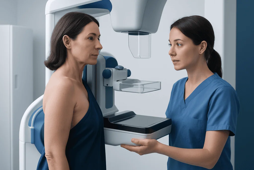
A diagnostic mammogram is done to investigate a breast change or to clarify a finding seen on a screening mammogram. It is more focused than screening and is designed to answer a specific clinical question. For example, if a screening study shows an asymmetry, calcifications, or a possible mass, diagnostic imaging can help determine whether the finding is benign or whether more testing is needed.
Doctors may also request this exam when a person notices symptoms between routine screenings. These symptoms can include:
- A new breast lump or thickened area
- Changes in breast size or shape
- Persistent pain in one part of the breast
- Nipple inversion or new nipple retraction
- Nipple discharge, especially if spontaneous or bloody
- Skin dimpling, redness, or scaling
People with a previous breast condition, prior surgery, or breast cancer treatment may also need diagnostic mammography for follow-up. In some cases, the radiologist combines it with breast ultrasound to better assess a lump or to distinguish a fluid-filled cyst from a solid area. The choice of tests depends on age, breast density, symptoms, and the appearance of the imaging finding.
What Happens During the Procedure
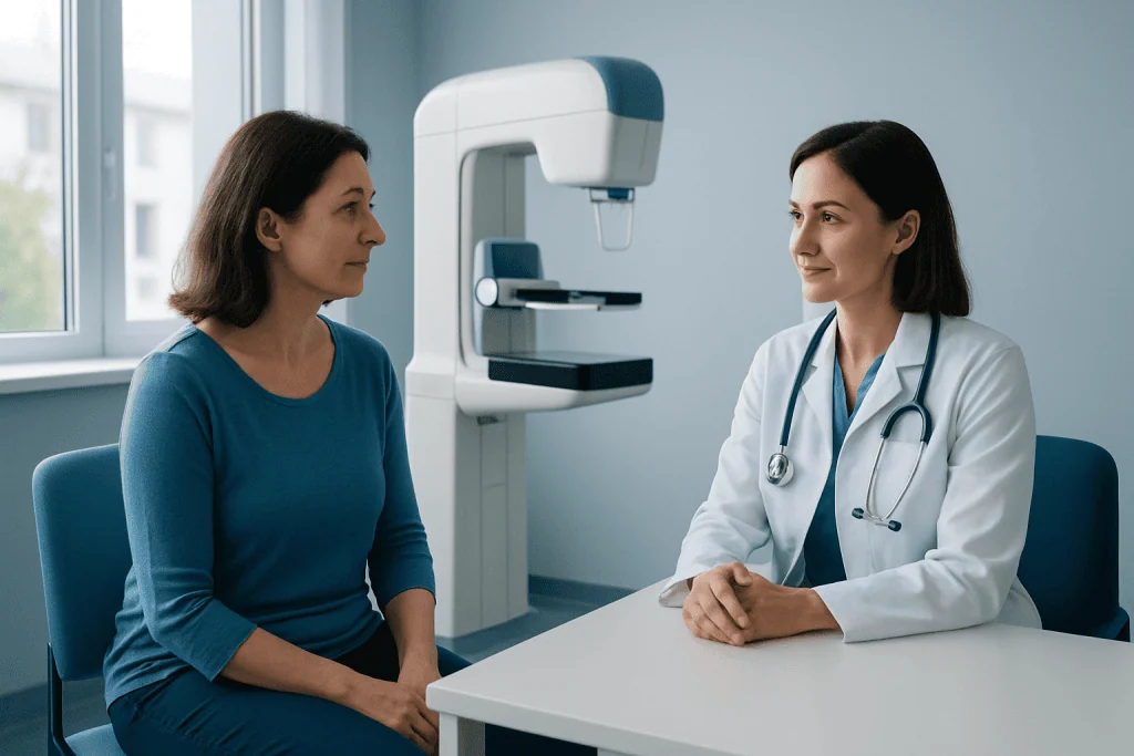
A diagnostic mammogram is usually performed in an imaging center or hospital radiology department. Before the exam, the patient will be asked to undress from the waist up and wear a gown. It is often helpful not to use deodorant, talcum powder, or lotions on the chest or underarm area on the day of the test, because some products can appear on the images.
During the exam, each breast is placed on the mammography machine and gently compressed with a clear plate. Compression lasts only a few seconds for each image. It can feel uncomfortable, but it helps spread the breast tissue, reduce motion, and improve image quality while keeping radiation dose as low as reasonably possible.
Compared with screening mammography, diagnostic mammography usually includes extra views. These may include magnification views, spot compression images, or images from different angles. Sometimes the radiologist reviews the pictures while the patient is still in the department and asks for more images. If needed, the same visit may include mammography follow-up views or additional breast imaging tests to complete the evaluation.
The appointment length can vary, but it is usually longer than a screening mammogram because of the extra imaging and possible same-day review. Many people are able to return to normal activities right away after the exam.
How to Prepare and What It Feels Like
Preparation for a diagnostic mammogram is simple. If possible, it can help to schedule the exam for a time when the breasts are less tender, such as a week after a menstrual period. Bringing prior mammogram images or making sure they are available to the radiology team is also useful, because comparison with earlier studies often helps clarify whether a finding is stable or new.
Patients should tell the imaging team if they are pregnant, breastfeeding, have breast implants, or have had prior breast surgery. They should also mention the exact location of any symptom, such as a lump or focal pain. This information helps the radiologist target the exam more precisely.
Most people describe the sensation as pressure or tightness during compression rather than pain. The discomfort is brief and usually resolves as soon as the compression is released. If someone has very sensitive breasts, they can let the technologist know, who can often work with them to make the experience as comfortable as possible while still obtaining clear images.
Understanding the Results
Diagnostic mammogram results may be available the same day or shortly afterward, depending on the facility. The radiologist evaluates the shape, margins, density, and pattern of any abnormality, as well as the presence of calcifications or architectural distortion. Findings may be described using the BI-RADS system, which helps standardize breast imaging reports and recommendations.
In many cases, the result is benign or probably benign. The radiologist may determine that the area is normal overlapping tissue, a simple cyst, stable calcifications, or another noncancerous change. Sometimes short-interval imaging follow-up is recommended to confirm stability over time rather than moving directly to a biopsy.
If the images are suspicious or incomplete, further tests may be recommended. These can include breast MRI in selected situations or a tissue sample with biopsy when imaging suggests that a closer diagnosis is needed. An abnormal result does not automatically mean cancer; it means the finding needs appropriate follow-up so the care team can reach a clear diagnosis.
Possible Next Steps After a Diagnostic Mammogram
The next step depends on what the mammogram shows. If the finding appears clearly benign, no immediate action may be needed beyond returning to routine screening. If the finding is probably benign, the radiologist may suggest repeat imaging in a few months to confirm that it stays unchanged.
If the area is not fully explained by mammography alone, another imaging test may help. Ultrasound is commonly used for a palpable lump, especially when the mammogram is inconclusive or when the breast tissue is dense. MRI may be useful in more complex cases, such as evaluating the extent of a known diagnosis or looking more closely at certain high-risk findings.
When tissue confirmation is needed, a biopsy is usually recommended. The type of biopsy depends on how the abnormality is best seen on imaging. For example, some areas are easiest to sample with ultrasound guidance, while others may need mammographic or MRI guidance. If cancer is diagnosed, the patient may then be referred for specialized care for conditions such as breast cancer.
For international patients who need coordinated evaluation, Acibadem International’s multidisciplinary specialists and JCI-accredited hospitals provide diagnosis and treatment pathways that may include advanced breast imaging and biopsy when appropriate.
Limitations, Safety, and Radiation
Diagnostic mammography is a valuable and widely used test, but like all imaging, it has limits. Some breast changes are easier to detect than others, and dense breast tissue can make interpretation more challenging. That is one reason why additional imaging may be recommended even after a mammogram has been performed.
The exam uses a low dose of ionizing radiation. For most patients, the benefit of evaluating a concerning breast finding outweighs the small radiation exposure. Imaging teams follow safety standards and use the lowest dose needed to produce high-quality images.
False-positive and false-negative results can happen. A false-positive result means imaging suggests a problem that later turns out to be benign. A false-negative result means a finding is missed on imaging. Because of this, patients should continue to report any ongoing or new breast symptoms to a doctor, even if imaging results are reassuring.
When to See a Doctor
A person should contact a doctor if they notice a new breast lump, persistent focal breast pain, nipple discharge, skin dimpling, redness that does not improve, or changes in the nipple or breast shape. These symptoms do not always indicate a serious condition, but they should be assessed so the right imaging or examination can be arranged.
It is also important to follow up promptly after an abnormal screening mammogram. Most call-backs do not lead to a cancer diagnosis, but timely diagnostic imaging helps avoid uncertainty and ensures that any issue is addressed early. Delaying follow-up can make care more complicated if a significant finding is present.
Patients should feel comfortable asking the care team what the images showed, whether additional tests are needed, and when results will be available. Clear communication can make the process easier and help patients understand the reason for each step in their care.
Frequently asked questions
What is the difference between a screening and a diagnostic mammogram?
A screening mammogram is a routine test for people without breast symptoms. A diagnostic mammogram is done when there is a symptom or when a screening mammogram finds an area that needs a closer look. It uses more targeted images to evaluate a specific concern.
Does a diagnostic mammogram mean cancer is likely?
No. A diagnostic mammogram means more detailed evaluation is needed, not that cancer is certain or even likely. Many findings turn out to be benign, such as cysts, overlapping tissue, or noncancerous calcifications.
How long does a diagnostic mammogram take?
It usually takes longer than a screening mammogram because extra images are often needed. The exact time varies, but many appointments last between 30 minutes and an hour, especially if the radiologist reviews the images during the visit.
Is a diagnostic mammogram painful?
Most people feel brief pressure or discomfort when the breast is compressed. The sensation lasts only a few seconds for each image. Telling the technologist about breast tenderness can help them make the exam as comfortable as possible.
Can a diagnostic mammogram be done with breast implants?
Yes. People with breast implants can still have diagnostic mammography. The imaging team may use special techniques and additional views to examine as much breast tissue as possible.
What happens if the diagnostic mammogram is abnormal?
The radiologist may recommend more imaging, such as ultrasound or MRI, or may advise a biopsy if tissue sampling is needed. An abnormal result does not by itself confirm cancer. It simply guides the next step in making an accurate diagnosis.
This article is for general information only and is not a substitute for professional medical advice. Please consult a qualified doctor about your individual situation.
Explore treatments in Turkey — costs, top hospitals & a free quote
JCI-accredited · board-certified surgeons · reply within 24h
Add us as a Preferred Source to see more of our trusted health content across Google Search, AI Overviews and Discover.

More from the Health Library

Neuropsychological Testing: Memory, Attention, and Brain Function Assessment

Understanding Abnormal Lab Results: Common Causes and Next Steps

Walk-In Diagnostic Testing: When You Can Skip an Appointment

Thyroid Ultrasound: Nodules, Goiter, and When Biopsy Is Needed

Blood Test or Imaging First? How Doctors Decide