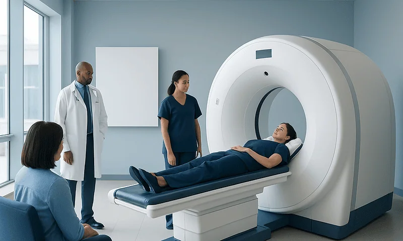
When to Get Imaging for Breast Symptoms: Lumps, Pain, and Nipple Changes

A new breast lump, persistent focal pain, or nipple changes may need imaging, even when symptoms seem mild. Mammography and breast ultrasound are the most common first-line imaging tests, and the best choice depends on age, symptoms, and exam findings.
Key Takeaways
- A new breast lump, persistent focal pain, or nipple changes may need imaging, even when symptoms seem mild.
- Mammography and breast ultrasound are the most common first-line imaging tests, and the best choice depends on age, symptoms, and exam findings.
- Most breast symptoms are caused by benign conditions, but imaging helps clarify whether follow-up, treatment, or biopsy is needed.
- Urgent medical review is important for bloody nipple discharge, skin dimpling, breast redness with fever, or a rapidly changing lump.
- Breast self-awareness is useful, but imaging decisions should be guided by a qualified clinician.
Breast symptoms are common, and many causes are not serious, but some changes should be assessed with imaging. Knowing when to seek evaluation for a lump, pain, or nipple change can help a person get the right care without unnecessary delay.
Overview: Why Breast Symptoms Sometimes Need Imaging
Breast symptoms are very common across different ages. A person may notice a lump, tenderness, nipple discharge, skin thickening, or a change in the shape of the breast. In many cases, these symptoms are linked to benign causes such as hormonal changes, cysts, or inflammation. Even so, imaging can be an important next step because symptoms alone cannot always show exactly what is happening inside the breast tissue.
Breast imaging for symptoms is used to look more closely at an area of concern and to help doctors decide whether the finding appears harmless, needs short-term follow-up, or requires additional testing. The main tools are mammography, breast ultrasound, and in selected cases breast MRI. These tests do not replace a clinical examination; rather, they work together with the medical history and physical exam.
Imaging is not only for suspected cancer. It is also used to evaluate pain that is limited to one spot, to assess nipple discharge, to confirm whether a lump is a fluid-filled cyst, and to investigate swelling or redness. For many people, imaging provides reassurance by showing that a symptom has a benign explanation.
Symptoms That Should Be Evaluated
Some breast symptoms deserve timely medical attention because they are new, persistent, or clearly different from a person’s usual pattern. A new lump is one of the most common reasons for imaging. Even when a lump feels smooth or mobile, it is still sensible to have it assessed, especially if it remains after a menstrual cycle or appears after menopause.
Breast pain is also common and is often related to hormones, chest wall strain, or benign tissue changes. Generalized pain in both breasts that comes and goes with the menstrual cycle is less likely to need imaging on its own. However, pain that is focused in one specific area, continues over time, or is associated with a lump, skin change, or nipple symptom is more likely to need evaluation.
Nipple changes should not be ignored. These include spontaneous discharge, especially if it is bloody or comes from one duct; new nipple inversion; crusting; scaling; or skin irritation that does not improve. Other symptoms that may need imaging include breast swelling, skin dimpling, warmth, redness, or a change in breast size or contour. These features can be caused by infection or other benign conditions, but they still need professional assessment.
- New or growing breast lump
- Persistent pain in one area of the breast
- Bloody, clear, or spontaneous nipple discharge
- New nipple inversion or visible nipple distortion
- Skin dimpling, thickening, redness, or swelling
- Changes that continue after a menstrual period or occur after menopause
What Can Cause Lumps, Pain, and Nipple Changes?

Many breast symptoms have benign explanations. Lumps may be caused by cysts, fibroadenomas, fat necrosis, or normal dense glandular tissue. Breast pain may come from cyclical hormonal changes, muscle strain, ill-fitting bras, or benign breast conditions. Nipple discharge can occur with hormonal shifts, benign intraductal growths, or breastfeeding-related changes.
Inflammation and infection can also cause breast symptoms. Mastitis or a breast abscess may lead to redness, warmth, pain, and swelling, particularly during breastfeeding, though they can occur outside lactation as well. Some people may have symptoms related to prior surgery, trauma, or implanted devices, which can alter breast tissue and create new palpable findings.
While many causes are not dangerous, breast cancer is one of the conditions doctors want to rule out when symptoms are new or concerning. Cancer may present as a hard or irregular lump, skin dimpling, nipple retraction, or unusual discharge, but it can also be subtle. Imaging helps distinguish between findings that look benign and those that need more investigation. In some situations, doctors may also consider related conditions such as breast cancer or inflammation of the breast tissue when planning evaluation.
Which Imaging Tests Are Used?
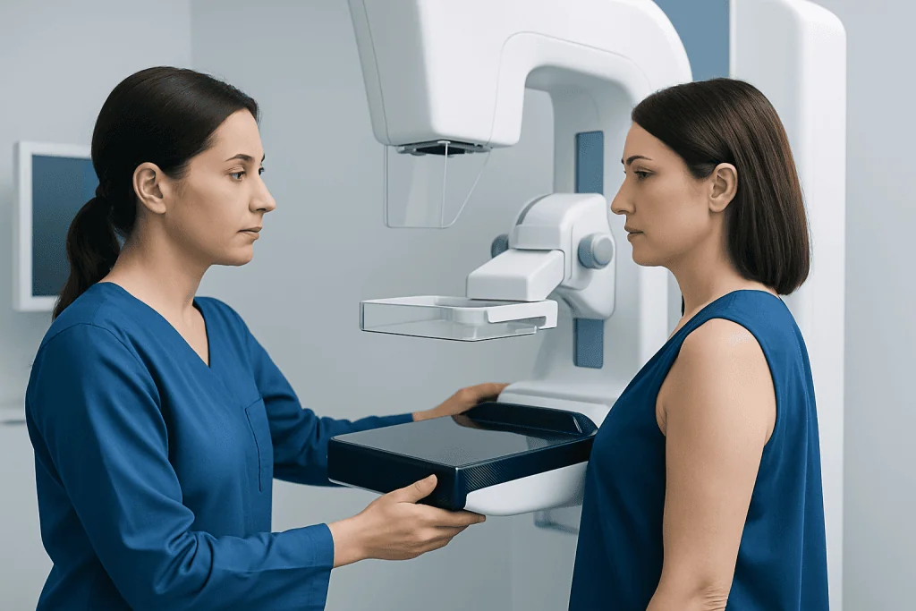
The most common tests used for breast imaging for symptoms are diagnostic mammography and breast ultrasound. Diagnostic mammography is an X-ray exam that focuses on the area of concern and may include additional views compared with routine screening. It is often used for adults over 30 to 40, depending on the symptom and local practice. Breast ultrasound uses sound waves and is especially helpful for distinguishing solid masses from cysts and for evaluating younger patients with denser breast tissue.
Ultrasound is often the first imaging test for younger people with a new lump, for pregnant patients, or for a very localized area of concern. Mammography may still be added when appropriate. When nipple discharge, skin changes, or a suspicious finding needs closer evaluation, doctors may recommend more than one imaging method because each test provides different information.
Breast MRI is not usually the first test for common symptoms, but it can be useful in selected situations. It may be considered when other imaging results are unclear, when there is very high risk, or when a doctor needs more detail about the extent of a finding. If a suspicious area is found, a tissue sample may be recommended with breast biopsy to provide a definite diagnosis. In some cases, patients may undergo diagnostic mammography or breast ultrasound as part of the initial workup.
How Doctors Decide When Imaging Is Needed
The decision to order imaging depends on several factors, including age, the type of symptom, whether the finding is new, how long it has been present, and what is felt on examination. A clinician will ask when the symptom started, whether it changes with the menstrual cycle, whether there has been trauma or breastfeeding, and whether there is a personal or family history of breast disease.
A new palpable lump usually requires imaging, even if it does not feel alarming. Persistent focal pain may also be evaluated, especially if it affects one small area or comes with another change. Spontaneous unilateral nipple discharge, nipple inversion that is new, or skin dimpling often leads to prompt imaging because these symptoms can signal an underlying structural problem.
By contrast, generalized breast tenderness affecting both breasts in a cyclical pattern may not always need imaging if the clinical exam is otherwise normal and the person is up to date with routine screening. Even then, the decision is individualized. If symptoms continue, worsen, or do not fit a typical benign pattern, further assessment is appropriate rather than waiting indefinitely.
What to Expect From Results and Next Steps
After imaging, results may show a clearly benign cause such as a simple cyst or stable fibrous tissue. In these situations, a doctor may recommend reassurance, symptom management, or routine follow-up. Sometimes the report suggests a short-interval repeat scan to confirm that an area remains stable over time.
If the findings are indeterminate or suspicious, additional imaging or biopsy may be advised. This does not automatically mean cancer; it means the radiologist has seen something that needs a closer look. Biopsy is the only way to confirm the exact nature of a concerning finding. Many biopsies ultimately show benign conditions.
For symptoms caused by infection, treatment may include antibiotics, drainage, or further evaluation if symptoms do not improve. If a benign lump is causing discomfort or growing, treatment options may vary depending on its type. When specialist care is needed, a breast clinic may coordinate imaging, pathology, surgery, and follow-up in one plan. Near the end of the care pathway, some patients may be referred for breast MRI if additional detail is needed.
Self-Care, Monitoring, and Routine Screening
Self-care can be helpful, but it should not replace medical assessment for a concerning symptom. Supportive bras, simple pain relief recommended by a doctor, and keeping a symptom diary can be useful for breast discomfort. A diary may help show whether pain is linked to the menstrual cycle, exercise, or a specific area in the breast.
Breast self-awareness is more useful than intensive self-examination routines for many people. This means knowing the usual look and feel of the breasts and reporting any new or persistent change. It is not necessary to panic over every variation, but changes that remain for more than one cycle or clearly stand out from the usual pattern should be discussed with a clinician.
Routine screening is still important even when no symptoms are present. Screening mammograms look for early changes before they can be felt, while symptom-based imaging investigates a specific concern. These are different but complementary parts of breast health. Acibadem International’s multidisciplinary specialists and JCI-accredited hospitals evaluate breast symptoms for international patients and coordinate imaging and treatment when needed.
When to See a Doctor Promptly
A person should arrange medical review promptly if they notice a new breast lump, a lump that is enlarging, or a change that persists beyond one menstrual cycle. Prompt review is also important for spontaneous nipple discharge, especially if it is bloody or only from one side, and for nipple inversion that has newly developed.
Redness, warmth, swelling, and pain with fever may suggest infection and should be assessed without delay, particularly if symptoms are worsening. Skin dimpling, orange-peel texture, or a breast that appears noticeably different in shape or size also deserves prompt evaluation. These signs do not always mean a serious illness, but they should not be ignored.
Immediate medical care may be needed for severe pain, rapidly progressing swelling, or signs of a significant infection. In general, it is better to seek assessment early than to wait and wonder. Early evaluation often leads either to reassurance or to timely treatment if something does need attention.
Frequently asked questions
Does every breast lump need imaging?
Most new breast lumps should be assessed by a clinician, and many will need imaging to identify the cause. Even when a lump feels benign, imaging helps determine whether it is a cyst, a solid benign mass, or something that needs biopsy.
Is breast pain alone a sign of cancer?
Breast pain by itself is more often related to benign causes such as hormonal changes or chest wall strain. However, pain that is persistent, limited to one area, or associated with a lump or skin change should be evaluated.
What is the difference between screening and diagnostic breast imaging?
Screening imaging is done routinely when a person has no symptoms, usually to detect early changes before they can be felt. Diagnostic imaging is performed to investigate a specific symptom or finding, such as a lump, focal pain, or nipple discharge.
Will a young person with a breast lump always need a mammogram?
Not always. In younger patients, ultrasound is often the first test because breast tissue tends to be denser, and ultrasound can be very helpful for evaluating lumps. A mammogram may still be added depending on the situation and the clinician’s judgment.
Is nipple discharge always serious?
No, nipple discharge can occur for several benign reasons, especially with hormonal changes or irritation. Still, discharge that is spontaneous, bloody, clear, one-sided, or coming from a single duct should be assessed promptly.
If imaging is abnormal, does that mean cancer?
No. An abnormal result means that the radiologist saw a finding that needs clarification or follow-up. Many abnormal breast imaging results are ultimately explained by benign conditions, but biopsy may be needed to be certain.
This article is for general information only and is not a substitute for professional medical advice. Please consult a qualified doctor about your individual situation.
Explore treatments in Turkey — costs, top hospitals & a free quote
JCI-accredited · board-certified surgeons · reply within 24h
Add us as a Preferred Source to see more of our trusted health content across Google Search, AI Overviews and Discover.

More from the Health Library

Contrast Dye Side Effects: What Is Normal and When to Seek Medical Help

What Is a Lab Test Panel and Why Are Tests Grouped Together?
Cerebral Angiography for Brain Vessel Disorders: Preparation, Procedure, and Recovery

How to Prepare for a Medical Imaging Test: Fasting, Contrast, and Medications

Quest Diagnostics Walk-In Visits: What Patients Should Know