

CT Scan vs MRI: Which Imaging Test Is Right for You?

CT uses X-rays and is often chosen for speed, emergencies, and imaging bones, lungs, or internal bleeding. MRI uses magnets and radio waves and is especially useful for soft tissues such as the brain, spine, joints, and organs.
Key Takeaways
- CT uses X-rays and is often chosen for speed, emergencies, and imaging bones, lungs, or internal bleeding.
- MRI uses magnets and radio waves and is especially useful for soft tissues such as the brain, spine, joints, and organs.
- MRI does not use ionizing radiation, while CT does involve radiation exposure.
- The best test depends on the symptoms, body area, medical history, and whether contrast dye can be used safely.
- People with certain implants, kidney problems, pregnancy concerns, or claustrophobia may need special planning before imaging.
CT scans and MRI scans are both important imaging tests, but they work in different ways and are used for different medical needs. The right choice depends on what the doctor is looking for, how quickly answers are needed, and the patient’s individual health situation.
Overview: How CT and MRI Differ
When a doctor needs a detailed look inside the body, a CT scan or an MRI scan may be recommended. Both tests create images of internal structures, but they do so in different ways and are used for different reasons. Understanding the difference can help patients feel more prepared and confident when discussing imaging options with their care team.
A CT scan, also called computed tomography, uses X-rays and computer processing to create cross-sectional images of the body. It is fast, widely available, and especially helpful when quick answers are needed. CT is commonly used in emergency care and for evaluating bones, the chest, the abdomen, and possible internal bleeding.
MRI stands for magnetic resonance imaging. Instead of X-rays, it uses a strong magnetic field and radio waves to produce highly detailed images, especially of soft tissues. MRI is often preferred when doctors need a closer look at the brain, spinal cord, muscles, ligaments, joints, or certain organs.
Neither test is universally “better” than the other. In many cases, the right imaging study depends on the body part being examined, the suspected condition, how urgently results are needed, and whether there are safety concerns such as metal implants or pregnancy. Sometimes both tests may be useful at different stages of diagnosis or treatment.
When a CT Scan May Be the Better Choice
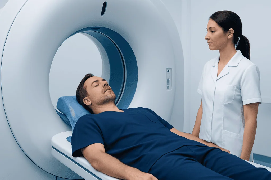
CT scans are often chosen when speed matters. The scan itself usually takes only a few minutes, which makes it especially valuable in emergency situations. Doctors may order CT when they need to quickly assess trauma, stroke symptoms, severe pain, suspected bleeding, or injuries after an accident.
CT is also very effective for showing bones, lung tissue, and many abdominal structures. It can help detect fractures, kidney stones, appendicitis, certain infections, and tumors. In some cases, a CT scan with contrast dye provides even clearer images of blood vessels and organs, helping doctors identify blockages, inflammation, or abnormal growths.
Because CT uses X-rays, it does involve exposure to ionizing radiation. For many patients, the benefit of getting an accurate diagnosis outweighs this risk, especially when the scan is medically necessary. However, doctors generally avoid unnecessary repeated CT scans and may consider other options, particularly for children or people who need frequent imaging.
CT may be especially useful in situations involving suspected stroke, chest emergencies, or urgent abdominal symptoms. It is also commonly used to guide procedures, monitor injuries, and support planning for surgery or other treatments.
When an MRI May Be the Better Choice

MRI is often preferred when the main goal is to examine soft tissues in greater detail. This includes the brain, spinal cord, nerves, muscles, tendons, ligaments, cartilage, and many internal organs. Because MRI can show subtle differences in tissue structure, it is especially valuable when doctors are evaluating chronic pain, neurological symptoms, joint injuries, or suspected tumors.
For example, MRI can be very helpful in assessing disc problems in the spine, ligament tears in the knee, or inflammation in the brain. It may be used when symptoms suggest conditions such as multiple sclerosis, certain liver or pelvic disorders, or joint damage not visible on standard X-rays. It is also commonly used for follow-up imaging when doctors need a more detailed view after other tests.
Unlike CT, MRI does not use ionizing radiation. This can make it an attractive choice for some patients, particularly when repeated imaging may be needed over time. However, MRI generally takes longer than CT and requires the patient to remain very still inside the scanner, which can be challenging for some people.
MRI may be chosen to evaluate problems such as a brain tumor or to assess the spine before spine surgery. It can also support care planning in orthopedic and neurological conditions where soft-tissue detail is essential.
Safety, Comfort, and Preparation
Before either test, patients are usually asked about allergies, kidney function, pregnancy, and any previous reactions to contrast dye. Contrast material may be given by vein, by mouth, or occasionally by other routes to make certain structures easier to see. Not every CT or MRI requires contrast, and the decision depends on the medical question being asked.
CT contrast often contains iodine, while MRI contrast commonly uses gadolinium-based agents. Both are generally used carefully and safely, but they may not be suitable for everyone. People with significant kidney disease, a history of contrast reactions, or certain other medical conditions may need special precautions or an alternative imaging approach.
MRI requires extra safety screening because the scanner’s magnetic field can interact with metal in or on the body. Patients should tell the imaging team about pacemakers, aneurysm clips, cochlear implants, metal fragments, insulin pumps, artificial joints, or any previous surgery. Many modern implants are MRI-compatible, but they still need to be checked in advance.
Comfort is another important factor. CT is quick and open, which may be easier for people with claustrophobia. MRI is noisier and usually longer, often taking 20 to 60 minutes depending on the study. Some patients benefit from reassurance, relaxation techniques, or mild sedation if approved by their doctor. Wearing comfortable clothing and following fasting instructions, if given, can also make the process smoother.
How Doctors Decide Which Imaging Test to Order
Choosing between CT scan vs MRI is rarely a simple matter of preference. Doctors select the test that is most likely to answer a specific clinical question. They consider the patient’s symptoms, age, medical history, physical exam findings, and how quickly a diagnosis is needed. Cost and availability may also play a role, but the main goal is to obtain the clearest and most useful information safely.
For example, a patient with sudden severe headache or head injury may first need CT because it is fast and effective for detecting acute bleeding. A patient with ongoing numbness, weakness, or seizures may be more likely to need MRI because it provides greater detail of the brain and nerves. In musculoskeletal care, CT may show fractures well, while MRI is often better for tendon, ligament, or cartilage injuries.
Doctors may also use imaging in sequence. An ultrasound, X-ray, or CT may be the first step, followed by MRI if more detail is needed. In some situations, imaging is paired with procedures such as interventional radiology or used to plan neurosurgery or other treatments.
Patients should feel comfortable asking why a particular test is being recommended. Helpful questions include what the scan is expected to show, whether contrast is needed, how to prepare, and whether another test could provide similar information. Clear communication can reduce anxiety and help patients take an active role in their care.
What to Expect During and After the Scan
For a CT scan, the patient usually lies on a narrow table that moves through a ring-shaped scanner. The test is painless, and the technologist may ask the patient to hold their breath briefly while images are taken. If contrast is used, there may be a warm sensation or a metallic taste for a short time, which usually passes quickly.
For an MRI, the patient lies on a table that slides into a tunnel-like machine. The scanner makes loud tapping or thumping sounds, so ear protection is usually provided. The patient needs to stay still so the images are sharp. In some cases, a contrast injection is given during the test.
Most people can return to normal activities soon after either scan unless they received sedation. Drinking fluids may be advised after contrast in some situations, depending on the doctor’s instructions. The images are reviewed by a radiologist, who sends a report to the referring doctor. That doctor then explains the results and what they mean for the next steps.
If a patient feels anxious about enclosed spaces, lying flat, or the possibility of contrast reactions, it is best to mention this ahead of time. Imaging teams are experienced in helping patients prepare and can often offer practical solutions to make the test more comfortable.
When to Speak With a Doctor and Getting the Right Support
A patient should speak with a doctor if they have symptoms that need investigation, such as severe or persistent pain, unexplained neurological changes, injury, or signs of a serious internal problem. Imaging tests are tools that help confirm or rule out a diagnosis, but the choice of test should always be guided by a qualified professional.
It is also important to seek medical advice before the scan if there is pregnancy, kidney disease, a history of contrast allergy, or an implanted medical device. Bringing previous imaging reports to appointments can help avoid unnecessary repeat testing and allow doctors to compare changes over time.
For international patients or those seeking coordinated care, multidisciplinary evaluation can be helpful when imaging findings are complex. Acibadem International’s multidisciplinary specialists and JCI-accredited hospitals diagnose and treat a wide range of conditions, helping patients understand which imaging tests are most appropriate and what the results may mean.
In general, CT is often best for speed and urgent assessment, while MRI is often best for detailed soft-tissue evaluation. The most suitable test is the one that answers the medical question clearly and safely. A doctor can explain the reasoning and guide each patient toward the right choice for their situation.
Frequently asked questions
Which is better, a CT scan or an MRI?
Neither test is always better in every situation. CT is often better for speed, emergencies, bones, lungs, and bleeding, while MRI is often better for soft tissues such as the brain, spine, joints, and nerves. The best choice depends on what the doctor needs to evaluate.
Does a CT scan use radiation?
Yes, a CT scan uses X-rays, which are a form of ionizing radiation. Doctors generally order CT only when the expected medical benefit outweighs the risk. They also try to keep radiation exposure as low as reasonably possible.
Does MRI use radiation?
No, MRI does not use ionizing radiation. It creates images using a magnetic field and radio waves. This is one reason MRI may be preferred when repeated imaging is needed, although it is not suitable for everyone.
Why would a doctor order an MRI after a CT scan?
A CT scan may provide a fast overview, but MRI can sometimes show soft tissues in greater detail. Doctors may order MRI if they need more information about the brain, spinal cord, joints, muscles, or certain organs. Using both tests can help create a clearer diagnosis.
Can someone with claustrophobia have an MRI?
Yes, many people with claustrophobia can still complete an MRI with support. Options may include reassurance, breathing techniques, music, a friend or family member nearby when allowed, or mild sedation if medically appropriate. It is important to tell the imaging center in advance.
How long do CT and MRI scans take?
CT scans are usually quick and may take only a few minutes, although preparation can add time. MRI scans are typically longer and may take 20 to 60 minutes or sometimes more, depending on the body area and whether contrast is used.
References
- World Health Organization
- Radiological Society of North America
- American College of Radiology
- National Institute of Biomedical Imaging and Bioengineering
- NHS
This article is for general information only and is not a substitute for professional medical advice. Please consult a qualified doctor about your individual situation.
Explore treatments in Turkey — costs, top hospitals & a free quote
JCI-accredited · board-certified surgeons · reply within 24h
Add us as a Preferred Source to see more of our trusted health content across Google Search, AI Overviews and Discover.

More from the Health Library



Related Specialists

Prof. Dr. Ercan Karaarslan
Radiology
Prof. Dr. Bora Peynircioğlu
Interventional Radiology
Prof. Dr. Koray Güven
Interventional Radiology
Assoc. Prof. Dr. Deniz Can Aliş
Radiology