Brain Tumor MRI: Preparation, Procedure and Results
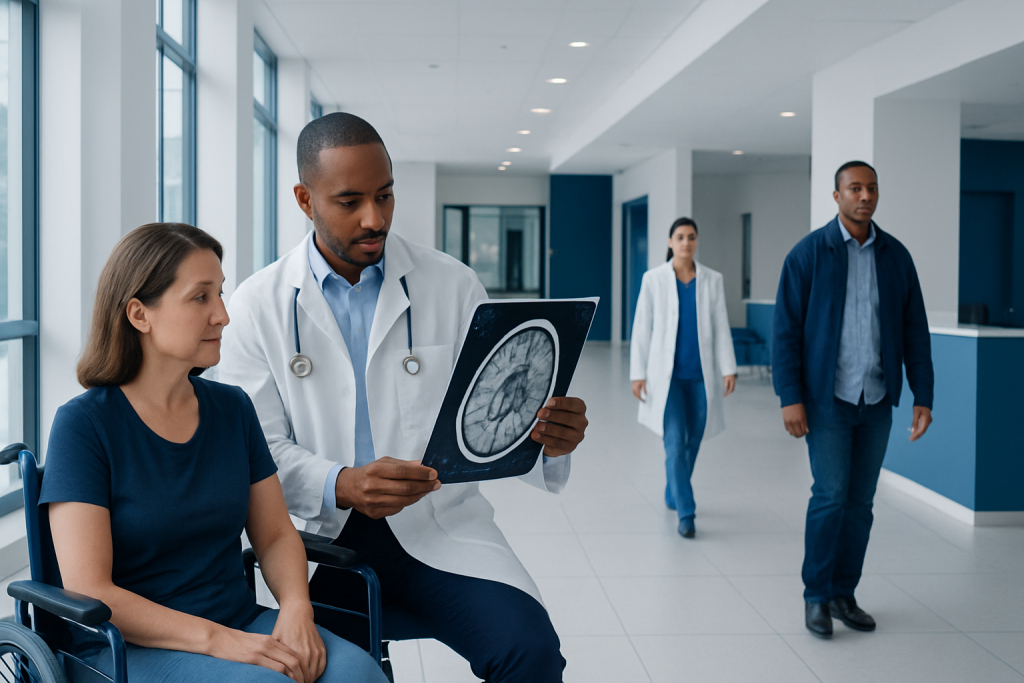
A brain MRI uses magnets and radio waves rather than ionising radiation. A contrast injection may be used to show blood vessels, inflammation and tumour-related changes more clearly.
Key Takeaways
- A brain MRI uses magnets and radio waves rather than ionising radiation.
- A contrast injection may be used to show blood vessels, inflammation and tumour-related changes more clearly.
- Most people can return to normal activities immediately after a routine MRI.
- MRI can strongly suggest tumour features, but it cannot always determine whether a tumour is cancerous without tissue testing.
- Patients should tell the MRI team about implants, metal fragments, kidney problems, pregnancy and prior contrast reactions.
A brain tumor MRI is a painless imaging examination that produces detailed pictures of the brain and surrounding structures. It can identify or characterize an abnormal area, but imaging findings may need to be combined with clinical assessment and, sometimes, biopsy results to confirm a diagnosis.
Overview: What a Brain Tumor MRI Shows
A brain tumor MRI is an imaging test that uses a powerful magnetic field and radio waves to create detailed pictures of the brain. It is commonly used when symptoms, examination findings or another scan suggest a possible brain abnormality. The examination helps doctors assess the location, size and appearance of a lesion and its relationship to nearby brain tissue, nerves and blood vessels.
MRI does not use X-rays or ionising radiation. Several image sequences are taken because each provides different information about brain anatomy, fluid, blood products, swelling and tissue characteristics. In many cases, a contrast agent called gadolinium is given through a small intravenous line to make certain tissues and blood vessels more visible.
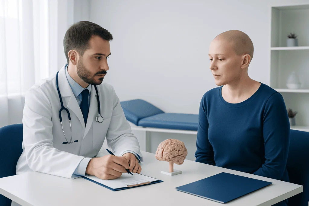
A scan result is one part of a wider assessment. Neurologists, radiologists, neurosurgeons and oncology specialists may consider a person’s symptoms, neurological examination, previous imaging and laboratory findings together. When a tumour is suspected, further evaluation may include monitoring scans, specialist consultation or a biopsy to examine tissue under a microscope.
How the Scan Works and Who May Need It
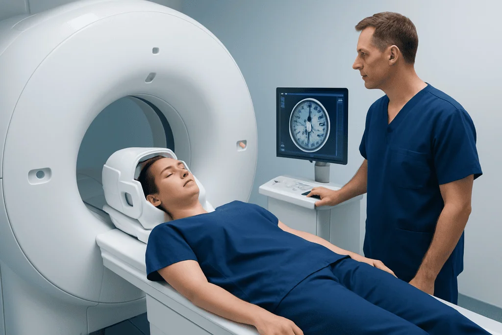
During MRI, the body is placed within a magnet and exposed to carefully controlled radiofrequency signals. The scanner detects signals from tissues in the body and a computer turns them into cross-sectional images. For brain imaging, the head is gently supported in a special holder to reduce movement and improve image quality.
A brain MRI may be recommended for persistent or unusual headaches, seizures, changes in vision, balance problems, weakness, numbness, speech changes or altered thinking. It may also be used to investigate a known lesion, monitor a diagnosed brain tumour, assess treatment response or plan surgery or radiotherapy.
Most people can have an MRI safely, but screening is essential. Some implanted devices, such as certain pacemakers, neurostimulators, cochlear implants, aneurysm clips or older metallic implants, may not be MRI-compatible. The imaging team will confirm the type of implant and decide whether MRI is appropriate or whether special arrangements are needed.
People with severe claustrophobia, difficulty lying flat or conditions that make it hard to remain still should discuss this before the appointment. Adjustments such as reassurance, relaxation strategies, an open MRI where available, or clinician-prescribed sedation may be considered when appropriate.
Preparation: What Should You Not Do Before a Brain MRI?
Before a brain MRI, patients should not bring metal objects into the scan room. Jewellery, watches, hairpins, hearing aids, removable dental appliances, coins, cards with magnetic strips and some clothing with metallic fasteners must be removed. Cosmetics, hair products and tattoos can occasionally contain metallic pigments, so it is helpful to mention them to the MRI staff.
Patients should not assume that an implant or previous surgery is safe for MRI without checking. They should tell the team about any pacemaker, implanted pump, clip, stent, joint replacement, dental implant, shrapnel injury or possible metal exposure at work. A history of metal fragments in the eyes is especially important and may require additional screening.
For most routine brain MRI examinations, people can eat, drink and take their usual medicines. However, fasting instructions may be given if sedation is planned or if another procedure is scheduled on the same day. Patients should follow the instructions provided by their imaging centre and inform the team about pregnancy, breastfeeding, kidney disease, dialysis, allergies and any previous reaction to MRI contrast.
If contrast may be used, kidney function may need to be reviewed in people with significant kidney impairment. The MRI team can explain whether blood testing is necessary and discuss the individual balance of benefits and risks.
What Happens During a Brain Tumor MRI?
On arrival, the patient completes a safety questionnaire and changes into suitable clothing if needed. An MRI radiographer reviews the answers, checks for metal and explains the process. If contrast is planned, a small intravenous cannula is usually placed in the hand or arm.
The patient lies on a padded table, usually on their back, with the head positioned comfortably in a head coil. Cushions, padding or light restraints may be used to help maintain stillness. Earplugs or headphones reduce the impact of the scanner’s loud tapping and knocking sounds, and the radiographer can communicate with the patient throughout the scan.
For a brain MRI, the head and upper body generally move into the scanner tunnel. The table position is adjusted so that the brain is centred within the imaging area. Although the space can feel enclosed, the scan is monitored continuously, and the patient is usually given an alarm device to call for assistance if necessary.
The examination often takes around 30 to 60 minutes, depending on the sequences needed and whether contrast is used. The most important task is remaining as still as possible, since movement can blur the images. If contrast is given, it is commonly injected partway through the scan, followed by additional image sequences.
Potential benefits include highly detailed images that can help distinguish tumour-related changes from other conditions and support treatment planning. MRI is generally safe, though some people experience temporary anxiety, noise discomfort or warmth. Contrast reactions are uncommon and usually mild, but staff are trained to identify and manage them.
Recovery and Results: How Soon After a Brain MRI Do You Get Results?
After a routine MRI, most people can leave soon after the examination and return to normal activities, including eating and drinking. If contrast was used, the body clears it naturally over time. Drinking fluids as normally advised by a clinician may be appropriate, unless the person has been told to limit fluids for another medical reason.
People who receive sedating medication need someone else to take them home and should avoid driving, alcohol, important decisions and operating machinery until the effects have fully worn off. The imaging centre will provide specific aftercare instructions. Any new rash, breathing difficulty or other concerning symptoms after contrast should be assessed urgently.
Image interpretation is performed by a radiologist, often a neuroradiologist with expertise in brain imaging. Timing varies by facility and clinical urgency. In an urgent hospital setting, preliminary findings may be available promptly; for a planned outpatient scan, a report may take several days. The referring clinician usually discusses the result and what it means in the context of the person’s health.
Some results identify a non-tumour explanation, such as inflammation, a vascular change or a benign structural finding. If an abnormal mass is seen, further tests may be needed. Care may involve specialists in brain tumours, neurology, neurosurgery, pathology and oncology. Acibadem International’s multidisciplinary specialists and JCI-accredited hospitals support diagnosis and treatment planning for international patients.
Can You Tell from an MRI if a Brain Tumour Is Cancerous?
MRI can provide important clues about whether a brain lesion may be more or less aggressive. Features such as its borders, growth pattern, surrounding swelling, blood supply, contrast enhancement and effect on nearby structures can help specialists estimate the likely type and grade of a tumour. Advanced MRI techniques may add information about tissue metabolism, cellularity and blood flow.
However, an MRI alone cannot always tell with certainty whether a brain tumour is cancerous. Some non-cancerous tumours can look concerning on imaging, while some cancerous tumours may have overlapping features. In addition, the term “cancerous” can be complex for brain tumours because their behaviour depends on tumour type, molecular features, location and grade.
When confirmation is needed, a neurosurgeon may recommend a biopsy or removal of part or all of the lesion where it is safe and appropriate. A pathologist examines the tissue and may perform molecular testing. These results help establish the diagnosis and guide decisions about observation, brain tumour surgery, radiotherapy, medicines or other treatment.
Not every lesion requires immediate biopsy. In selected situations, specialists may advise repeat imaging over time, particularly when a finding appears low risk or when surgery would carry substantial risk. The best approach is individualised through expert review.
When to Seek Medical Care
Persistent headaches are common and are not usually caused by a brain tumour. Still, medical assessment is appropriate for a new headache pattern that is worsening, unusually severe, associated with neurological symptoms or different from a person’s typical headaches. A clinician can assess symptoms and decide whether imaging, including MRI, is needed.
Urgent medical attention is important for a first seizure, sudden weakness or numbness on one side, new difficulty speaking, sudden confusion, loss of consciousness, abrupt severe headache, significant vision loss or repeated vomiting with worsening neurological symptoms. These symptoms can have several causes, some of which require prompt treatment.
Anyone who has been advised to have a brain MRI should arrange it within the recommended timeframe and share relevant medical history with the imaging team. If the scan identifies an abnormality, a follow-up appointment with the referring doctor is important even if symptoms improve.
When a confirmed or strongly suspected tumour requires treatment, a multidisciplinary discussion can help clarify choices. Depending on the diagnosis, care may include stereotactic radiosurgery or other targeted treatments alongside surgery, systemic therapy, rehabilitation and supportive care.
Frequently asked questions
Do you go all the way in the machine for a brain MRI?
For a brain MRI, the head and upper body usually need to be positioned inside the scanner tunnel so the brain is in the centre of the magnetic field. The legs may remain closer to the opening, depending on the scanner and the person’s height. Staff can communicate throughout the examination and can pause the scan if needed.
Is a brain tumor MRI painful?
A brain MRI is not painful. The person must lie still, and the scanner makes loud knocking or tapping noises, but ear protection is provided. If contrast is used, there may be a brief pinprick when the intravenous line is inserted.
How long does a brain MRI take?
Many brain MRI examinations take about 30 to 60 minutes. The exact duration depends on the number of image sequences, whether contrast is used and whether additional specialised imaging is needed. The radiographer can give a more specific estimate before the appointment.
Do I need contrast for a brain tumour MRI?
Contrast is often useful when investigating a possible brain tumour because it can make certain tissue changes and blood vessels easier to see. It is not necessary for every scan, and the decision depends on the clinical question and the person’s health history. The radiologist or referring clinician will determine whether it is appropriate.
Can a brain MRI miss a tumour?
MRI is one of the most detailed imaging tests for assessing the brain, but no test is perfect. Very small lesions, unusual tumour types or changes that develop after the scan may be harder to identify. If symptoms persist or change, a doctor may recommend further assessment or repeat imaging.
Can I drive home after a brain MRI?
Most people can drive and return home after a routine brain MRI, including one with contrast, if they feel well. Driving is not safe after sedation or anti-anxiety medicine until the medication’s effects have resolved. In that situation, a responsible adult should provide transport home.
References
- World Health Organization
- National Cancer Institute
- Radiological Society of North America
- American College of Radiology
- National Institute of Neurological Disorders and Stroke
This article is for general information only and is not a substitute for professional medical advice. Please consult a qualified doctor about your individual situation.
Oncology care in Turkey — second opinion and treatment plan
JCI-accredited · board-certified surgeons · reply within 24h
Add us as a Preferred Source to see more of our trusted health content across Google Search, AI Overviews and Discover.