Breast Biopsy: A Complete Clinical Guide for Patients

A breast biopsy helps explain an abnormal breast finding and does not automatically mean cancer. Most breast biopsies are minimally invasive needle procedures done with local anesthesia.
Key Takeaways
- A breast biopsy helps explain an abnormal breast finding and does not automatically mean cancer.
- Most breast biopsies are minimally invasive needle procedures done with local anesthesia.
- Imaging such as ultrasound, mammography, or MRI may be used to guide the biopsy accurately.
- Results usually describe whether tissue is benign, high-risk, or cancerous and may guide next steps.
- Mild bruising, soreness, and swelling are common after a biopsy and usually improve within days.
- Prompt medical review is important for a new lump, nipple discharge, skin changes, or abnormal breast imaging.
A breast biopsy is a test that removes a small sample of breast tissue so it can be examined under a microscope. It is the most reliable way to find out whether a breast lump, abnormal imaging finding, or other breast change is benign or needs further treatment.
Overview: What a Breast Biopsy Is and Why It Is Done
A breast biopsy is a procedure used to remove a small sample of breast tissue for laboratory examination. It is usually recommended after a breast lump, change in breast tissue, nipple discharge, skin change, or an abnormal mammogram, ultrasound, or MRI. The goal is to find out exactly what the tissue is, because imaging can suggest a problem but cannot always confirm the diagnosis.
For many patients, the word “biopsy” immediately raises concern about cancer. In practice, a breast biopsy is a diagnostic step, not a diagnosis by itself. Many biopsies show noncancerous conditions such as cysts, fibroadenomas, inflammation, fat necrosis, or other benign breast changes. Even so, biopsy remains important because it gives the most reliable tissue-based answer.
Breast biopsy can also help doctors classify abnormalities more precisely. In some cases, it identifies changes that are not cancer but may increase future risk or require closer follow-up. In others, it confirms breast cancer and provides information that helps plan treatment safely and accurately.
When a Breast Biopsy May Be Recommended
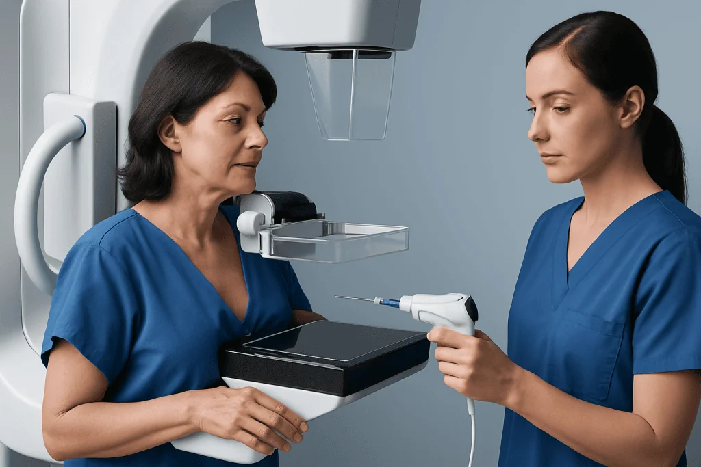
A doctor may suggest a breast biopsy when there is a finding that cannot be fully explained by physical examination or imaging alone. This includes a new breast lump, an area of thickening, a suspicious calcification pattern on mammography, a mass seen on ultrasound, or an abnormal area detected on MRI. A biopsy may also be recommended if a previously known lump has changed over time.
Symptoms can also lead to biopsy when they are persistent or concerning. These may include spontaneous nipple discharge, especially if it is bloody; dimpling or puckering of the skin; redness that does not improve; nipple inversion that is new; or a change in breast shape without a clear reason. Not all of these symptoms mean cancer, but they should be evaluated carefully.
Doctors use the biopsy result together with examination findings and imaging reports to make a complete assessment. If those three pieces of information match, planning is more straightforward. If they do not match, additional imaging or another biopsy may be needed to be sure the right area was sampled.
Types of Breast Biopsy
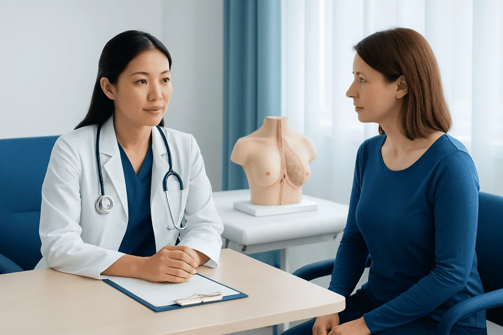
There is more than one kind of breast biopsy, and the best option depends on the size, location, and appearance of the breast change. The most common approach is a needle biopsy, which is less invasive than surgery and is often enough to make a diagnosis. It is usually done with local anesthesia, meaning the breast is numbed while the patient remains awake.
Fine-needle aspiration uses a very thin needle to remove fluid or cells. It may be used for some cysts or selected masses, but it gives less tissue than other methods. Core needle biopsy uses a larger hollow needle to remove small cylinders of tissue and is often preferred because it provides more detail for the pathologist. Vacuum-assisted biopsy is a form of needle biopsy that can collect multiple tissue samples through one small opening.
Some biopsies are guided by imaging. Ultrasound-guided biopsy is commonly used for masses visible on ultrasound. Stereotactic biopsy uses mammography to target calcifications or areas seen best on mammogram. MRI-guided biopsy is used when a finding is visible mainly on breast MRI. If a needle biopsy cannot reach the area or does not provide a clear answer, a surgical biopsy may be advised, sometimes as part of breast surgery.
- Fine-needle aspiration: removes fluid or cells through a very thin needle.
- Core needle biopsy: removes small tissue samples and is widely used.
- Vacuum-assisted biopsy: collects several samples through one incision.
- Surgical biopsy: removes part or all of a suspicious area when needed.
How to Prepare and What to Expect During the Procedure
Before a breast biopsy, the care team usually reviews symptoms, prior imaging, medical history, allergies, and current medicines. Patients may be asked about blood thinners or supplements that can increase bleeding risk. Specific instructions vary, so it is important to follow the advice given by the treating doctor or radiology team. Comfortable clothing and a supportive bra are often recommended for after the procedure.
During most needle biopsies, the skin is cleaned and numbed with local anesthesia. Imaging may be used to guide the needle precisely into the target area. The patient may feel pressure or a brief pushing sensation, but significant pain is uncommon. Several samples are usually taken to improve diagnostic accuracy, and a tiny marker clip may be placed to identify the biopsied spot for future imaging.
After the tissue is collected, pressure is applied to reduce bleeding, and the area is covered with a dressing. Stitches are usually not needed for needle biopsies. Patients generally go home the same day and receive instructions about rest, showering, exercise, pain relief, and when to call the clinic if symptoms such as increasing swelling or fever occur.
Understanding Breast Biopsy Results
Breast biopsy results are reviewed by a pathologist, a doctor who examines tissue under a microscope. Results may show benign tissue, high-risk or atypical changes, infection or inflammation, or cancer. A benign result may include fibroadenoma, cyst-related changes, papilloma, scar-like changes, or other noncancerous findings. When the pathology matches the imaging and examination findings, the next step may simply be follow-up.
Sometimes the report describes atypia or a high-risk lesion, such as atypical ductal hyperplasia, lobular neoplasia, radial scar, or papilloma with atypia. These findings are not always cancer, but they can increase the chance that more significant disease is nearby or may develop later. In such cases, the doctor may recommend additional surgery, closer imaging surveillance, or referral to a breast specialist.
If cancer is found, the pathology report usually provides details that help guide treatment planning. These may include the cancer type, whether it is invasive or noninvasive, and sometimes hormone receptor or HER2 testing. Further treatment may involve breast cancer treatment and, when appropriate, coordinated care with medical oncology. In some patients, advanced imaging such as MRI is part of staging or treatment planning.
Recovery, Risks, and Self-Care After a Breast Biopsy
Most people recover quickly from a breast biopsy, especially when it is done with a needle. Mild soreness, bruising, swelling, and tenderness are common for a few days. A cool pack and the pain relief recommended by the treating doctor are often enough to control discomfort. Heavy lifting and strenuous upper-body activity may need to be limited briefly, depending on the type of biopsy.
Complications are uncommon, but every procedure carries some risk. These include bleeding, hematoma, infection, temporary discomfort, and rarely a false-negative result if the sampled tissue does not fully represent the abnormal area. This is why doctors compare the pathology with the imaging findings. If the results do not fit the scan or the physical exam, repeat biopsy or surgical removal may be considered.
Simple self-care can support recovery. Keeping the dressing clean and dry as instructed, wearing a supportive bra, avoiding trauma to the area, and watching for changes can help. Patients should contact their medical team if pain is worsening rather than improving, if there is increasing redness or warmth, if fever develops, or if there is bleeding that does not stop with gentle pressure.
When to Seek Medical Care
Medical evaluation is important for any new breast change that lasts, recurs, or feels unusual. A person should arrange prompt assessment for a new lump, thickening, spontaneous nipple discharge, skin dimpling, a change in breast shape, persistent redness, or a nipple that has newly turned inward. Regular screening should also continue as recommended, even when no symptoms are present.
After a biopsy, urgent advice should be sought if there is heavy bleeding, rapidly increasing swelling, severe pain, fever, chills, or signs of infection such as spreading redness or pus. Shortness of breath, fainting, or any severe unexpected symptom also needs urgent medical attention. These problems are not common, but they should not be ignored.
If the diagnosis is unclear or the next steps feel confusing, a consultation with a breast specialist can help. Acibadem International’s multidisciplinary specialists and JCI-accredited hospitals evaluate breast conditions and provide diagnostic and treatment planning for international patients. Clear communication about pathology results, follow-up imaging, and whether any further procedure is needed is an important part of care.
Frequently asked questions
Does a breast biopsy mean cancer?
No. A breast biopsy is a test used to find out what an abnormal breast finding represents. Many breast biopsies show benign conditions rather than cancer.
Is a breast biopsy painful?
Most breast biopsies are done with local anesthesia, so the area is numbed first. Patients often feel pressure or brief discomfort, but significant pain is usually limited and short-lived.
How long does it take to recover from a breast biopsy?
Recovery after a needle breast biopsy is often quick, with mild soreness or bruising for a few days. Surgical biopsy may require a longer recovery, depending on how much tissue is removed.
What is the difference between a core needle biopsy and a surgical biopsy?
A core needle biopsy uses a hollow needle to remove small tissue samples through a tiny skin opening. A surgical biopsy removes part or all of the suspicious area through an incision and is usually used when needle biopsy is not enough or results are unclear.
How long do breast biopsy results take?
Timing varies by hospital and by the type of analysis needed. In many cases, basic pathology results are available within several days, while additional specialized testing can take longer.
Can a breast biopsy miss cancer?
Although breast biopsy is highly useful, no test is perfect. Doctors reduce this risk by using imaging guidance, taking enough tissue samples, and checking that the pathology result matches the imaging and examination findings.
References
- World Health Organization
- American Cancer Society
- National Cancer Institute
- American College of Radiology
- Centers for Disease Control and Prevention
This article is for general information only and is not a substitute for professional medical advice. Please consult a qualified doctor about your individual situation.
Oncology care in Turkey — second opinion and treatment plan
JCI-accredited · board-certified surgeons · reply within 24h
Add us as a Preferred Source to see more of our trusted health content across Google Search, AI Overviews and Discover.

More from the Health Library



Related Specialists

Prof. Dr. Mehmet Cıncık
Vitro Fertilization and Reproductive Medicine Center
Dr. Burcu Sena Günaydın Şahin
Pediatrics
Dr. Orkhan Bairamov
Dermatology
Dr. Pelin Açık
Oral & Dental Health