Breast Cancer Screening Guidelines: How It Works, Results and What to Expect

Mammography is the main screening test for people at average risk of breast cancer. Screening recommendations vary between health organizations, so an individual plan should be discussed with a clinician.
Key Takeaways
- Mammography is the main screening test for people at average risk of breast cancer.
- Screening recommendations vary between health organizations, so an individual plan should be discussed with a clinician.
- A screening result that needs further imaging is common and does not mean cancer is present.
- People at higher risk may need earlier screening, breast MRI, genetic counseling, or a more tailored schedule.
- New breast symptoms should be assessed promptly, even after a recent normal screening test.
Breast cancer screening guidelines recommend regular, risk-based screening to help detect changes in the breast before they cause symptoms. The most appropriate starting age and testing schedule depend on age, personal history, family history, breast density, and inherited cancer risk.
Overview: Breast Cancer Screening Guidelines
Breast cancer screening guidelines are recommendations for finding breast cancer early in people who do not have breast symptoms. For most people at average risk, mammography is the main screening test. It uses low-dose X-rays to look for very small cancers or changes, such as calcifications, that cannot be felt during a breast examination.
Recommendations differ slightly among professional organizations. Many advise beginning regular mammograms in the 40s, while the timing and interval may vary according to personal preferences, local guidance, and individual risk. A clinician can help determine whether screening every year or every two years is suitable and when it should begin.
Screening is different from diagnostic assessment. Screening is planned testing for someone without symptoms. Diagnostic breast imaging is used when there is a lump, nipple change, skin change, unexplained breast pain, or an abnormal screening result. Screening lowers the chance that breast cancer will be found only at a later stage, but it cannot prevent every breast cancer or detect every cancer.
Who Should Have Breast Cancer Screening?
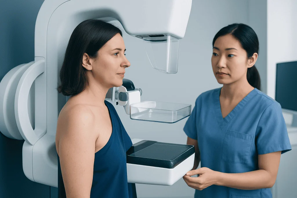
People considered at average risk generally have no personal history of breast cancer, no known high-risk gene mutation, no previous chest radiation at a young age, and no particularly strong family history of breast or ovarian cancer. For this group, mammography is usually the starting point for screening. The decision about when to start and how often to screen should take account of age, overall health, expected benefits, and the possibility of false-positive results.
Some people have a higher-than-average chance of breast cancer and may benefit from screening at an earlier age or with additional tests. Factors that can increase risk include a BRCA1 or BRCA2 mutation or another cancer-related gene change, a close relative with breast or ovarian cancer, a previous breast biopsy showing certain high-risk changes, a personal history of breast cancer, or radiation therapy to the chest during childhood or young adulthood.
For people at high risk, a care team may recommend annual breast MRI in addition to mammography. MRI is more sensitive in some higher-risk situations, but it can also identify areas that require more testing and are not cancer. Genetic counseling can help clarify whether inherited testing and a personalized surveillance plan are appropriate.
- Average-risk screening: mammography is usually the principal test.
- Higher-risk screening: mammography, MRI, and specialist follow-up may be advised.
- Breast density: may affect how easily mammograms identify changes and should be discussed after results are available.
How Mammography Screening Works
A mammogram creates detailed X-ray images of the breast. During the test, each breast is placed briefly between two plates and gently compressed. Compression spreads breast tissue so the images are clearer and helps keep the radiation dose low. Although it can feel uncomfortable or cause short-lived pressure, the image-taking portion is usually brief.
Most screening programs use digital mammography. Some centers also offer digital breast tomosynthesis, often called 3D mammography, which captures images from several angles to create thin image slices. This can be particularly helpful in some people with dense breast tissue, although the best approach depends on local practice and individual circumstances.
Mammography uses a low dose of radiation. The potential benefit of identifying breast cancer early is considered to outweigh this small exposure for people who are advised to screen. Ultrasound is not generally a replacement for mammography in average-risk screening, but it may be used to investigate a specific finding or alongside other imaging in selected cases.
What Happens During the Procedure and Afterward?
Before a screening mammogram, the patient may be asked about previous breast surgery, implants, pregnancy or possible pregnancy, breastfeeding, medicines, symptoms, and family history. It is helpful to tell the imaging team about breast implants or prior mammograms performed elsewhere, as comparison images can make interpretation more accurate.
On the day of the examination, the patient undresses from the waist up and stands at the mammography machine. A radiographer positions one breast at a time and takes standard views. Usually, several images are taken of each breast. The appointment often takes less than 30 minutes, though timing varies by center and whether additional images are needed.
There is no physical recovery period after a routine mammogram. Most people return to their normal activities immediately. Mild tenderness can occur for a short time, particularly if the breasts are sensitive. Scheduling the test when breasts are less tender, where possible, and avoiding deodorant, powder, lotion, or perfume on the chest and underarms on the day of imaging may help avoid artifacts on the images.
Results may be communicated by letter, electronic portal, telephone, or a follow-up appointment. If further imaging is requested, this commonly means the radiologist needs clearer views or wants to examine an area more closely. It is not a diagnosis of cancer.
Understanding Results, Benefits and Limitations
Mammogram reports may describe a result as normal, benign, probably benign, incomplete, suspicious, or highly suggestive of malignancy. In many healthcare systems, radiologists use the BI-RADS assessment system to communicate the level of concern and the recommended next step. A normal result usually means routine screening can continue, while an incomplete result means more imaging is needed before a final assessment can be made.
The main benefit of screening is earlier detection, when treatment may be less extensive and outcomes may be better. However, screening also has limitations. Some cancers are not visible on mammography, especially in dense breast tissue, and some findings lead to additional imaging or biopsy but prove to be benign. Screening can also detect slow-growing cancers that might never have caused symptoms, a concern known as overdiagnosis.
If an abnormality needs evaluation, the next steps may include diagnostic mammography, targeted ultrasound, breast MRI, or a biopsy. A biopsy removes a small sample of tissue for laboratory examination and is the only way to confirm whether a suspicious area is cancer. Information about breast cancer can help patients understand the diagnostic pathway and possible treatment planning if a diagnosis is confirmed.
Breast Awareness and Everyday Breast Health
Breast awareness means knowing what is usual for the individual’s breasts and reporting changes rather than performing a rigid self-examination routine. Breasts naturally vary in size, shape, texture, and sensitivity. Hormonal changes during the menstrual cycle, pregnancy, breastfeeding, and menopause can also affect how the breasts feel.
Healthy breasts do not have one standard appearance. It is common for one breast to be slightly different in size or shape from the other, and breast tissue may feel nodular or firmer in some areas. What matters most is a new or persistent change, such as a new lump, focal thickening, skin dimpling, a nipple that turns inward for the first time, unusual nipple discharge, persistent redness, or an unexplained change in breast contour.
Healthy habits may support overall cancer risk reduction, including staying physically active, maintaining a weight that is appropriate for the individual, limiting alcohol, and avoiding smoking. These steps do not replace screening. People should continue scheduled screening even if they feel well and should not wait for the next mammogram if a new symptom appears.
When to Seek Medical Care
A person should arrange a medical assessment promptly if they notice a new breast lump, a lump in the armpit, persistent one-sided breast or nipple pain, skin puckering, nipple retraction that is new, spontaneous bloody or clear nipple discharge, redness that does not settle, or a change in breast size or shape that is not explained by normal hormonal changes. Most breast changes are not cancer, but they deserve careful assessment.
Anyone with a strong family history of breast, ovarian, pancreatic, or prostate cancer should discuss risk assessment with a clinician, even if they have no breast symptoms. This may lead to genetic counseling, earlier screening, or additional imaging when appropriate.
Acibadem International’s multidisciplinary specialists and JCI-accredited hospitals support international patients with breast imaging, diagnostic assessment, and individualized care plans. When cancer treatment is needed, options may include breast cancer treatment based on the cancer type, stage, and the patient’s preferences and health needs.
Can I breastfeed my bf?
Breastfeeding is intended to feed an infant. An adult partner touching, kissing, or sucking the breasts is generally a personal and consensual matter, but it is not the same as breastfeeding a baby and should never be pressured. Clear communication, mutual consent, and comfort are important.
During lactation, breast stimulation may trigger milk release. A partner should avoid contact if it causes pain, irritation, cracked skin, or discomfort. If there is mastitis, an open wound, bleeding nipple, or a concern about infection or medication exposure, it is sensible to discuss the situation with a healthcare professional.
What does a healthy breast look like?
A healthy breast can look and feel different from person to person. There is no single “normal” size, shape, symmetry, skin tone, nipple direction, or texture. Mild asymmetry and changes linked to menstrual cycles, pregnancy, breastfeeding, weight changes, or aging are common.
The most useful approach is to notice what is normal for the individual. A new lump, persistent localized thickening, skin dimpling, a new nipple change, unexplained discharge, or an ongoing change in shape should be evaluated by a clinician rather than judged from appearance alone.
Can my bf touch my breasts?
Yes, a boyfriend or other partner can touch someone’s breasts when there is clear, freely given consent and the contact is comfortable for both people. Consent can be withdrawn at any time, including in an established relationship. Communication about pressure, sensitivity, and boundaries is important.
Breasts may be more tender during menstruation, pregnancy, breastfeeding, after surgery, or during some medical treatments. If touching causes persistent pain or reveals a new lump or other change, the person should arrange a medical assessment.
What is the female breast called?
The female breast is simply called the breast. Anatomically, it includes skin, fatty tissue, connective tissue, milk-producing glands called lobules, milk ducts, blood vessels, nerves, the nipple, and the areola, which is the darker area around the nipple.
Breast tissue extends toward the underarm area, which is one reason clinicians may examine both the breast and armpit when assessing symptoms. People of all sexes have breast tissue and can develop breast conditions, although the structure and amount of glandular tissue vary.
Frequently asked questions
At what age should breast cancer screening begin?
For many people at average risk, discussions about mammography begin in the 40s. Exact starting age and screening interval vary between guidelines and individual circumstances, so a clinician can help create a plan based on personal risk and preferences.
How often should I have a mammogram?
Many recommendations support mammography every one to two years for average-risk adults in the recommended age range. Annual screening may be chosen by some people, while others may screen every two years. Higher-risk individuals may need a different schedule and additional imaging.
Does a call back after a mammogram mean I have cancer?
No. A callback means that the radiologist would like additional images, comparison with older studies, or sometimes an ultrasound to clarify an area. Most callbacks do not result in a cancer diagnosis.
Can breast cancer occur after a normal mammogram?
Yes. Mammograms are valuable but do not find every cancer, especially when breast tissue is dense or a cancer develops between screening tests. New breast symptoms should be assessed even if a recent mammogram was normal.
Are mammograms painful?
Mammograms can cause brief pressure or discomfort because the breast is compressed for imaging. The compression lasts only a few seconds for each image. People with breast tenderness can discuss ways to make the appointment more comfortable with the imaging center.
Do dense breasts mean breast cancer?
No. Dense breasts are common and describe the proportion of fibrous and glandular tissue compared with fatty tissue on a mammogram. Density can make mammograms harder to interpret and is one factor a clinician may consider when discussing an individualized screening plan.
References
- World Health Organization
- U.S. Preventive Services Task Force
- American Cancer Society
- National Cancer Institute
- American College of Radiology
This article is for general information only and is not a substitute for professional medical advice. Please consult a qualified doctor about your individual situation.
Breast Cancer in Turkey — costs, top hospitals & a free quote
JCI-accredited · board-certified surgeons · reply within 24h
Add us as a Preferred Source to see more of our trusted health content across Google Search, AI Overviews and Discover.

Medical Oncology Department
Medical treatment of cancer with chemotherapy, immunotherapy and targeted therapies under a multidisciplinary tumor board.
60 specialists in this unit