Calcific Tendonitis Treatment: How It Works, Results and What to Expect

Calcific tendonitis is caused by calcium deposits developing inside a tendon, most often in the shoulder rotator cuff. Many cases improve without surgery, although recovery can take weeks to months and symptoms may fluctuate.
Key Takeaways
- Calcific tendonitis is caused by calcium deposits developing inside a tendon, most often in the shoulder rotator cuff.
- Many cases improve without surgery, although recovery can take weeks to months and symptoms may fluctuate.
- Ultrasound-guided barbotage, also called needling and lavage, can break up and remove suitable deposits with minimal incisions.
- Surgery is generally reserved for ongoing pain or restricted function after appropriate nonsurgical treatment.
- Sudden severe shoulder pain, fever, marked swelling or new weakness should be assessed promptly by a clinician.
Calcific tendonitis treatment usually starts with pain relief, activity modification and guided rehabilitation, while persistent or severe symptoms may benefit from ultrasound-guided needling, shockwave therapy or arthroscopic surgery. The best approach depends on the location and size of the calcium deposit, the stage of the condition and how much it limits daily life.
Calcific Tendonitis Treatment: How It Works
Calcific tendonitis treatment aims to control pain, restore comfortable movement and, when necessary, reduce or remove calcium deposits within a tendon. It most commonly affects the rotator cuff tendons of the shoulder, but deposits can also occur around the hip, elbow, wrist, knee or ankle. A clinician will tailor treatment to the person’s symptoms, examination findings, imaging results and day-to-day needs.
Calcium deposits can pass through different stages. They may be quiet for a long time, then become inflamed and painful as the body begins to break them down. This helps explain why one person may have a deposit seen on an X-ray with little discomfort, while another has sudden, intense pain and limited movement.
Most people do not need an operation. Initial care often combines relative rest from painful overhead or repetitive tasks, pain-relieving measures and a structured rehabilitation plan. When pain remains disruptive, image-guided procedures or other specialist treatments may be considered before surgery.
Symptoms, Root Cause and Diagnosis
Calcific tendonitis can cause aching, sharp pain with movement, pain at night, stiffness and reduced strength. In the shoulder, lifting the arm, reaching overhead, dressing or lying on the affected side may be difficult. During an active inflammatory phase, pain may become severe over a short period, even without a new injury.
What is the root cause of calcific tendonitis? The exact cause is not fully understood. It is not simply calcium from food “building up” in the tendon. Research suggests that tendon-cell changes, local blood supply, repetitive mechanical stress and the body’s healing response may contribute. It is more common in adults and may occur alongside metabolic conditions such as diabetes or thyroid disease, but many affected people have no clear risk factor.
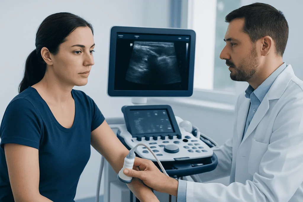
Diagnosis starts with a medical history and physical examination. X-rays commonly show calcium deposits, while ultrasound can assess the tendon, surrounding bursa and deposit characteristics in real time. Magnetic resonance imaging may be useful when clinicians need to evaluate other causes of shoulder symptoms, such as a rotator cuff tear or joint condition.
Who May Benefit From Calcific Tendonitis Treatment?
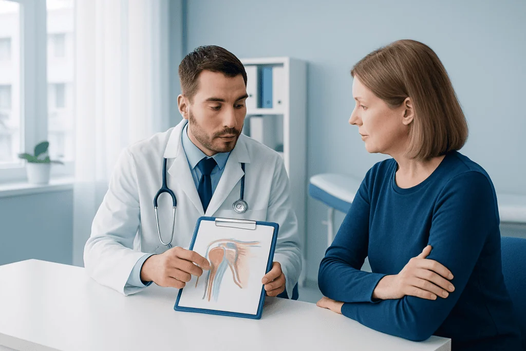
People are usually candidates for active treatment when pain affects sleep, work, self-care, sport or rehabilitation, or when movement remains limited despite a reasonable period of conservative care. The treatment choice is not based on deposit size alone. Symptoms, functional restriction and the appearance of the deposit on ultrasound or X-ray are all important.
Conservative treatment may be appropriate for mild or improving symptoms. It can include temporary activity adjustment, ice or heat according to comfort, and nonsteroidal anti-inflammatory medicines when a clinician considers them safe. People with kidney disease, stomach ulcers, cardiovascular disease, bleeding risk, pregnancy or certain medication interactions should ask a clinician or pharmacist before using anti-inflammatory medicines.
Ultrasound-guided procedures may be considered for a painful deposit that is accessible and has not responded sufficiently to first-line care. Shockwave therapy can be another nonsurgical option in selected cases. Surgery may be discussed if symptoms persist for months, function remains significantly limited, or another shoulder problem also requires treatment.
Nonsurgical and Minimally Invasive Treatment Options
Rehabilitation is an important part of calcific tendonitis treatment. A physiotherapist may guide gentle range-of-motion work first, followed by exercises that gradually improve rotator cuff and shoulder-blade strength. Exercises should be adapted to pain levels; forcing painful movement can aggravate symptoms, while avoiding all movement for too long can increase stiffness.
Corticosteroid injection into the inflamed bursa may provide short-term relief for some people, particularly when bursitis is contributing to pain. It does not directly remove the calcium deposit, and repeated injections are considered carefully because they may affect tendon health. Clinicians may also recommend extracorporeal shockwave therapy, which uses externally delivered acoustic waves to support pain reduction and deposit resorption in selected patients.
For suitable deposits, ultrasound-guided needling and lavage—often called barbotage—can be a useful minimally invasive option. Under local anesthetic and ultrasound guidance, a clinician places fine needles into the deposit, repeatedly flushes it with sterile fluid and aspirates loosened calcium material. The procedure may be followed by an injection to calm nearby bursal inflammation when clinically appropriate.
Benefits of image-guided needling may include targeted treatment, no large incision and relatively quick return to light daily activities. Possible risks include temporary soreness, bruising, incomplete removal, infection, bleeding and continued or recurrent symptoms. The treating team will explain whether the expected benefit outweighs these risks for the individual.
What Happens During Surgery and What Results Can Be Expected?
Arthroscopic surgery is usually considered only after well-delivered nonsurgical care has not provided enough relief, or when there is an associated problem that needs repair. Arthroscopy uses a small camera and fine instruments inserted through small skin openings. The surgeon identifies and removes or decompresses the calcium deposit while assessing the surrounding tendon and shoulder joint.
If removing the deposit leaves a meaningful tendon defect, a repair may be needed. This can change the rehabilitation plan and recovery period. The aim is to relieve pain and improve function while preserving as much healthy tendon as possible; complete removal of every trace of calcium is not always necessary for a good clinical outcome.
Arthroscopy generally has a favorable outlook when carefully selected patients follow their rehabilitation plan, but it still carries risks. These include infection, bleeding, anesthesia-related complications, stiffness, persistent pain, tendon injury and the possible need for further treatment. A surgeon can discuss the individual findings, alternatives and realistic recovery expectations before any operation.
Recovery Timeline and What to Expect
How long does it take for calcific tendonitis to go away? The timeline varies considerably. A painful flare may settle over days to weeks, while the deposit and tendon irritation can take several weeks to months to improve. Some deposits gradually resorb without an invasive procedure, but persistent symptoms deserve reassessment rather than simply waiting indefinitely.
After ultrasound-guided needling, soreness commonly improves over the first few days, though the shoulder may remain sensitive for one to two weeks. Light activities can often resume as comfort allows, while heavy lifting and demanding overhead activity are reintroduced gradually. Follow-up rehabilitation supports recovery of movement and strength.
Recovery after arthroscopic treatment is usually longer. Gentle movement often starts early under the surgical team’s guidance, but return to strenuous work, repetitive overhead use or sport may take several months, especially if tendon repair was required. Progress is best judged by pain, range of motion and strength rather than by a single deadline.
How bad is the pain from calcific tendonitis? Pain ranges from mild discomfort to severe pain that interferes with sleep and basic arm movement. Severe pain can occur during the resorptive inflammatory phase and may feel disproportionate to the size of the deposit. Effective pain management and timely assessment can help a person stay mobile while the condition is treated.
What Is the Fastest Way to Cure Calcific Tendonitis?
What is the fastest way to cure calcific tendonitis? There is no single fastest treatment that is right for every person. The quickest safe path is an accurate diagnosis followed by treatment matched to the deposit, stage of inflammation and functional limitations. For severe, persistent symptoms with an appropriate deposit, ultrasound-guided needling or shockwave therapy may offer faster relief than watchful waiting for some people, while others improve with medication and rehabilitation alone.
Trying to “push through” severe pain or using unproven methods to dissolve calcium is unlikely to speed recovery. Continuing gentle, clinician-approved movement is usually preferable to complete immobilization. A physiotherapist can help modify work, household and exercise tasks while recovery is underway.
Acibadem International’s multidisciplinary specialists and JCI-accredited hospitals can assess shoulder and tendon conditions for international patients, coordinating orthopedic evaluation, imaging, rehabilitation and image-guided care when needed.
When to Seek Medical Care
A person should arrange medical assessment for shoulder or tendon pain that persists, repeatedly returns, disrupts sleep, limits normal activities or does not improve with simple self-care. Assessment is also sensible before returning to demanding work or sport when strength or range of motion has not fully recovered.
Prompt medical care is important for fever, redness or warmth with significant swelling, a visibly deformed joint, sudden inability to move the arm after injury, numbness, coldness or color change in the arm, chest pain, shortness of breath, or symptoms of a serious allergic reaction after medication. These features may indicate a problem other than uncomplicated calcific tendonitis.
People with diabetes, thyroid disease, inflammatory arthritis or a history of shoulder surgery should mention this during assessment. They may need an individualized plan and closer attention to factors that can influence healing, medication choices and rehabilitation.
Frequently asked questions
Can calcific tendonitis heal on its own?
Yes. Many calcium deposits gradually resorb and symptoms improve with time, pain management and rehabilitation. However, recovery may be slow, and persistent pain or restricted movement should be reviewed by a qualified clinician.
Is calcific tendonitis the same as a rotator cuff tear?
No. Calcific tendonitis means calcium has formed within a tendon, commonly a rotator cuff tendon in the shoulder. A rotator cuff tear is a partial or complete disruption of tendon fibers, although both conditions can cause similar pain and weakness.
Does ultrasound-guided needling hurt?
The area is usually numbed with local anesthetic, so people may feel pressure or brief discomfort rather than sharp pain during the procedure. It is common to have temporary soreness afterward, and the care team will explain how to manage it safely.
Will the calcium deposit come back after treatment?
Recurrence is possible but not inevitable. Ongoing symptoms can also result from tendon irritation, bursitis, stiffness or another shoulder condition rather than a new deposit. Follow-up assessment helps determine the cause if symptoms return.
Should exercise be avoided with calcific tendonitis?
Painful or heavy movements may need to be reduced temporarily, particularly repetitive overhead activity. Complete rest for a prolonged period is usually not advised because it can worsen stiffness; a clinician or physiotherapist can recommend safe, gradual exercises.
When is surgery needed for calcific tendonitis?
Surgery is generally reserved for ongoing, function-limiting symptoms after appropriate nonsurgical treatment or when another shoulder problem also needs surgical management. An orthopedic surgeon will consider imaging, symptoms, examination findings and the person’s goals before recommending it.
References
- American Academy of Orthopaedic Surgeons
- American College of Radiology
- National Institute of Arthritis and Musculoskeletal and Skin Diseases
- Mayo Clinic
- Society of Radiologists in Ultrasound
This article is for general information only and is not a substitute for professional medical advice. Please consult a qualified doctor about your individual situation.
Joint and spine care in Turkey — expert assessment & treatment
JCI-accredited · board-certified surgeons · reply within 24h
Add us as a Preferred Source to see more of our trusted health content across Google Search, AI Overviews and Discover.