

Calcification in Breast: A Complete Clinical Guide for Patients

Breast calcifications are common and are often benign, especially with age. They usually do not cause pain or a lump and are most often seen on mammography.
Key Takeaways
- Breast calcifications are common and are often benign, especially with age.
- They usually do not cause pain or a lump and are most often seen on mammography.
- The size, shape, and pattern of calcifications help doctors decide whether follow-up is needed.
- Suspicious calcifications do not mean cancer is certain, but they may need diagnostic imaging or biopsy.
- Regular breast screening and timely follow-up are the best ways to manage calcifications safely.
Calcification in breast refers to tiny calcium deposits within breast tissue, usually found on a mammogram rather than felt on examination. Most breast calcifications are harmless, but certain shapes or patterns can require follow-up imaging or a biopsy to exclude an early cancer or precancerous change.
What calcification in breast means
Calcification in breast means small deposits of calcium have formed within the breast tissue. These deposits are very common, especially after age 40, and they are usually found during a screening or diagnostic mammogram. In most cases, they are benign and do not turn into cancer.
Breast calcifications are not the same as calcium from diet or supplements collecting in the breast. They are microscopic changes within tissue that can happen as part of normal aging, after inflammation, or due to other non-cancerous breast conditions. Their appearance on imaging helps radiologists decide whether they are clearly harmless or whether closer evaluation is needed.
Doctors usually describe calcifications as either macrocalcifications or microcalcifications. Macrocalcifications are larger, coarser deposits and are almost always benign. Microcalcifications are tiny specks of calcium; many are still harmless, but certain clustered or irregular patterns can be associated with ductal changes, including early breast cancer.
Types and why the pattern matters
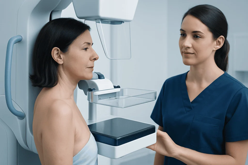
The most important part of evaluating calcification in breast is not simply whether calcifications are present, but how they look and where they appear. Radiologists assess the size, shape, distribution, and number of deposits. This imaging pattern helps estimate whether the finding is benign, probably benign, or suspicious.
Macrocalcifications typically appear as large white dots or coarse deposits on a mammogram. They are commonly linked to aging changes in the breast arteries, old injuries, past inflammation, or benign growths. When this pattern is classic, no special treatment is needed.
Microcalcifications are much smaller and may appear scattered, grouped, linear, or branching. Many grouped microcalcifications are still non-cancerous, but some clustered or irregular forms can suggest abnormal cells in the milk ducts. In these situations, the radiologist may recommend additional mammographic views, short-term imaging follow-up, or a tissue sample through breast biopsy.
- Benign pattern: coarse, round, scattered, or stable over time
- Probably benign pattern: low-risk appearance that often needs short-interval follow-up
- Suspicious pattern: fine, irregular, tightly clustered, or linear/branching calcifications
Symptoms and what a person may notice

Most people with breast calcifications have no symptoms at all. The deposits are usually too small to feel and do not cause pain. That is why calcifications are most often discovered on routine mammography rather than through self-examination.
When symptoms are present, they usually come from the underlying breast condition rather than from the calcifications themselves. For example, fibrocystic changes, an old injury, inflammation, or a benign cyst can cause tenderness or lumpiness. Calcifications seen in those settings are often incidental findings.
If calcifications are linked to a more serious condition, a person might also notice a breast lump, skin changes, nipple inversion, or nipple discharge, although many early cancers associated with calcifications still cause no symptoms. For this reason, screening remains important even when the breasts feel normal.
Causes and risk factors
There are many possible causes of calcification in breast, and most are not dangerous. Common benign causes include natural aging of the breast, prior inflammation, healed injury, fat necrosis, skin or blood vessel calcification, and deposits within benign cysts or fibroadenomas. Calcifications can also appear after surgery, radiation therapy, or infection as tissue heals.
Sometimes calcifications are related to abnormal cell changes within the milk ducts. These changes can include atypical cells, ductal carcinoma in situ, or invasive breast cancer. This does not mean every calcification is cancerous. Rather, it explains why suspicious imaging patterns should be investigated carefully.
Risk factors for concerning calcifications often overlap with general breast cancer risk factors. These may include increasing age, a personal or family history of breast cancer, certain inherited genetic mutations, prior chest radiation, and some high-risk breast lesions. A radiologist may compare current images with older mammograms to see whether calcifications are new or stable over time, which can be very helpful in judging risk.
People who already have known benign breast conditions may also see calcifications on imaging. In some cases, doctors may explain findings in the context of breast cancer screening or evaluation of fibrocystic breast changes if these are part of the broader clinical picture.
How doctors diagnose and classify breast calcifications
The first step is usually a mammogram. Calcifications often appear as white specks on the image. If they are found on routine screening, the person may be called back for diagnostic mammography, which uses additional magnified or focused views to show the deposits in greater detail. This helps the radiologist decide whether the pattern is benign or needs further study.
Radiologists commonly use the BI-RADS system to report breast imaging results. Benign calcifications may be classified as routine findings, while probably benign ones may be assigned short-term follow-up. Suspicious calcifications may lead to a recommendation for biopsy. Ultrasound is not always able to show calcifications clearly, but it may be used to assess other associated findings in the breast.
If tissue sampling is needed, a stereotactic or image-guided biopsy is often used because it can target the calcifications precisely. The pathology result then confirms whether the deposits are associated with benign tissue, atypical cells, or cancer. If cancer is identified, additional evaluation may include mammography, breast ultrasound, or breast MRI depending on the case.
Treatment and follow-up options
There is usually no treatment needed for benign breast calcifications. When the imaging appearance is clearly non-cancerous, doctors may simply advise routine breast screening according to age and risk. Calcifications themselves do not need to be removed if they are harmless and are not causing any problem.
If the finding is probably benign, the doctor may recommend a repeat mammogram in a short interval, often to confirm that the pattern remains stable. This approach helps avoid unnecessary procedures while still monitoring the area carefully. Stability over time is reassuring.
When calcifications look suspicious, biopsy is the standard next step. If pathology shows a benign result, follow-up imaging may still be advised to match the imaging and tissue findings. If precancerous changes or cancer are detected, treatment depends on the diagnosis and may involve surgery and, in some cases, radiation therapy, hormone therapy, or other oncology care.
Management is individualized. The person’s age, family history, prior imaging, breast density, and pathology results all influence next steps. Near the end of the care pathway, some patients may seek multidisciplinary evaluation; Acibadem International’s specialists in JCI-accredited hospitals also diagnose and treat breast conditions for international patients.
Prevention, self-care, and breast health habits
There is no guaranteed way to prevent breast calcifications because many occur as a normal part of breast tissue aging or healing. However, consistent breast health habits can help ensure that any changes are detected early and interpreted accurately. The most practical step is to attend recommended screening mammograms and keep copies of prior imaging available for comparison.
General lifestyle choices that support breast health include maintaining a healthy weight, staying physically active, limiting alcohol, and not smoking. These measures may not prevent calcifications directly, but they can contribute to overall health and may help reduce breast cancer risk in some people.
It is also helpful for a person to become familiar with the normal look and feel of their breasts. Self-awareness is different from trying to diagnose calcifications at home, since calcifications usually cannot be felt. Instead, it helps a person notice new lumps, skin dimpling, nipple changes, or discharge and report them promptly.
When to seek medical care
A person should seek medical care if they are called back after a screening mammogram for additional views. This is common and does not automatically mean cancer, but the recommended follow-up should not be delayed. Timely imaging gives the clearest picture of whether the calcifications are harmless or need more evaluation.
Medical review is also important if there are breast symptoms such as a new lump, persistent focal pain, skin thickening, redness that does not improve, nipple discharge, or a change in breast shape. These symptoms often have benign causes, but they deserve professional assessment.
People with a strong family history of breast or ovarian cancer, previous abnormal breast biopsies, or inherited cancer risk may benefit from more personalized screening advice. A qualified doctor can explain whether standard mammography is enough or whether closer surveillance is appropriate.
Frequently asked questions
Is calcification in breast a sign of cancer?
Not usually. Most breast calcifications are benign and are related to normal aging or non-cancerous breast changes. Some patterns, especially certain clusters of microcalcifications, may need more testing to rule out precancerous change or cancer.
Can breast calcifications be felt as a lump?
In most cases, no. Calcifications are usually too small to feel and are found only on a mammogram. If a lump is present, it may be due to another breast condition that is being evaluated at the same time.
Do breast calcifications need to be removed?
Benign calcifications do not need to be removed. They are usually monitored with routine screening or short-term follow-up if the radiologist wants to confirm stability. Removal is not the goal unless biopsy is needed to clarify a suspicious pattern.
What is the difference between macrocalcifications and microcalcifications?
Macrocalcifications are larger calcium deposits and are almost always benign. Microcalcifications are tiny specks that are also often benign, but their arrangement and shape sometimes raise concern. That is why radiologists pay close attention to the imaging pattern.
Can calcium supplements cause calcification in breast?
Breast calcifications are not generally caused by calcium in food or supplements. They form as part of tissue changes within the breast, not because calcium is building up from diet. A doctor can advise on supplements based on overall health, but breast calcifications alone are not usually a reason to stop them.
If I am called back after a mammogram, should I worry?
A callback is common and often leads to a benign explanation. It usually means the radiologist wants extra images to examine the calcifications more closely. The best step is to attend the follow-up promptly so the finding can be classified accurately.
References
- American Cancer Society
- Centers for Disease Control and Prevention
- National Cancer Institute
- Radiological Society of North America
- World Health Organization
This article is for general information only and is not a substitute for professional medical advice. Please consult a qualified doctor about your individual situation.
Oncology care in Turkey — second opinion and treatment plan
JCI-accredited · board-certified surgeons · reply within 24h
Add us as a Preferred Source to see more of our trusted health content across Google Search, AI Overviews and Discover.

More from the Health Library



Related Specialists

Prof. Dr. Erdal Okur
Thoracic Surgery
Assoc. Prof. Dr. Mehmet Altiparmak
Aesthetic Plastic & Reconstructive Surgery
Dt. Kerim Muhammed Aktaş
Oral & Dental Health
Dr. İpek Betül Özçivit Erkan
Gynecology & Obstetrics