Ccam Ultrasound: Preparation, Procedure and Results

CCAM is an older term for CPAM, a developmental lung malformation present before birth. Ultrasound is the main tool for finding and monitoring CPAM during pregnancy.
Key Takeaways
- CCAM is an older term for CPAM, a developmental lung malformation present before birth.
- Ultrasound is the main tool for finding and monitoring CPAM during pregnancy.
- Most pregnancies with an isolated CPAM can be monitored closely and result in a stable newborn.
- The scan is painless and does not use radiation; it is safe for the pregnant person and fetus when medically indicated.
- Follow-up may include detailed ultrasound, fetal echocardiography, MRI, and postnatal imaging depending on findings.
A CCAM ultrasound is a prenatal imaging examination that evaluates a congenital lung lesion in a developing baby. It helps the care team monitor lesion size, assess effects on the heart and surrounding structures, and plan appropriate follow-up during pregnancy and after birth.
CCAM ultrasound overview
A CCAM ultrasound is a prenatal ultrasound used to identify and monitor a lung abnormality called congenital cystic adenomatoid malformation (CCAM), now more commonly called congenital pulmonary airway malformation (CPAM). The examination uses sound waves to create images of the fetus and can show whether an area of lung contains cysts, appears unusually solid, or is affecting nearby structures.
CCAM ultrasound fetal assessments are usually performed during the detailed mid-pregnancy anatomy scan or when a routine scan raises a question about the fetal chest. The findings help fetal medicine, obstetric, neonatal, and pediatric surgery teams decide how often to monitor the pregnancy and where delivery should take place.
Although learning about a fetal lung finding can be worrying, many CPAM lesions remain stable or become less visible later in pregnancy. A scan result is interpreted in context, including the lesion’s appearance, size, growth pattern, blood supply, and whether there are signs that the fetus is under strain.
Are CPAM and CCAM the same?
Yes. CPAM and CCAM describe the same general group of congenital lung malformations. CCAM, or congenital cystic adenomatoid malformation, is the older term. CPAM, or congenital pulmonary airway malformation, is now preferred because it more accurately describes the abnormal development of the airways within part of the lung.
The condition develops before birth and is not caused by anything a pregnant person did or did not do. CPAM may involve one section of a lung and can contain fluid-filled cysts, tissue that looks solid on ultrasound, or a combination of both. Its appearance may change as pregnancy progresses.
Doctors may also assess whether the lesion has an unusual blood vessel supply, because some lung lesions can overlap with another congenital finding called pulmonary sequestration. Detailed imaging helps clarify these features and supports individualized care planning.
How a CCAM ultrasound works and who may need one

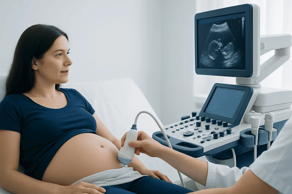
CCAM sonography works like other prenatal ultrasound examinations. A trained sonographer places a handheld probe on the abdomen after applying gel. The probe sends sound waves into the body and receives returning echoes, which are converted into real-time images. It does not involve X-rays or radiation.
A detailed scan may be recommended after a routine prenatal ultrasound identifies an unusual area in the fetal chest, a shifted heart position, excess amniotic fluid, or a possible lung cyst. It may also be requested for follow-up when CPAM has already been diagnosed. Some families are referred to a maternal-fetal medicine specialist for a more detailed evaluation.
The assessment often includes measurements of the lesion and the fetus, evaluation of the heart’s position and function, amniotic fluid assessment, and Doppler ultrasound to examine blood flow. Clinicians may calculate a CPAM volume ratio, which is one of several tools used to estimate the likelihood that closer monitoring may be needed.
CCAM procedure: preparation and step-by-step process
Most people do not need special preparation for a CCAM prenatal ultrasound. The imaging center may provide instructions based on the stage of pregnancy and the type of scan planned. Comfortable two-piece clothing can make the abdominal examination easier. A full bladder is generally not required later in pregnancy, but patients should follow the appointment-specific guidance they receive.
During the ccam procedure, the patient lies on an examination couch, usually on the back with positioning adjustments if needed for comfort. Gel is applied to the abdomen, and the sonographer moves the probe over the skin to obtain images from several angles. Pressure may be felt, but the examination should not be painful.
A focused fetal chest assessment can take longer than a routine scan because the sonographer needs to record detailed measurements and evaluate multiple organs. The team may ask the patient to change position, walk briefly, or return for additional images if fetal position limits the view. The scan itself does not require needles, sedation, or recovery time.
In selected cases, further assessment may include fetal echocardiography to examine the heart, or fetal MRI to provide additional information about the lungs and nearby structures. These tests complement rather than replace ultrasound, and the recommended plan depends on the individual findings.
What are the symptoms of CCAM?
Before birth, CPAM usually does not cause symptoms that the pregnant person can feel. It is most often detected incidentally on a prenatal ultrasound. The important prenatal question is whether the lesion is large enough to compress normal lung tissue or shift the heart, which can be evaluated through serial imaging.
After birth, some babies with CPAM have no immediate symptoms. Others may develop fast breathing, increased work of breathing, low oxygen levels, feeding difficulty, or recurrent chest infections later in infancy or childhood. Symptoms depend on the lesion’s size, location, and effect on normal lung function.
Not all newborn symptoms are caused by CPAM, and a baby with a prenatal diagnosis should be assessed by the neonatal and pediatric team after delivery. Even when a lesion appears to shrink during pregnancy, postnatal imaging is often advised because ultrasound findings before birth may not show the full anatomy after the lungs fill with air.
Can CPAM resolve on its own?
Some CPAM lesions appear to become smaller or less visible on prenatal ultrasound, especially during the later stages of pregnancy. This is often called regression. However, a lesion that is difficult to see before birth may still be present after delivery, so apparent prenatal improvement does not always mean the malformation has completely disappeared.
For this reason, doctors commonly recommend imaging after birth, often with chest X-ray and, when appropriate, CT or MRI. The timing and type of imaging depend on the baby’s symptoms, the prenatal findings, and local clinical practice. The care team will explain why follow-up is recommended in each situation.
When a child has an ongoing lesion, management may involve careful observation or surgery to remove the affected lung segment or lobe. Decisions are made by considering respiratory symptoms, infection risk, imaging results, and the lesion’s characteristics. Pediatric surgical consultation can help families understand the options.
Results, benefits, risks and recovery after the scan
The results of a CCAM ultrasound describe the lesion’s location, size, appearance, blood flow, and effect on the fetal chest. The report may also comment on fetal growth, amniotic fluid, and any signs of fluid buildup in the fetus, called hydrops. Most findings require planned follow-up rather than emergency treatment.
The main benefit of the scan is that it allows the pregnancy to be monitored and delivery care to be organized in advance if needed. In many cases, serial ultrasounds provide reassurance that the lesion is stable. If the lesion grows quickly or affects the fetus, a specialist team can discuss closer surveillance and, in uncommon severe situations, treatment options during pregnancy.
Diagnostic ultrasound has an excellent safety record when used appropriately by trained professionals. There is no radiation exposure, and no physical recovery period is needed after the scan. Patients may return to usual activities immediately unless their obstetric team has given other advice.
At Acibadem International, multidisciplinary fetal medicine, obstetric, neonatal, radiology, and pediatric surgery specialists in JCI-accredited hospitals can assess congenital lung findings and coordinate care for international patients.
Is CPAM a rare disease?
CPAM is considered an uncommon congenital lung malformation. Because it is often detected during routine prenatal imaging, many cases are identified before birth rather than after symptoms develop. The condition can vary widely in size and clinical significance.
Most CPAM cases occur as isolated findings, meaning no other major structural difference is identified on imaging. However, a detailed fetal assessment is important because the outlook and management depend on the specific features of the lesion and whether any associated concerns are present.
Families benefit from care that includes maternal-fetal medicine specialists and pediatric specialists familiar with congenital lung conditions. A clear monitoring plan can reduce uncertainty and ensure that appropriate expertise is available around the time of birth.
When to seek medical care
Anyone who has been told that a fetal chest or lung finding was seen on ultrasound should arrange timely follow-up with their obstetrician or maternal-fetal medicine specialist. Prompt specialist review is particularly important if the report mentions rapid lesion growth, a displaced heart, excess amniotic fluid, fluid around the fetus, or concerns about fetal well-being.
After birth, urgent medical assessment is needed if a baby has difficulty breathing, persistent bluish lips or skin, poor feeding with breathing effort, unusual sleepiness, or signs of respiratory distress such as grunting or pronounced chest retractions. Parents should seek emergency care rather than wait for a scheduled appointment when these symptoms occur.
For babies who appear well, families should still attend recommended pediatric, imaging, and surgical follow-up appointments. These visits help clinicians confirm the diagnosis, monitor lung development, and discuss whether observation or planned treatment is most appropriate.
Frequently asked questions
What is a CCAM ultrasound?
A CCAM ultrasound is a prenatal scan that evaluates a suspected congenital lung malformation in a fetus. It uses sound waves to assess the lesion’s size, appearance, blood flow, and effect on nearby structures such as the heart.
Is a CCAM ultrasound safe during pregnancy?
Ultrasound does not use ionizing radiation and is widely used during pregnancy when medically indicated. It is performed by trained professionals, who use the examination to obtain the information needed for safe monitoring and care planning.
How often is CCAM monitored during pregnancy?
The frequency of follow-up depends on the lesion’s size, growth pattern, and any effects on the fetus. Some pregnancies need periodic scans, while others require more frequent surveillance during the period when lesions are most likely to change.
Can a baby with CPAM be born normally?
Many babies with a small or stable CPAM can be delivered as planned, with neonatal evaluation after birth. Delivery recommendations may change if imaging suggests a large lesion or a need for specialized support immediately after delivery.
Does CPAM always require surgery?
Not always. Surgery may be recommended for symptomatic children or for lesions with features that create future concerns, while some children can be monitored carefully. The decision should be made with a pediatric surgical and respiratory team after postnatal imaging and clinical assessment.
What happens after a prenatal CCAM diagnosis?
The next step is usually detailed review by a maternal-fetal medicine specialist, followed by scheduled ultrasound monitoring. After birth, the baby may have clinical assessment and imaging to define the lung anatomy and guide longer-term care.
References
- American College of Obstetricians and Gynecologists
- International Society of Ultrasound in Obstetrics and Gynecology
- Children's Hospital of Philadelphia
- Society for Maternal-Fetal Medicine
- National Organization for Rare Disorders
This article is for general information only and is not a substitute for professional medical advice. Please consult a qualified doctor about your individual situation.
Ultrasound in Turkey — costs, top hospitals & a free quote
JCI-accredited · board-certified surgeons · reply within 24h
Add us as a Preferred Source to see more of our trusted health content across Google Search, AI Overviews and Discover.

Check-up & Preventive Medicine
Comprehensive health screenings designed for early detection and prevention.
3 specialists in this unit