Cholangiography: What Patients Need to Know

Cholangiography examines the bile ducts, which carry bile from the liver and gallbladder to the small intestine. Different techniques include ERCP, percutaneous transhepatic cholangiography, intraoperative cholangiography, and magnetic resonance cholangiography.
Key Takeaways
- Cholangiography examines the bile ducts, which carry bile from the liver and gallbladder to the small intestine.
- Different techniques include ERCP, percutaneous transhepatic cholangiography, intraoperative cholangiography, and magnetic resonance cholangiography.
- The most suitable test depends on the suspected problem, symptoms, anatomy, and whether treatment may be needed.
- Some forms of cholangiography are diagnostic only, while ERCP and certain percutaneous procedures can remove stones or relieve a blockage.
- Patients should promptly report fever, worsening abdominal pain, vomiting, jaundice, or black stools after an invasive bile duct procedure.
Cholangiography is a type of bile duct imaging that uses contrast dye and X-rays or other imaging methods to show how bile flows from the liver to the intestine. It can help clinicians find conditions such as gallstones, narrowed bile ducts, bile leaks, inflammation, or tumors, and some forms can also allow treatment during the same procedure.
What Is Cholangiography?
Cholangiography is an examination that creates detailed images of the biliary system, also called the bile duct system. Bile is a fluid made by the liver that helps the body digest fats and remove certain waste products. It travels through small channels in the liver and larger bile ducts before entering the first part of the small intestine.
During many types of cholangiography, a contrast dye is introduced into the bile ducts. The dye makes the ducts visible on X-ray imaging, allowing the clinical team to assess their shape, drainage, and any areas of obstruction. The test may be recommended when symptoms, blood tests, ultrasound, or other scans suggest that bile is not flowing normally.
Cholangiography is not one single procedure. The word describes several ways of imaging the bile ducts. Some are minimally invasive imaging tests, while others involve an endoscope or a small needle placed through the skin. The care team will explain which approach is appropriate and why it has been recommended.
Why a Cholangiogram May Be Recommended
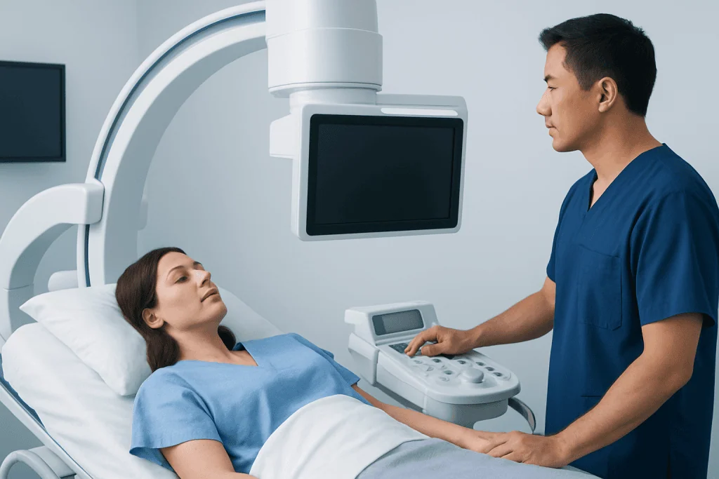
A cholangiogram can help clarify the cause of jaundice, which is yellowing of the skin or eyes caused by a buildup of bilirubin. It may also be used to investigate upper abdominal pain, fever with suspected bile duct infection, unexplained abnormal liver blood tests, pale stools, dark urine, or recurrent inflammation of the pancreas.
The images can identify gallstones that have moved into the common bile duct, narrowing caused by inflammation or scarring, bile duct injury, or a leak after surgery or trauma. They may also help evaluate a mass affecting the pancreas, gallbladder, liver, or bile ducts. A finding on cholangiography does not automatically mean cancer; further imaging, blood tests, or tissue sampling may be needed to establish the cause.
Cholangiography is often part of a broader assessment rather than a standalone answer. Ultrasound, CT, MRI, liver function tests, and clinical symptoms all provide important information. In selected situations, the test can move directly from diagnosis to treatment, such as removing a stone or placing a small tube called a stent to support bile drainage.
Types of Cholangiography

Endoscopic retrograde cholangiopancreatography (ERCP) is performed using a flexible camera tube passed through the mouth into the small intestine under sedation or anesthesia. Contrast dye is injected through a small opening where the bile duct enters the intestine, and X-ray images are taken. ERCP is generally used when treatment is likely to be needed, because stones can sometimes be removed and narrowed areas can be treated during the procedure.
Percutaneous transhepatic cholangiography (PTC) involves placing a fine needle through the skin and liver into a bile duct, usually with local anesthetic and sedation. It may be considered when ERCP is not possible or has not provided access to the blocked area. A drainage catheter or stent can sometimes be placed through this route to reduce bile buildup.
Intraoperative cholangiography is performed during gallbladder surgery. Contrast is injected into the bile ducts while X-ray images are taken, helping the surgeon assess the duct anatomy and look for stones in the main bile duct. This can be particularly useful when there is concern about bile duct stones or when the anatomy is not fully clear.
Magnetic resonance cholangiography, commonly performed as part of MRCP, uses MRI technology to produce images of the bile ducts without placing dye directly into them. It is noninvasive and can be useful for diagnosis, but it cannot remove stones, collect tissue samples, or place a stent. The choice between MRCP and an invasive cholangiogram depends on the individual clinical question.
How to Prepare and What Happens During the Test
Preparation depends on the technique being used. For ERCP, PTC, or surgery-related cholangiography, patients are usually asked not to eat or drink for a period beforehand because sedation, anesthesia, or contrast injection may be involved. The care team should be told about all medicines, including blood thinners, diabetes medicines, supplements, and any previous reactions to contrast agents or anesthesia.
Before an invasive procedure, clinicians may check blood tests, particularly liver function, kidney function, and blood clotting. Patients should also tell their clinician if they are pregnant, may be pregnant, have a history of kidney disease, or have an implanted medical device. These details help the team select the safest imaging method and make appropriate adjustments to care.
During ERCP, patients commonly receive sedation and are monitored throughout the procedure. During PTC, the skin is cleaned and numbed before the needle is inserted, and imaging guidance is used to access a bile duct. Intraoperative cholangiography is carried out while the patient is already under anesthesia for surgery. The procedure length varies according to the method and whether treatment is required.
After sedation, patients generally need someone to take them home and should avoid driving, alcohol, signing important documents, or operating machinery for the rest of the day. The care team will provide specific eating, activity, medication, and follow-up instructions based on the procedure performed.
Understanding Results and Possible Next Steps
A normal cholangiogram shows bile ducts with a smooth outline and normal flow of contrast toward the intestine. An abnormal result may show a filling defect, which can suggest a stone or debris, or a narrowed segment known as a stricture. It may also reveal a leak, a complete blockage, or changes in the ducts related to previous surgery or inflammatory disease.
Results should always be interpreted in context. For example, a bile duct may appear wider in some older adults or after gallbladder removal without indicating a dangerous blockage. Conversely, symptoms and blood test abnormalities may require further assessment even if initial imaging is not conclusive.
If a stone is found during ERCP, the endoscopist may be able to remove it at the same visit. If a narrowing is identified, a small brush sample or biopsy may be collected, and a stent may be inserted to improve bile flow. Some patients may need follow-up imaging, planned stent replacement, surgery, or consultation with hepatobiliary, gastroenterology, pancreatic, or oncology specialists.
Benefits, Risks, and Recovery
The main benefit of cholangiography is that it can provide a direct view of the bile ducts when a blockage or leak is suspected. Invasive approaches may avoid delays by allowing diagnosis and treatment in one session. This can be particularly helpful when obstruction is causing significant jaundice, pain, infection, or impaired liver function.
Noninvasive MRCP generally has fewer procedure-related risks, although it may not answer every question or provide treatment. ERCP and PTC are more invasive and have recognized complications. These can include bleeding, infection, inflammation of the pancreas after ERCP, contrast reactions, injury to the bile ducts or nearby organs, and sedation-related problems. Serious complications are uncommon, but the individual risk varies according to health status and the complexity of the procedure.
Most people have mild temporary throat discomfort, bloating, fatigue, or soreness at an access site after an invasive test. Recovery instructions may include rest, drinking fluids as advised, and watching for symptoms that could indicate a complication. The clinical team will discuss the expected benefits, alternatives, and risks before asking for consent.
When to Seek Medical Care
Anyone with new jaundice, severe or persistent upper abdominal pain, fever or chills, repeated vomiting, confusion, or very dark urine with pale stools should seek urgent medical assessment. These symptoms can occur when bile flow is blocked or when an infection develops in the bile ducts, and prompt evaluation is important.
After ERCP, PTC, or another invasive cholangiography procedure, patients should contact the treating team urgently if they develop worsening abdominal pain, fever, persistent vomiting, chest pain, shortness of breath, fainting, bleeding, black stools, or redness and swelling around a drainage site. Mild discomfort can be expected, but symptoms that intensify or do not settle should not be ignored.
For planned investigation of ongoing symptoms or abnormal liver tests, a gastroenterologist or hepatobiliary specialist can help determine whether cholangiography is needed and which method is most appropriate. Acibadem International’s multidisciplinary specialists and JCI-accredited hospitals diagnose and treat biliary conditions for international patients, with care plans tailored to the individual clinical situation.
Frequently asked questions
Is cholangiography painful?
The experience depends on the type of cholangiography. MRCP is noninvasive and should not be painful, although some people find lying still in an MRI scanner uncomfortable. ERCP and PTC are performed with sedation, anesthesia, local anesthetic, or a combination of these to improve comfort.
Is cholangiography the same as ERCP?
ERCP is one method of performing cholangiography. It uses an endoscope and X-ray contrast dye to image the bile ducts and can also be used for treatment. Cholangiography can also be performed during surgery, through the skin, or with MRI-based techniques.
How long does it take to recover after ERCP cholangiography?
Many people return home on the same day after an uncomplicated ERCP, although they need to rest because of sedation. Mild bloating, throat irritation, or tiredness may occur briefly. Recovery can take longer when a stent is placed, an infection is treated, or another procedure is required.
Can a cholangiogram remove gallstones?
Imaging alone cannot remove stones, but ERCP can often identify and remove stones from the common bile duct during the same procedure. This is different from removing the gallbladder, which may be recommended separately for people with recurrent gallbladder stones or complications. The best approach depends on where the stones are located and the person's overall health.
What is the difference between MRCP and cholangiography?
MRCP is an MRI-based method for viewing the bile and pancreatic ducts without inserting instruments into the ducts. It is often used as a diagnostic test because it is noninvasive. Conventional cholangiography usually involves injecting contrast dye directly into the duct system and may allow treatment at the same time.
Are there alternatives to cholangiography?
Yes. Depending on the reason for testing, clinicians may use abdominal ultrasound, CT scanning, MRI with MRCP, endoscopic ultrasound, or blood tests. These alternatives may provide enough information in some situations, while invasive cholangiography may be preferred when a blockage needs immediate treatment.
References
- National Institute of Diabetes and Digestive and Kidney Diseases
- American Society for Gastrointestinal Endoscopy
- American College of Radiology
- National Health Service
- European Society of Gastrointestinal Endoscopy
This article is for general information only and is not a substitute for professional medical advice. Please consult a qualified doctor about your individual situation.
Persistent digestive symptoms? Get evaluated in Turkey
JCI-accredited · board-certified surgeons · reply within 24h
Add us as a Preferred Source to see more of our trusted health content across Google Search, AI Overviews and Discover.