Club Foot on Ultrasound: Preparation, Procedure and Results

A clubfoot ultrasound scan is usually performed during the detailed anatomy scan in mid-pregnancy. The key ultrasound finding is persistent inward turning of the foot relative to the lower leg, often seen in more than one imaging view.
Key Takeaways
- A clubfoot ultrasound scan is usually performed during the detailed anatomy scan in mid-pregnancy.
- The key ultrasound finding is persistent inward turning of the foot relative to the lower leg, often seen in more than one imaging view.
- An isolated finding often has a favorable outlook, and clubfoot can usually be effectively treated after birth.
- Ultrasound cannot always confirm severity or distinguish a temporarily positioned foot from true clubfoot.
- Early referral to fetal medicine and pediatric orthopedics supports clear planning and timely treatment.
Club foot on ultrasound is a prenatal finding that suggests one or both fetal feet may be positioned inward and downward. Ultrasound can identify many cases during pregnancy, but confirmation may require repeat views, specialist assessment and an examination after birth.
Overview: What Does Club Foot on Ultrasound Mean?
Club foot on ultrasound means that the sonographer has seen a fetal foot that appears turned inward, downward or both. The medical term is congenital talipes equinovarus, often shortened to clubfoot. It can affect one foot or both feet and may be identified during a routine detailed pregnancy ultrasound, commonly performed around the middle of pregnancy.
This finding does not usually cause discomfort during pregnancy and does not mean that the baby will be unable to walk. A clubfoot ultrasound scan is a screening and assessment tool rather than a final diagnosis in every case. The care team may recommend additional views or a follow-up scan because fetal position can sometimes make a normal foot look unusually turned.
When clubfoot is confirmed, care is planned after delivery with a pediatric orthopedic team. Most children respond very well to early non-surgical treatment, particularly serial casting using the Ponseti method.
How Ultrasound Assesses the Fetal Feet
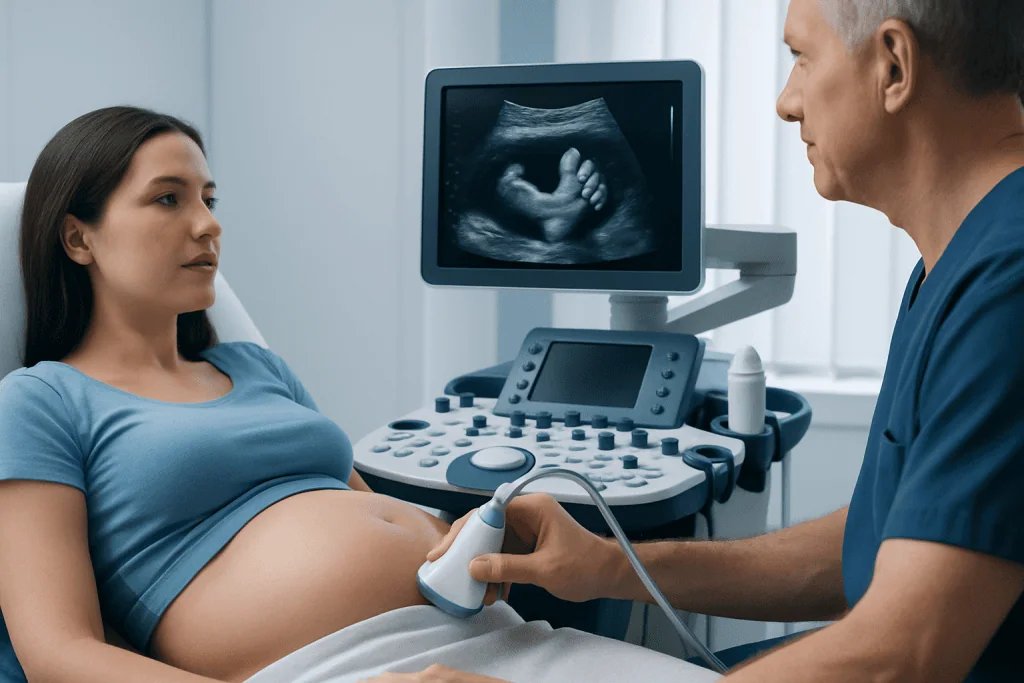
Ultrasound uses sound waves to create moving images of the fetus. It does not use radiation, and standard prenatal ultrasound is considered safe when performed for appropriate medical reasons by trained professionals. During a club foot ultrasound pregnancy assessment, the sonographer examines the feet, ankles, lower legs, hands, spine, brain, heart and other developing structures.
A normal foot can usually be seen at a near-right angle to the lower leg. With suspected clubfoot, the sole may appear to face inward toward the shin rather than outward in the usual position. The examiner aims to view the foot in several planes and observes whether this alignment remains present as the fetus moves.
Preparation is generally simple. The pregnant person can usually eat and drink normally unless the imaging center provides different instructions. Wearing comfortable two-piece clothing may make the abdominal scan easier. The appointment often includes time for careful imaging if the fetal position is not favorable.
A maternal-fetal medicine specialist may be involved when images are unclear, when both feet appear affected, or when there are other ultrasound findings. This review helps families understand what is known, what remains uncertain and whether any further testing is appropriate.
What Are the Ultrasound Findings for Clubfoot?
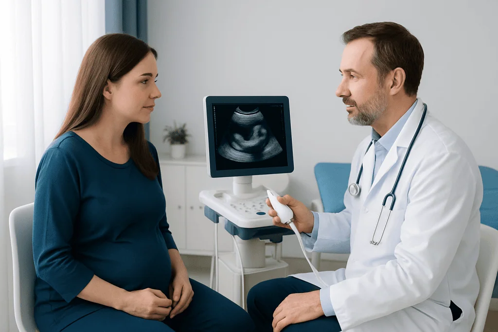
The typical clubfoot ultrasound appearance is a foot that remains abnormally aligned with the lower leg. In classic clubfoot, the foot may point downward at the ankle, turn inward, and have the front of the foot drawn toward the midline. Sonographers look for this pattern repeatedly rather than relying on one still image.
Another important observation is whether the sole of the foot appears in the same imaging plane as the tibia and fibula, the two bones of the lower leg. The foot may look as though it is lying alongside the lower leg rather than at its usual angle. A clubfoot ultrasound versus normal comparison is therefore based mainly on persistent alignment and movement, not foot size alone.
The examination also checks whether the finding is isolated. This means there are no other visible structural differences. When other concerns are seen, the clinician may discuss a more detailed ultrasound, genetic counseling or diagnostic testing. These options are individualized; an ultrasound finding alone does not establish a genetic condition.
Ultrasound is less able to predict how stiff the foot will be after birth. The newborn physical examination remains essential because it distinguishes a structural clubfoot from positional molding, in which a flexible foot was temporarily held in an unusual position in the uterus.
Candidacy, Procedure Steps and Ultrasound Accuracy
Any pregnancy can be assessed for fetal foot position as part of routine prenatal care. A targeted examination may be advised if a previous scan raised a question, there is a family history of clubfoot, or the routine anatomy scan did not provide clear views. The scan is noninvasive and does not require needles, anesthesia or a recovery period.
In a typical appointment, gel is applied to the abdomen and a handheld probe is moved across the skin. The sonographer obtains images of each foot and lower leg, checks the position from different angles, and records measurements and views for the reporting clinician. If the baby is curled up, moving actively or facing a difficult direction, the team may ask the pregnant person to change position, return later that day, or schedule a repeat scan.
Ultrasound clubfoot accuracy is good when the foot position can be clearly seen in multiple views, but it is not perfect. False-positive results can occur when the foot is temporarily positioned inward or images are limited. Conversely, a mild deformity can be difficult to recognize before birth. A repeat scan and postnatal examination improve diagnostic confidence.
The main benefits are earlier understanding, reassurance when the finding is isolated, and time to arrange appropriate postnatal care. There are no special physical risks from the ultrasound itself, although waiting for clarification can understandably feel stressful. A clinician can explain the report in the context of the whole pregnancy.
Can Clubfoot Be Misdiagnosed on Ultrasound?
Yes. Clubfoot can be misdiagnosed on ultrasound because fetal feet may be temporarily crossed, pressed against the uterine wall or held inward during the scan. Image quality may also be affected by fetal position, gestational age, amniotic fluid volume and other technical factors. For these reasons, suspected clubfoot is often checked with repeat imaging or specialist review.
A foot that looks abnormal on ultrasound may be flexible and normally aligned after birth. This is sometimes called positional foot deformity and often differs from true structural clubfoot. In contrast, some cases that are subtle before birth become clearer during the newborn examination.
Families should ask whether the finding appears isolated, whether one or both feet are involved, and whether follow-up imaging is recommended. It is helpful to remember that prenatal imaging guides planning; it does not replace assessment by a clinician who examines the baby after delivery.
Is Clubfoot 100% Curable?
Clubfoot is not usually described as “100% curable,” because response depends on the type and stiffness of the deformity, whether it is isolated, and adherence to treatment and follow-up. However, modern treatment is highly effective for most children. With early specialist care, many children develop comfortable, functional feet and can participate in everyday activities, including walking, running and sports.
The usual treatment begins soon after birth with gentle weekly manipulation and casts that gradually guide the foot into a better position. Many babies then have a small procedure to release a tight Achilles tendon, followed by a brace worn according to the orthopedic team’s instructions. Bracing is important because clubfoot can recur as a child grows.
Some children require additional treatment, particularly when clubfoot is rigid, recurrent or associated with another neuromuscular or genetic condition. Ongoing follow-up allows the team to recognize relapse early and adjust treatment. Surgery is not the starting treatment for most typical cases.
Pediatric orthopedic specialists work alongside obstetric, fetal medicine, neonatal and rehabilitation teams when needed. Acibadem International’s multidisciplinary specialists and JCI-accredited hospitals support diagnosis and treatment planning for international patients.
How Long Does It Take to Treat Club Foot?
Initial clubfoot correction often starts within the first weeks after birth. The casting phase commonly takes several weeks, with casts changed regularly as the foot position improves. The exact timeline varies, and the orthopedic clinician can explain the expected schedule after examining the baby.
After initial correction, a brace is used for a longer period to help maintain the result while the child grows. It is often worn full-time at first and later during sleep, according to the individual treatment plan. This maintenance phase is a major part of successful care and may continue for several years.
Although treatment takes commitment over time, children are generally able to feed, be held and develop normally during casting and bracing. Parents receive practical guidance on cast care, skin checks, bathing and when to contact the care team. Regular visits help ensure that the foot continues to develop well.
When to Seek Medical Care
During pregnancy, medical advice should be sought promptly if an ultrasound report mentions suspected clubfoot or another fetal structural finding. The obstetric clinician can arrange follow-up imaging, discuss the result and coordinate referral to fetal medicine or pediatric orthopedics when appropriate. There is usually time to assess the finding carefully and plan ahead.
After birth, a baby should be examined by a pediatric clinician if either foot appears persistently turned inward or downward, seems stiff, or cannot be gently moved into a more typical position. Parents should also contact the treating team if a cast becomes wet, damaged, unusually tight, slips off, or if toes look swollen, pale, blue or increasingly red.
Early assessment is valuable, but families do not need to manage this alone. Clear prenatal communication and timely postnatal orthopedic care give children with clubfoot the best opportunity for effective correction and healthy mobility.
Frequently asked questions
When can clubfoot be seen on ultrasound?
Clubfoot may be suspected during the detailed anatomy scan, often performed in the middle of pregnancy. It can sometimes be seen earlier or later, depending on fetal position and image quality. A repeat scan may be recommended if the feet cannot be assessed clearly.
Does a club foot on ultrasound mean the baby has other health problems?
Not necessarily. Clubfoot can occur as an isolated finding, meaning no other differences are identified on ultrasound. The care team will carefully review the full scan and may recommend further assessment if there are additional findings or relevant family history.
What are the ultrasound findings for clubfoot?
The main finding is persistent inward and downward positioning of the foot relative to the lower leg. The sonographer looks at both feet in multiple views and checks whether the alignment remains unusual as the fetus moves. The rest of the anatomy scan helps determine whether the finding appears isolated.
Can clubfoot be misdiagnosed on ultrasound?
Yes. A temporarily inward-facing foot, limited imaging views or fetal position can create an appearance similar to clubfoot. Follow-up imaging and examination after birth help confirm whether a structural clubfoot is present.
Is clubfoot 100% curable?
No treatment can be promised to work identically for every child, so clubfoot is not considered 100% curable. Still, early treatment is effective for most children, and many achieve comfortable, functional feet. Continued brace use and follow-up are important to reduce the chance of recurrence.
How long does it take to treat club foot?
The first casting phase often takes several weeks and begins soon after birth. Bracing then continues for a longer period, commonly over several years during growth. The precise schedule depends on the child’s foot and response to treatment.
References
- American College of Obstetricians and Gynecologists
- International Federation for Spina Bifida and Hydrocephalus
- National Health Service
- OrthoInfo – American Academy of Orthopaedic Surgeons
- Society for Maternal-Fetal Medicine
This article is for general information only and is not a substitute for professional medical advice. Please consult a qualified doctor about your individual situation.
Ultrasound in Turkey — costs, top hospitals & a free quote
JCI-accredited · board-certified surgeons · reply within 24h
Add us as a Preferred Source to see more of our trusted health content across Google Search, AI Overviews and Discover.

Check-up & Preventive Medicine
Comprehensive health screenings designed for early detection and prevention.
2 specialists in this unit