Congenital Nevi Birthmark Removal: Procedure, Recovery and Results

A congenital melanocytic nevus is a mole-like birthmark present at birth or appearing shortly afterward. The choice to remove a nevus depends on its size, location, changes over time, symptoms, cosmetic concerns and the person’s overall health.
Key Takeaways
- A congenital melanocytic nevus is a mole-like birthmark present at birth or appearing shortly afterward.
- The choice to remove a nevus depends on its size, location, changes over time, symptoms, cosmetic concerns and the person’s overall health.
- Surgical excision is the main option when complete removal and tissue examination are needed; larger lesions may require staged reconstruction.
- Most congenital nevi do not become cancerous, but larger lesions and concerning changes deserve specialist review.
- Recovery varies with the size and location of the treatment area, and scars are an expected part of surgical removal.
- Regular skin checks, photographs and sun protection are important whether or not a nevus is removed.
Congenital nevi birthmark removal is an individualized decision made with a dermatologist and, when needed, a plastic surgeon. Removal can improve comfort or appearance and may allow complete laboratory assessment, but not every congenital nevus needs treatment.
Overview: congenital nevi birthmark removal
Congenital nevi birthmark removal is a medical or cosmetic procedure used to remove or reduce a congenital melanocytic nevus (CMN), a pigmented mole-like mark that is present at birth or develops in the first weeks of life. The best approach depends on the nevus size, body location, depth, appearance, symptoms and whether it can be removed safely while achieving an acceptable scar.
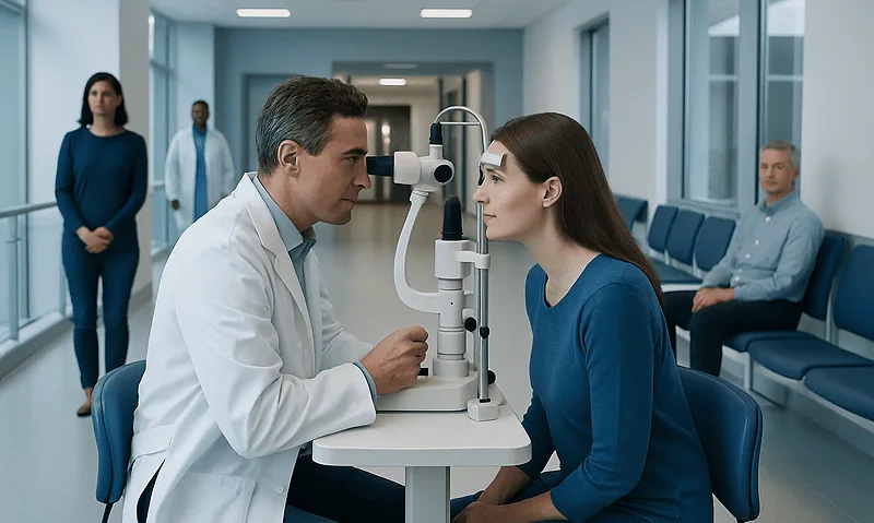
Some people choose removal because the lesion is difficult to monitor, repeatedly irritated by clothing, affects movement or appearance, or has developed features that require assessment. Others choose regular observation instead. A dermatologist can examine the birthmark with dermoscopy, discuss its future behavior, and help weigh the benefits of removal against anesthesia, healing time and scarring.
Congenital nevi vary from small, well-defined marks to extensive lesions that may cover a larger region of the body. They can be tan, brown or nearly black and may become more raised, textured or hairy as a child grows. These changes can be normal, but new or unexpected changes should still be checked.
Should I get a congenital nevus removed?
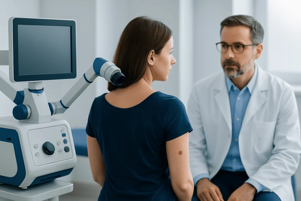
Whether a congenital nevus should be removed is a personal and clinical decision rather than a routine requirement. A doctor may recommend removal or a biopsy when a lesion develops a new firm lump, ulceration, persistent bleeding, unusual pain, rapid change, marked color variation, or other features that cannot be confidently explained by normal development.
Removal may also be reasonable when a nevus is in a location where it is frequently rubbed, injured or difficult to observe, such as the scalp, back, genital area or skin folds. Cosmetic and emotional considerations are valid as well, particularly when a visible lesion affects confidence or daily life. For children, timing is planned carefully around growth, the likely complexity of repair and the need for local or general anesthesia.
Not all lesions can or need to be completely removed. For some larger congenital nevi, ongoing dermatology follow-up and family education about skin changes may be safer or more practical than extensive surgery. A consultation should include discussion of realistic outcomes, including the likelihood of a permanent scar.
How the procedure works and who may be a candidate
The most definitive method of congenital melanocytic nevus removal is surgical excision. The surgeon removes the visible nevus and a small margin of surrounding skin when appropriate, then closes the wound with sutures. The removed tissue can be examined by a pathologist, which is particularly useful when the diagnosis is uncertain or a lesion has concerning features.
Small lesions can often be removed in one procedure. Medium or larger lesions may need staged excisions, where sections are removed over more than one operation as surrounding skin gradually relaxes. Depending on location and size, reconstruction can involve local skin flaps, tissue expansion or skin grafting. These techniques aim to close the area safely and preserve function, but they cannot guarantee an invisible scar.
Laser treatment, dermabrasion or curettage may sometimes lighten superficial pigment or improve texture, especially where full excision is not feasible. However, pigment cells may remain deeper in the skin, so these approaches do not provide the same tissue analysis or assurance of complete removal as surgery. Suitability is assessed by a dermatologist and plastic surgeon based on the individual lesion and treatment goals.
Good candidates are people whose health allows wound healing and anesthesia when needed, and who understand the expected trade-off between the birthmark and a surgical scar. In children, the care team also considers developmental needs, family preferences and whether treatment can be safely delayed.
Step-by-step: what happens during removal
Before treatment, the specialist reviews medical history, medications, allergies and previous scars or wound-healing problems. The nevus is measured and photographed, and the clinician may use dermoscopy or recommend a biopsy if there is diagnostic uncertainty. The procedure plan includes the expected scar direction, closure method, anesthesia type and any need for staged treatment.
On the day of surgery, the skin is cleaned and anesthetized. Small excisions may be performed with local anesthesia, sometimes with sedation. Young children and people having larger or more complex reconstructions may require general anesthesia. The surgeon removes the lesion, controls bleeding, closes the wound with stitches and applies a dressing.
If the area is too large to close directly, a planned reconstructive technique may be used. Tissue expansion involves placing a temporary inflatable device under nearby skin and gradually filling it over time to create extra skin for closure. Skin grafts transfer healthy skin from another area, while local flaps reposition neighboring tissue to cover the wound.
Excised tissue is usually sent for laboratory examination. The treating clinician explains when results are expected and whether any further follow-up is needed. Detailed wound-care instructions should be followed closely, especially for lesions on the face, scalp, joints or other areas exposed to movement and friction.
Recovery timeline, results, benefits and risks
Recovery after a straightforward excision commonly involves tenderness, swelling and mild bruising for several days. The wound should be kept clean and protected as instructed. Stitches may be removed after a period determined by the body site and closure type, while some stitches dissolve on their own. Children should avoid activities that could pull on the wound until the clinical team says it is safe.
Over the first weeks, the incision usually becomes stronger, although redness and firmness may persist. Scar maturation takes many months and sometimes longer than a year. Scar-care recommendations may include silicone products, moisturizing once the wound has healed, massage when advised, and careful sun protection. A scar can fade over time but cannot be fully predicted or eliminated.
Potential benefits include removal of an irritating or cosmetically troubling lesion, easier observation of the surrounding skin and the ability to examine removed tissue. Risks include bleeding, infection, wound separation, altered sensation, pigment changes, poor scarring, asymmetry, recurrence of pigment after partial treatment, and anesthesia-related complications. Larger procedures may have additional reconstructive risks.
It is important to contact the treatment team for increasing redness, warmth, drainage, fever, worsening pain, wound opening or significant bleeding. A follow-up plan is still useful after removal, because people may have other moles or residual pigment requiring routine skin assessment.
Can a congenital nevus become cancerous?
Yes, a congenital melanocytic nevus can rarely develop melanoma, a type of skin cancer. The degree of risk is not the same for every person: it is generally influenced by the nevus size, number of associated satellite nevi, location and individual clinical features. Larger congenital nevi have a greater concern than small lesions, but specialist monitoring is appropriate for any lesion that changes unexpectedly.
Removing a nevus may reduce concern in some situations, but it does not always remove every pigment cell, particularly in very large or deep lesions. It also does not replace regular medical review when a person has multiple nevi or other risk factors. Decisions about preventive removal should therefore be based on a full discussion rather than fear of cancer alone.
Warning signs that warrant prompt assessment include a new or enlarging nodule, persistent ulcer or sore, bleeding without an obvious injury, a clear change in color or shape, or ongoing itching or pain. Normal growth and textural change can occur as a child grows, so parents and caregivers should seek professional advice rather than trying to judge changes alone.
How rare is a nevus birthmark?
Congenital melanocytic nevi are relatively uncommon birthmarks, but they are well-recognized in dermatology. Small congenital nevi are much more common than large or giant congenital nevi. A clinician classifies them mainly by their predicted adult size, rather than only by their size at birth, because the skin grows as a child grows.
They occur in people of all backgrounds and are caused by changes in pigment-producing cells during fetal development. They are not caused by something a parent did or did not do during pregnancy, and they are usually not inherited in a simple predictable pattern. Most occur as an isolated skin finding.
Because appearance alone cannot always confirm the exact type of pigmented lesion, medical assessment is useful, especially for a large, atypical or changing birthmark. Baseline clinical photographs can support safe observation by making changes easier to identify over time.
When to seek medical care
A congenital nevus should be assessed by a dermatologist if it is new to the family’s attention, very large, located in a difficult-to-monitor area, associated with multiple smaller pigmented spots, or causing distress. An early review can clarify the diagnosis, establish a monitoring plan and discuss whether referral for surgery or reconstruction may be helpful.
Seek timely medical care for bleeding, ulceration, infection, a rapidly enlarging lump, persistent pain, a striking new color change or any change that concerns the patient or caregiver. These findings often have non-cancerous explanations, but they should not be ignored. Avoid home removal products, cutting, burning or attempting to treat a nevus without professional guidance.
Ongoing self-care includes protecting the skin from intense sun exposure with shade, clothing and appropriate sunscreen, while avoiding sunburn. Regularly checking the lesion and taking photographs in consistent lighting can make follow-up discussions more informative. Acibadem International’s multidisciplinary dermatology, plastic surgery and pathology teams in JCI-accredited hospitals can assess and treat congenital nevi for international patients when specialist care is needed.
Frequently asked questions
Should I get a congenital nevus removed?
Removal is not necessary for every congenital nevus. A dermatologist may advise observation, biopsy or removal based on size, location, symptoms, changes over time, cancer-related concerns and personal preferences. The expected scar and recovery should be considered alongside potential benefits.
How much does it cost to remove a congenital melanocytic nevus?
The cost varies substantially by country, clinic, lesion size and location, anesthesia needs, pathology testing and whether reconstruction or multiple procedures are required. A small outpatient excision is different from staged surgery for a large lesion. A surgical center can provide an individualized estimate after examination and treatment planning.
How rare is a nevus birthmark?
Congenital melanocytic nevi are uncommon but familiar birthmarks seen by dermatologists. Small lesions are considerably more common than large or giant lesions. A clinician can confirm whether a particular birthmark is a congenital nevus and explain its likely course.
Can a congenital nevus become cancerous?
A congenital nevus can rarely develop melanoma. Risk varies, with larger lesions generally receiving closer specialist attention than small lesions. New lumps, bleeding, ulceration, persistent pain or notable changes in color or shape should be assessed promptly.
Will congenital nevus removal leave a scar?
Surgical removal creates a scar because skin must be cut and closed. The scar often changes and softens over many months, but its final appearance depends on the lesion location, procedure, skin type, healing and individual tendency to scar. The surgeon can discuss scar placement and aftercare before treatment.
Can laser treatment completely remove a congenital nevus?
Laser treatment may lighten pigment or improve the surface appearance of selected lesions, but it may not reach deeper nevus cells. Therefore, it is usually not considered equivalent to complete surgical excision. The best option depends on the nevus characteristics and the person’s goals.
References
- American Academy of Dermatology
- American Academy of Pediatrics
- DermNet
- National Cancer Institute
- British Association of Dermatologists
This article is for general information only and is not a substitute for professional medical advice. Please consult a qualified doctor about your individual situation.
Congenital Nevi in Turkey — costs, top hospitals & a free quote
JCI-accredited · board-certified surgeons · reply within 24h
Add us as a Preferred Source to see more of our trusted health content across Google Search, AI Overviews and Discover.