

Contrast Dye in MRI and CT: Benefits, Risks, and Safety Checks
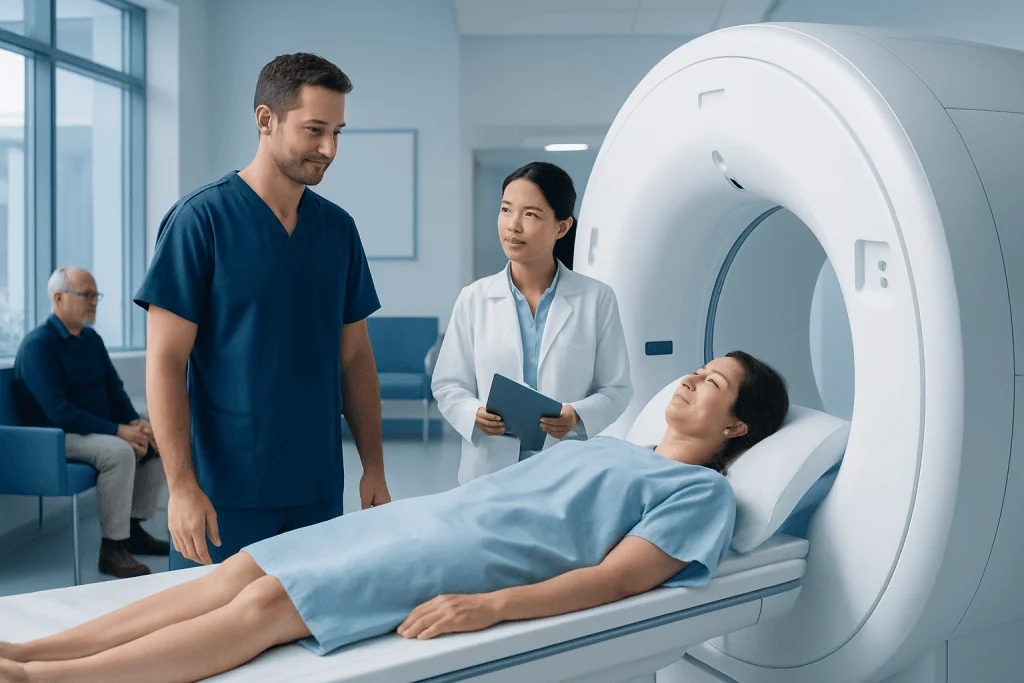
Contrast dye in MRI and CT can improve the visibility of organs, blood vessels, inflammation, tumors, and injuries. MRI and CT use different types of contrast agents, and they are not interchangeable.
Key Takeaways
- Contrast dye in MRI and CT can improve the visibility of organs, blood vessels, inflammation, tumors, and injuries.
- MRI and CT use different types of contrast agents, and they are not interchangeable.
- Safety checks usually include questions about allergies, kidney function, pregnancy, and past reactions to contrast.
- Most side effects are mild and short-lived, but serious reactions can happen and require prompt medical care.
- Hydration, clear communication with the imaging team, and following preparation instructions can help make contrast imaging safer.
Contrast agents used in MRI and CT scans can make important details easier to see, helping doctors diagnose many conditions more accurately. Most people tolerate them well, and careful screening helps reduce the risk of allergic reactions, kidney problems, and other complications.
Overview: What contrast dye does in MRI and CT
Contrast dye in MRI and CT refers to special substances used to make certain body structures stand out more clearly on imaging. Although people often call all of them “dye,” they do not color the body in the usual sense. Instead, they change how the scan captures tissues, blood vessels, or organs, so a radiologist can see important details that may not be visible on a non-contrast exam.
CT scans usually use iodine-based contrast material, most often given through a vein, although some CT studies also use oral or rectal contrast. MRI scans typically use gadolinium-based contrast agents, which affect magnetic signals rather than X-rays. Because MRI and CT work in different ways, the contrast agents used for each test are also different.
Doctors may request contrast when they need a clearer view of infection, inflammation, tumors, active bleeding, blood flow, blockages, or organ injury. In many cases, contrast helps answer a specific clinical question more confidently. For some people, however, a non-contrast study is enough, and the decision depends on the reason for the scan, medical history, and safety considerations.
Why contrast is used and what it can help show
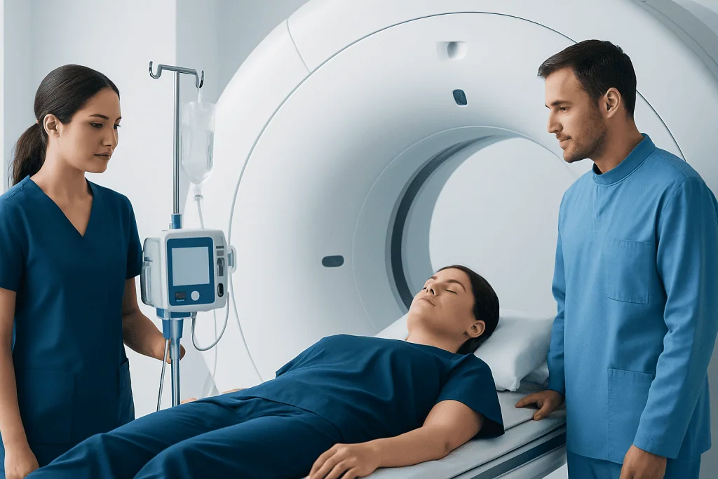
The main benefit of contrast-enhanced imaging is better detail. It can help distinguish normal tissue from abnormal tissue, show whether a mass has a blood supply, and reveal narrowing or leakage in blood vessels. This added clarity can support earlier diagnosis, better treatment planning, and more accurate follow-up after treatment.
In CT, contrast is often useful for examining the chest, abdomen, pelvis, and blood vessels. It may help doctors evaluate infections, injuries, kidney or liver problems, bowel conditions, and suspected clots. In emergency situations, contrast CT can be especially valuable because it is fast and can quickly highlight urgent problems.
In MRI, contrast is commonly used to assess the brain, spine, joints, liver, and soft tissues. It may help identify inflammation, active disease, scar tissue, or certain tumors. In some cases, a doctor may recommend a detailed MRI scan or a CT scan with contrast as part of a broader diagnostic workup, depending on the symptoms and the area being studied.
- Better visibility of blood vessels and blood flow
- Improved detection of inflammation and infection
- Clearer assessment of tumors or suspicious masses
- More precise evaluation of injuries or internal bleeding
- Greater confidence in treatment planning and follow-up
Types of contrast agents and how they differ

CT contrast agents are usually iodine-based. When injected into a vein, they circulate through the bloodstream and help X-ray images show differences between tissues more clearly. Some CT exams also use oral contrast to outline the stomach or intestines. People sometimes notice a warm feeling, a metallic taste, or a brief flushed sensation during injection; these effects are common and usually pass quickly.
MRI contrast agents are usually gadolinium-based. These substances change magnetic properties in the body, which can improve the visibility of certain tissues on MRI images. They are not the same as iodine contrast, and an allergy or reaction to one type does not automatically mean a person will react to the other. MRI contrast is often used when the doctor needs additional information that a standard MRI cannot provide clearly enough.
The choice to use contrast depends on the clinical question, the body part being examined, and the patient’s health status. Sometimes doctors may combine imaging methods for a fuller picture. For example, a person may have MRI for soft tissue detail or computed tomography for rapid evaluation of internal structures, with or without contrast depending on what needs to be seen.
Possible risks and side effects
Most people have no serious problems with contrast agents, but side effects and complications are possible. Mild effects are the most common and may include nausea, a warm sensation, headache, dizziness, or a strange taste in the mouth. These symptoms are usually temporary and resolve without treatment.
Allergic-like reactions can happen with both CT and MRI contrast agents, though they are more often discussed with iodine-based CT contrast. Reactions may range from mild itching or hives to more serious breathing difficulty or swelling. Imaging teams are trained to recognize and manage these reactions quickly, which is one reason patients are monitored during and after injection.
Kidney-related concerns are another important safety issue. Iodine-based CT contrast may be used cautiously in people with reduced kidney function, especially if the scan is not urgent or if alternatives exist. Gadolinium-based MRI contrast also requires extra care in people with severe kidney disease, because certain agents may rarely be linked to a serious condition called nephrogenic systemic fibrosis. In addition, a small amount of gadolinium can remain in the body after some scans, but the clinical significance of this in people with normal kidney function remains under study.
A less common risk occurs if contrast leaks outside the vein into nearby tissue during injection, a problem called extravasation. This can cause pain, swelling, or skin irritation around the injection site. It is usually managed with local care, but the imaging team should be told immediately if burning, tightness, or worsening discomfort develops during the injection.
Safety checks before the scan
Before giving contrast, the imaging team usually asks about past reactions to contrast agents, asthma, allergies, kidney disease, diabetes, pregnancy, breastfeeding, and current medications. These questions help determine whether contrast is appropriate and whether extra precautions are needed. Patients should answer as fully as possible, even if a previous reaction seemed mild.
Kidney function may be checked with a blood test, especially in older adults or people with known kidney disease, diabetes, dehydration, or other risk factors. This is a routine safety step that helps doctors decide whether contrast can be used safely or whether the imaging plan should be changed. In some situations, hydration or a different type of exam may be recommended.
People who have had a prior contrast reaction may need a different contrast agent, a non-contrast study, or premedication in selected cases. Pregnant patients should always inform the radiology team, because imaging choices are made carefully during pregnancy. Breastfeeding questions can also be discussed individually, since guidance may vary depending on the contrast agent and the clinical situation.
Preparation instructions may include fasting for a few hours, drinking fluids, or temporarily adjusting certain medicines if advised by a doctor. Patients should not stop prescription medications on their own unless they have received clear instructions. If there are concerns about claustrophobia, needle anxiety, or language barriers, it is helpful to mention them in advance so the team can plan support.
What to expect during and after contrast imaging
For an intravenous contrast study, a healthcare professional places a small cannula into a vein, usually in the arm. The contrast is then injected by hand or with an automatic injector at a controlled speed. Depending on the type of scan, the images may be taken immediately, in phases, or after a short delay to capture how the contrast moves through tissues and blood vessels.
During a CT scan, the person typically lies still on a table that moves through the scanner. During MRI, the person lies inside the magnet while the machine takes images; this can take longer than CT and may involve loud sounds, but ear protection is usually provided. If a patient feels unwell at any point, they should tell the staff right away.
After the scan, most people can return to their usual activities unless told otherwise. Drinking water may be recommended to support normal hydration, especially after intravenous contrast, although instructions can vary with medical conditions such as heart or kidney disease. The body removes most contrast through the kidneys over time.
Specialist radiology teams may also use contrast in advanced procedures such as angiography when detailed imaging of blood vessels is needed. Near the end of a patient’s diagnostic journey, they may choose a center such as Acibadem International, where multidisciplinary specialists and JCI-accredited hospitals evaluate and treat international patients with coordinated imaging and follow-up care.
How risks can be reduced
Several practical steps can help lower the chance of complications. Good communication is one of the most important. Patients should tell the imaging team about any previous contrast reaction, kidney disease, asthma, allergies, pregnancy, or recent illness. Bringing a current medication list can also be useful.
Staying appropriately hydrated before and after the exam may support kidney function, unless a doctor has advised fluid restriction. Following fasting or preparation instructions carefully can also help the exam run smoothly. If a patient is anxious, discussing concerns in advance may allow time for reassurance, scheduling adjustments, or other supportive measures.
Doctors may choose the lowest necessary dose, a different contrast agent, or a non-contrast scan when appropriate. In some cases, another imaging method such as ultrasound may be considered if it can answer the same medical question. The goal is to balance the diagnostic benefit with the smallest reasonable risk.
- Share any history of prior contrast reactions
- Report kidney problems, diabetes, asthma, or pregnancy
- Follow instructions about eating, drinking, and medicines
- Tell staff immediately about pain, itching, or breathing symptoms during the injection
- Ask questions if anything about the exam is unclear
When to contact a doctor after the scan
Most mild effects settle quickly, but medical advice should be sought if symptoms continue or worsen after going home. Warning signs include increasing rash, trouble breathing, swelling of the face or throat, severe vomiting, fainting, or significant pain and swelling at the injection site. These may need urgent evaluation.
People with kidney disease or other complex medical conditions should follow any specific aftercare advice given by their doctor. If urine output changes noticeably, swelling develops, or a person feels unusually unwell after contrast imaging, it is sensible to contact a healthcare professional. While serious delayed problems are uncommon, it is always better to ask if there is uncertainty.
The scan report itself is also an important part of follow-up. Patients should discuss the results with the doctor who ordered the test, since the meaning of a finding depends on symptoms, examination, and other tests. Contrast imaging is a tool to support diagnosis, and the next step is based on the full clinical picture.
Frequently asked questions
Is contrast dye in MRI and CT safe?
For most people, contrast agents used in MRI and CT are safe and well tolerated. The imaging team reduces risk by reviewing allergies, kidney function, pregnancy status, and prior reactions before the scan.
What is the difference between MRI contrast and CT contrast?
CT usually uses iodine-based contrast, while MRI usually uses gadolinium-based contrast. They work in different ways and are chosen based on the type of scan and the medical question being investigated.
Can contrast dye cause an allergic reaction?
Yes, allergic-like reactions can happen, although serious reactions are uncommon. Symptoms may include itching, rash, swelling, or breathing difficulty, which is why patients are monitored and should report symptoms right away.
Why do doctors check kidney function before giving contrast?
The kidneys help clear many contrast agents from the body. If kidney function is reduced, the care team may need to take extra precautions, choose a different contrast agent, or use another imaging approach.
Should a person drink water after a contrast scan?
Many people are advised to stay well hydrated after contrast imaging, because this supports normal fluid balance and kidney function. However, people with conditions such as heart failure or advanced kidney disease should follow their doctor’s specific instructions.
Can a person have an MRI or CT without contrast?
Yes, many scans are performed without contrast and still provide useful information. Whether contrast is needed depends on the symptoms, the body area being examined, and the level of detail required for diagnosis.
References
- World Health Organization
- Radiological Society of North America
- American College of Radiology
- European Society of Urogenital Radiology
- National Institute of Diabetes and Digestive and Kidney Diseases
This article is for general information only and is not a substitute for professional medical advice. Please consult a qualified doctor about your individual situation.
Explore treatments in Turkey — costs, top hospitals & a free quote
JCI-accredited · board-certified surgeons · reply within 24h
Add us as a Preferred Source to see more of our trusted health content across Google Search, AI Overviews and Discover.

More from the Health Library



Related Specialists

Prof. Dr. Güralp Onur Ceyhan
General Surgery
Dyt. Zübeyde Demet Cerit
Nutrition & Diet
Dr. Sirri Baştürk
Orthopedic Surgery & Traumatology
Assoc. Prof. Dr. Bertan Cengiz
Orthopedic Surgery & Traumatology