

Coronary CT Angiography: Imaging Heart Arteries Without a Catheter

Coronary CT angiography can show narrowing, plaque buildup, and some structural problems in the coronary arteries without an invasive catheter procedure. The test usually involves an intravenous contrast dye, ECG monitoring, and sometimes medication to slow the heart rate for clearer images.
Key Takeaways
- Coronary CT angiography can show narrowing, plaque buildup, and some structural problems in the coronary arteries without an invasive catheter procedure.
- The test usually involves an intravenous contrast dye, ECG monitoring, and sometimes medication to slow the heart rate for clearer images.
- It is most useful for selected patients, especially those with stable symptoms or an intermediate likelihood of coronary artery disease.
- Preparation may include avoiding caffeine, sharing kidney or allergy history, and following instructions about medications.
- Results should be interpreted by qualified specialists and discussed with a doctor in the context of symptoms, risk factors, and other tests.
Coronary CT angiography is a noninvasive imaging test that uses CT scanning and contrast dye to create detailed pictures of the heart arteries. It can help doctors evaluate chest pain, coronary artery disease, and certain heart anatomy concerns without placing a catheter into the heart.
Overview
Coronary CT angiography, often called CCTA or a CT coronary angiogram, is a specialized computed tomography scan that creates detailed images of the coronary arteries. These are the blood vessels that supply the heart muscle with oxygen-rich blood. Unlike traditional catheter coronary angiography, CCTA does not require a tube to be inserted through an artery in the wrist or groin and advanced to the heart.
During the test, a contrast dye is injected through a vein, usually in the arm. A CT scanner then takes rapid images timed to the heartbeat, allowing doctors to see the artery walls, the vessel openings, and areas where plaque may be present. Modern CT technology can capture very thin image slices and reconstruct them into three-dimensional views.
CCTA is mainly used as a diagnostic imaging test. It can help rule out or detect coronary artery disease, evaluate certain causes of chest discomfort, assess unusual coronary anatomy, and support planning for further care. It is not the right test for every patient, but when used appropriately, it can provide valuable information in a noninvasive way.
What Coronary CT Angiography Can Show
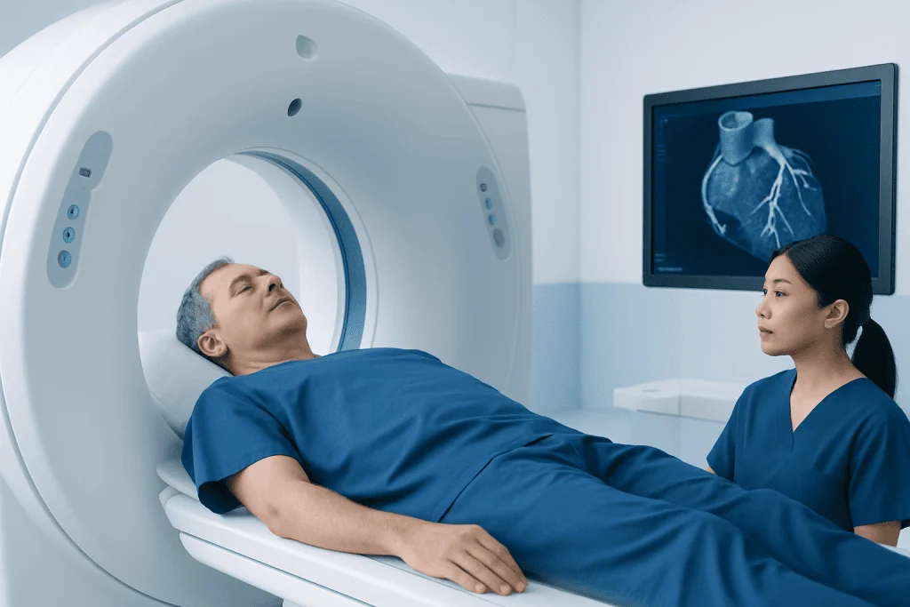
Coronary CT angiography can show whether the coronary arteries are open, narrowed, or affected by plaque buildup. Plaque may be calcified, non-calcified, or mixed. This is important because some plaques may not be visible on a standard calcium score alone, while CCTA can provide more detailed information about the vessel wall and the artery channel.
The scan may help doctors evaluate symptoms such as chest pain, pressure, shortness of breath with exertion, or unexplained discomfort when a heart-related cause is being considered. It can also be used to assess certain congenital coronary artery anomalies, check selected bypass grafts, or help clarify findings from other tests such as stress testing.
Common clinical questions CCTA may help answer include:
- Are there signs of coronary artery narrowing or blockage?
- Is plaque present, and if so, where is it located?
- Is a coronary artery following an unusual pathway?
- Is further testing, medication, or an invasive procedure likely to be needed?
Who May Be a Candidate

Doctors most often consider coronary CT angiography for people with stable symptoms and a low-to-intermediate likelihood of coronary artery disease. It may be especially useful when the goal is to look directly at the coronary arteries and determine whether symptoms are likely related to plaque or narrowing.
CCTA may not be ideal for everyone. Image quality can be affected by a very fast or irregular heartbeat, difficulty holding the breath, severe coronary calcification, or certain body and technical factors. People with advanced kidney disease, a history of significant reaction to iodinated contrast, or certain thyroid conditions may need special assessment before contrast is given.
The decision to use CCTA should be individualized. A doctor will usually consider the person’s age, symptoms, medical history, cardiovascular risk factors, kidney function, heart rhythm, and previous test results. In some cases, a stress test, echocardiogram, cardiac MRI, invasive angiography, or another approach may be more suitable.
How to Prepare for the Test
Preparation instructions can vary by hospital and by the patient’s health status. Many patients are asked to avoid caffeine for several hours before the scan because caffeine can increase the heart rate and reduce image clarity. They may also be asked not to eat for a short period before the examination, while clear fluids may be allowed depending on local instructions.
Patients should tell the medical team about kidney disease, diabetes, asthma, thyroid disease, pregnancy or possible pregnancy, breastfeeding, previous reactions to contrast dye, and all current medications or supplements. Blood tests to check kidney function may be requested before the scan, particularly for older adults or people with known kidney problems.
Some patients receive medication before or during the scan to slow the heart rate, because a steadier, slower rhythm can improve image quality. A medication that temporarily widens the coronary arteries may also be used in some cases. The care team will check whether these medicines are appropriate and monitor the patient during the examination.
What Happens During Coronary CT Angiography
On the day of the scan, the patient usually changes into a gown and lies on the CT table. Small ECG patches are placed on the chest to record the heartbeat and synchronize image capture. An intravenous line is inserted, usually into a vein in the arm, for the contrast injection.
The CT table moves through the scanner, which is shaped like a short, wide ring rather than a tunnel. The scan itself is usually quick, although preparation and heart rate control may take longer. The patient may be asked to hold the breath for a few seconds while images are taken, because chest movement can blur the pictures.
When contrast dye is injected, it is common to feel a warm sensation, a flushed feeling, or a temporary metallic taste. These sensations usually pass quickly. The medical team remains nearby, communicates with the patient, and monitors for any unexpected reaction throughout the process.
After the scan, most people can return to normal activities unless they received medication that requires short-term observation or specific precautions. Drinking fluids may be encouraged if appropriate, to help the body clear the contrast dye. The images are then processed and interpreted by specialists trained in cardiac imaging.
Benefits, Limitations, and Safety
The main benefit of coronary CT angiography is that it provides detailed information about the coronary arteries without an invasive catheter procedure. It can be particularly helpful for ruling out significant coronary artery disease in appropriately selected patients. When the scan is normal or shows no important narrowing, it may reduce the need for additional invasive testing in some cases.
Like all medical tests, CCTA has limitations. It may show a narrowing that later proves not to significantly reduce blood flow, or it may be difficult to interpret in heavily calcified arteries. Very small vessels, rapid heart rhythms, motion, and certain stents can also limit accuracy. For this reason, results should always be interpreted in the context of symptoms and other clinical information.
CCTA uses ionizing radiation and iodinated contrast dye. Modern scanners and protocols aim to keep radiation exposure as low as reasonably achievable while maintaining image quality. Contrast dye is generally well tolerated, but it can pose risks for people with significant kidney disease or previous contrast reactions. The care team reviews these risks before the test and takes precautions when needed.
Results, Follow-Up, and When to See a Doctor
Results may describe whether the coronary arteries are normal, whether plaque is present, and whether any narrowing appears mild, moderate, or severe. The report may also include comments about calcium, vessel anatomy, image quality, and sometimes other findings seen in the scanned area of the chest. A cardiologist or referring doctor explains what the findings mean for the individual patient.
Follow-up depends on the result and the person’s overall risk profile. Some patients may be advised to focus on lifestyle changes and preventive medications. Others may need additional tests to evaluate blood flow, or referral for invasive coronary angiography if a significant narrowing is suspected and treatment planning is needed.
Medical attention should be sought promptly for new, severe, persistent, or worsening chest pain; chest discomfort with shortness of breath, sweating, fainting, or pain spreading to the arm, jaw, back, or neck; or symptoms that feel unusual or concerning. For stable or recurring symptoms, patients should arrange a medical evaluation rather than trying to interpret symptoms on their own.
Acibadem International’s multidisciplinary specialists and JCI-accredited hospitals diagnose and treat coronary artery disease and related heart conditions for international patients, including when advanced cardiac imaging is part of the evaluation. Patients should consult a qualified doctor to determine whether coronary CT angiography is appropriate for their situation.
Frequently asked questions
Is coronary CT angiography the same as a traditional angiogram?
No. Coronary CT angiography is noninvasive and uses a CT scanner with contrast dye injected through a vein. A traditional coronary angiogram is invasive and uses a catheter inserted through an artery to reach the heart, which also allows certain treatments such as stent placement if needed.
Does coronary CT angiography hurt?
The scan itself is not painful. Patients may feel a brief pinch when the intravenous line is placed and a warm or flushed sensation when the contrast dye is injected. These feelings are usually short-lived.
How long does the test take?
The image acquisition is often very quick, sometimes taking only seconds. However, the full appointment may take longer because of preparation, heart rate monitoring, possible medication, IV placement, and observation after the scan.
Is the contrast dye safe?
Iodinated contrast dye is commonly used and is generally well tolerated. Before the test, the medical team checks for kidney problems, previous contrast reactions, and other factors that may require precautions or an alternative imaging plan.
Can CCTA detect all heart problems?
No single test detects every heart condition. Coronary CT angiography is designed mainly to evaluate the coronary arteries and certain anatomic details. Other tests may be needed to assess heart rhythm, valve disease, heart muscle function, or how well blood flows during exercise or stress.
What happens if the scan shows a blockage?
The next step depends on how severe the narrowing appears, the patient’s symptoms, and overall risk. A doctor may recommend lifestyle changes, medications, additional functional testing, or invasive coronary angiography to confirm findings and consider treatment options.
References
- American College of Cardiology
- American Heart Association
- European Society of Cardiology
- Radiological Society of North America
- Society of Cardiovascular Computed Tomography
This article is for general information only and is not a substitute for professional medical advice. Please consult a qualified doctor about your individual situation.
Coronary CT Angiography in Turkey — costs, top hospitals & a free quote
JCI-accredited · board-certified surgeons · reply within 24h
Add us as a Preferred Source to see more of our trusted health content across Google Search, AI Overviews and Discover.
Check your numbers in seconds
BMI, calories, due date, blood pressure and 30+ more clinical calculators — free, instant, doctor-reviewed ranges.

More from the Health Library



Related Specialists

Prof. Dr. Ercan Karaarslan
Radiology
Prof. Dr. Bora Peynircioğlu
Interventional Radiology
Prof. Dr. Koray Güven
Interventional Radiology
Assoc. Prof. Dr. Deniz Can Aliş
Radiology