Cranial Nerve Palsies: Why Eye Movement Problems and Double Vision Happen

Cranial nerve palsies most often involve the third, fourth, or sixth cranial nerves, which control eye movements. Common symptoms include double vision, drooping eyelid, abnormal head posture, and limited eye movement.
Key Takeaways
- Cranial nerve palsies most often involve the third, fourth, or sixth cranial nerves, which control eye movements.
- Common symptoms include double vision, drooping eyelid, abnormal head posture, and limited eye movement.
- Causes include diabetes, high blood pressure, trauma, infection, inflammation, aneurysm, stroke, and tumors.
- Some cases improve on their own, but sudden symptoms always need prompt medical assessment.
- Diagnosis may involve an eye exam, neurological evaluation, blood tests, and brain or orbit imaging.
- Treatment depends on the cause and may include observation, prism glasses, medication, or surgery.
Cranial nerve palsies affect the nerves that control eye muscles, eyelid position, and sometimes pupil function. They often lead to double vision, misaligned eyes, or trouble moving one eye in certain directions, and the cause can range from temporary inflammation to conditions needing urgent care.
Overview
Cranial nerve palsies are conditions in which one or more of the nerves controlling eye movement do not work normally. The nerves most commonly involved are the third cranial nerve (oculomotor nerve), fourth cranial nerve (trochlear nerve), and sixth cranial nerve (abducens nerve). These nerves help the eyes move together, keep vision aligned, and in some cases control the eyelid and pupil.
When one of these nerves is weakened, irritated, compressed, or injured, the eyes may no longer move in coordination. This can lead to double vision, an eye that appears turned in or out, difficulty focusing, or a compensatory head tilt. Symptoms may begin suddenly or develop gradually, depending on the underlying cause.
Cranial nerve palsies can happen in children or adults, but the causes differ by age and medical history. In adults, blood vessel disease related to diabetes or high blood pressure is common. In other situations, doctors must also consider inflammation, infection, head injury, tumors, stroke, or pressure on the nerve from an aneurysm.
Because some causes are harmless and others require urgent treatment, careful evaluation is important. A neuroophthalmologist, neurologist, or ophthalmologist can help determine whether the problem is isolated to the eye movement nerves or is part of a broader neurological condition.
Symptoms of Cranial Nerve Palsies
The most common symptom is double vision, also called diplopia. People may notice two side-by-side images, one image above the other, or a tilted appearance. Double vision often becomes worse when looking in a particular direction because the affected eye muscle cannot move fully.
Eye movement limitation is another common sign. One eye may not move outward, inward, up, or down as expected. Some people notice that one eye looks misaligned in photographs or in the mirror. Others may not see obvious misalignment but develop eye strain, headache, or trouble reading.
Depending on which nerve is involved, other symptoms may appear. A third nerve palsy may cause a drooping eyelid and, in some cases, a large or poorly reactive pupil. A fourth nerve palsy often causes vertical or tilted double vision and may lead a person to tilt the head to reduce symptoms. A sixth nerve palsy typically makes it hard to move the eye outward, causing horizontal double vision.
- Double vision that improves when one eye is covered
- Abnormal head turn or head tilt
- Drooping eyelid
- Eye pain or headache in some cases
- Blurred vision from poor alignment
- Dizziness or imbalance related to visual confusion
Symptoms can be constant or intermittent. Any sudden onset of double vision, especially if accompanied by severe headache, facial numbness, weakness, trouble speaking, or changes in pupil size, should be assessed urgently.
Causes and Risk Factors

Cranial nerve palsies happen when the nerve itself or its pathway is affected. In older adults, reduced blood flow to the nerve due to small vessel disease is a frequent cause. This is more likely in people with diabetes, high blood pressure, high cholesterol, or smoking history. These cases are often called microvascular cranial nerve palsies.
Trauma is another important cause. Even a relatively mild head injury can stretch or damage the fourth or sixth cranial nerve because of the way these nerves travel through the skull. In children, trauma, viral illness, congenital differences, and increased pressure inside the skull may all be considered.
Other possible causes include inflammation, autoimmune disease, infections, tumors, and disorders that affect the brainstem, cavernous sinus, or eye socket. Conditions such as multiple sclerosis, myasthenia gravis, thyroid eye disease, and stroke can sometimes mimic or contribute to eye movement abnormalities, so doctors may evaluate for related neurological or orbital disorders such as brain tumors or stroke.
One cause that needs urgent exclusion is compression of the third cranial nerve by an aneurysm. This is especially important when a third nerve palsy is painful or involves the pupil. Risk factors for serious causes include cancer history, immune suppression, significant headache, recent infection, new neurological symptoms, or progressive worsening rather than steady improvement.
How Doctors Diagnose the Problem
Diagnosis starts with a detailed medical history and examination. The doctor asks when symptoms began, whether they appeared suddenly or gradually, and whether there is pain, drooping eyelid, pupil change, trauma, or other neurological symptoms. Existing conditions such as diabetes, high blood pressure, migraine, autoimmune disease, or prior cancer are also relevant.
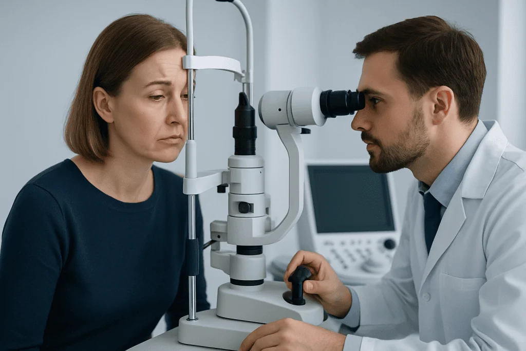
A careful eye and neurological examination helps identify which nerve is affected. The clinician checks eye alignment in different directions of gaze, eyelid position, pupil responses, head posture, and whether double vision changes when one eye is covered. Vision, color vision, and the health of the optic nerve are also evaluated to rule out broader neuro-ophthalmic disease.
Imaging is often needed, especially in children, younger adults, people with pain, pupil involvement, trauma, or additional neurological signs. Brain or orbit imaging may include MRI scanning or, in urgent situations, other forms of advanced neuroradiological evaluation. Blood tests may be ordered to check for diabetes, inflammation, infection, thyroid disease, or autoimmune conditions.
Sometimes the diagnosis is straightforward, but in other cases follow-up is part of the process. If an isolated microvascular palsy is suspected, doctors may monitor for recovery over weeks to months while still reassessing if symptoms change. Persistent, recurrent, or unexplained cases usually need broader evaluation.
Treatment Options
Treatment depends on the underlying cause rather than the eye movement problem alone. If the palsy is related to diabetes or high blood pressure, management focuses on controlling those conditions and monitoring recovery. If the cause is infection, inflammation, autoimmune disease, tumor, aneurysm, or stroke, treatment is directed at that specific problem and may involve urgent specialist care.
Symptom relief is also important. Temporary measures may include patching one eye, using a temporary opaque lens, or prism glasses to reduce double vision. These options do not cure the nerve problem but can make daily activities more comfortable while recovery is taking place.
Some patients need procedure-based treatment. For example, if symptoms remain stable after the recovery period, eye muscle surgery may help improve alignment, and some people benefit from strabismus surgery. In selected cases, treatment of the underlying neurological condition may require coordinated care through neurology services.
Recovery varies. Many microvascular palsies improve within a few months, while traumatic or compressive palsies may take longer or may not fully resolve. A doctor may recommend repeated examinations to confirm that the eye alignment is improving and that no new warning signs are developing.
Prevention and Self-care
Not all cranial nerve palsies can be prevented, but some risk factors can be reduced. Good control of diabetes, blood pressure, and cholesterol may lower the risk of small vessel nerve injury. Regular medical follow-up, not smoking, physical activity, and a balanced diet all support vascular health.
Protecting the head and eyes is also helpful. Seat belts, helmets for sports and cycling, and workplace safety measures can reduce the chance of traumatic nerve injury. People with a history of falls or balance problems may benefit from home safety adjustments and medical review.
At home, symptom management can make daily life easier. Covering one eye temporarily may reduce double vision enough for reading or walking, but patients should ask a clinician which eye to cover and when to use this strategy. Driving can be unsafe with active double vision, so medical advice should be followed before returning to the road.
Self-care should never replace assessment when symptoms are new. Even if double vision seems mild, it is important to identify whether the cause is a benign temporary nerve problem or a condition requiring prompt treatment.
When to See a Doctor
Anyone with new double vision or a sudden change in eye movement should seek medical assessment promptly. It is especially important to be seen quickly if symptoms began suddenly, are getting worse, or are associated with headache, eye pain, drooping eyelid, imbalance, weakness, numbness, or speech changes.
Urgent evaluation is particularly important when one pupil becomes larger than the other, when a third nerve palsy is suspected, or when symptoms follow head trauma. Children with a new head tilt, eye turn, or suspected double vision should also be examined without delay.
If symptoms have already been diagnosed as a microvascular palsy, follow-up remains important. Patients should contact their doctor sooner if the double vision worsens, recovery does not begin as expected, or new neurological symptoms appear. Ongoing review helps confirm the diagnosis and adjust treatment if needed.
For international patients needing coordinated assessment, Acibadem International’s multidisciplinary specialists in JCI-accredited hospitals evaluate and treat neuro-ophthalmic conditions, including cranial nerve palsies, with input from ophthalmology, neurology, and imaging teams.
Frequently asked questions
What are cranial nerve palsies?
Cranial nerve palsies are disorders affecting the nerves that control the eye muscles, eyelid, or pupil. When one of these nerves does not function normally, the eyes may become misaligned, causing double vision or limited eye movement.
Which cranial nerves most often cause double vision?
The third, fourth, and sixth cranial nerves are the ones most commonly involved. These nerves control most of the eye muscles, so damage to any of them can prevent the eyes from moving together normally.
Can a cranial nerve palsy go away on its own?
Some can, especially microvascular palsies related to diabetes or high blood pressure. However, recovery is not guaranteed, and medical follow-up is important to confirm improvement and rule out more serious causes.
Is double vision from a cranial nerve palsy an emergency?
Double vision should always be assessed promptly, because the cause is not always obvious without an examination. It is more urgent if there is severe headache, pupil change, drooping eyelid, weakness, numbness, speech difficulty, or recent head injury.
How is a cranial nerve palsy diagnosed?
Doctors use a detailed history, eye movement testing, and a neurological examination to identify the affected nerve. Depending on the situation, they may also order blood tests and imaging such as MRI to look for structural or inflammatory causes.
What treatments help with double vision while healing occurs?
Temporary treatments may include covering one eye, using an opaque lens, or wearing prism glasses. These approaches can improve comfort and daily function while doctors monitor recovery or treat the underlying cause.
References
- American Academy of Ophthalmology
- National Eye Institute
- National Institute of Neurological Disorders and Stroke
- Mayo Clinic
- Merck Manual Professional Edition
This article is for general information only and is not a substitute for professional medical advice. Please consult a qualified doctor about your individual situation.
Explore treatments in Turkey — costs, top hospitals & a free quote
JCI-accredited · board-certified surgeons · reply within 24h
Add us as a Preferred Source to see more of our trusted health content across Google Search, AI Overviews and Discover.
Check your numbers in seconds
BMI, calories, due date, blood pressure and 30+ more clinical calculators — free, instant, doctor-reviewed ranges.