Neuroradiology
Neuroradiology uses advanced imaging to diagnose and guide treatment of brain, spine, head, and neck conditions. It supports neurologists and neurosurgeons with precise MRI, CT, and angiographic evaluation.
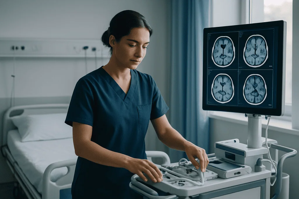
Quick answer
Neuroradiology is the radiology subspecialty that uses MRI, CT, ultrasound and angiography to diagnose conditions of the brain, spinal cord, nerves, head, neck and their blood vessels. A neuroradiologist selects the right imaging protocol for the clinical question, interprets the findings, and reports them to the treating team. Most examinations are non-invasive and need little or no recovery time.
Neuroradiology: Imaging the Brain, Spine, Head and Neck
Neuroradiology is the subspecialty of radiology that uses advanced imaging — chiefly MRI, CT and catheter-based angiography — to diagnose conditions of the brain, spinal cord, nerves, blood vessels, skull base, face, sinuses and neck. Its job is to answer clinical questions: is there a lesion, what is it, where exactly does it sit, and what does that mean for treatment. It is relevant to anyone whose symptoms point to the nervous system, from suspected stroke to back pain with nerve involvement.
When symptoms involve the brain or spine, uncertainty is hard to live with. A new headache that behaves differently from every previous headache, sudden weakness, memory changes, seizures, dizziness, or pain radiating down an arm or leg all raise the same questions: is this serious, does it need surgery, can treatment wait, and is the diagnosis precise enough to make the right decision? Neuroradiology sits at the centre of those answers.
Modern neuroradiology is not simply “taking a scan”. It means choosing the right imaging method for the question, performing the study with a protocol designed for the suspected condition, interpreting subtle findings against the clinical picture, and communicating the result clearly to neurologists, neurosurgeons, oncologists, ear, nose and throat specialists, emergency physicians and the rest of the care team. A technically flawless image acquired with the wrong protocol can still leave the real question unanswered, which is why protocol selection is treated as a medical decision in its own right.
Many patients arrive with previous MRI or CT scans from different hospitals and different medical systems, sometimes with conflicting opinions attached. A neuroradiology review can reconcile those findings, decide whether new imaging would actually change anything, and support a treatment plan built on current evidence rather than on an accumulation of reports.
At Acibadem, neuroradiology supports a broad range of clinical services: emergency stroke evaluation, brain tumour planning, spine disorders, vascular malformations, paediatric neurological conditions, epilepsy assessment, head and neck tumours, and structured follow-up after treatment. The purpose is always the same — to give you and your physicians information solid enough to act on, at the right time, with a clear next step.
Is a neuroradiologist a medical doctor?
Yes. A neuroradiologist is a fully qualified medical doctor who completed medical school, then specialist training in diagnostic radiology, then additional subspecialty training focused on imaging of the nervous system. Neuroradiologists do not usually run outpatient clinics the way a neurologist does, but they are physicians in every sense: they take clinical responsibility for the studies they interpret, they consult with treating doctors about difficult findings, and some perform image-guided procedures. In many hospitals the neuroradiologist is the specialist most likely to notice the small detail — an early stroke sign, a subtle cord lesion, a lymph node in an unusual position — that changes the direction of care.
What is the difference between a radiologist and a neuroradiologist?
A radiologist interprets medical imaging across the whole body; a neuroradiologist has completed further dedicated training in imaging of the brain, spine, head and neck. Both work within a hospital’s radiology department, and both are essential. The difference matters most when anatomy is complex and the margin for interpretation is narrow — the skull base, the inner ear, the spinal cord, the pituitary region, the vessels of the brain. Radiology has other organ-focused subspecialties built on the same logic: renal radiology concentrates on the kidneys and urinary tract, and musculoskeletal radiology on bones, joints and soft tissues of the limbs. For nervous system disease, subspecialty reading reduces the chance that a subtle but decisive finding is missed or that a harmless variant is mistaken for disease.
Neuroradiology, neurorad or neuroradiologi — which is correct?
All three refer to the same specialty. Neurorad is the clinical shorthand you may hear in hospital corridors or see in scheduling systems and staff rotas. Neuroradiologi is the spelling used in Scandinavian and several other European languages, so it appears often when patients research from outside the English-speaking world. If you are searching in English, “neuroradiology” is the term that will lead you to the most reliable sources.
The Imaging Methods Neuroradiology Uses
Neuroradiology draws on four main tools — MRI, CT, ultrasound in selected head and neck applications, and angiographic imaging — and the skill lies in matching the tool to the question. Each method shows something the others cannot, and each has practical limits worth understanding before your appointment.
Magnetic resonance imaging (MRI) uses a strong magnetic field and radiofrequency signals to build detailed images without ionising radiation. It is the method of choice for soft tissue: the brain itself, the spinal cord, nerves, tumours, inflammation, infection, demyelination, and many degenerative or developmental conditions. Different MRI sequences answer different questions — diffusion imaging can reveal acute stroke within minutes of onset, contrast-enhanced sequences highlight tumours and inflammation, and dedicated protocols exist for epilepsy, the pituitary gland, the inner ear and the skull base. The trade-off is time: MRI examinations take longer than CT and require the patient to remain still.
Computed tomography (CT) uses X-rays and computer processing to produce cross-sectional images quickly. Speed makes it the usual first test in emergencies — head trauma, suspected bleeding, acute stroke assessment — and its clarity for bone makes it valuable for skull, facial, sinus and spine anatomy, calcification, and evaluation after operations involving bone or implants. Contrast material injected into a vein can extend CT to show blood vessels, tumours, inflammation or infection more clearly.
Ultrasound has a narrower but genuine role in the head and neck: assessing neck masses, lymph nodes, salivary glands and the thyroid region, guiding needle sampling of superficial lesions, and evaluating blood flow in neck vessels. It involves no radiation and can be repeated freely, though it cannot see through bone and therefore contributes little to imaging the brain in adults.
Angiographic imaging focuses on blood vessels. Non-invasive versions — CT angiography and MR angiography — map the arteries and veins of the brain and neck without a catheter and are sufficient for many questions. Catheter angiography is more specialised: a thin catheter is guided through the blood vessels, commonly from the groin or wrist, and images are taken as contrast is injected directly into the vessels of interest. It offers the most detailed vascular pictures available and is used when non-invasive imaging cannot settle the question, or when an image-guided treatment is being considered at the same time.
Method selection also involves radiation stewardship. Because CT and angiography use X-rays, the examination is designed so that the dose stays as low as the diagnostic question allows, and MRI or ultrasound is preferred where either can answer the question equally well. This matters most in children, whose imaging pathways are planned with particular care: paediatric protocols use adjusted parameters, shorter acquisitions where possible, and preparation designed around the child — play-based explanation, a parent nearby during suitable examinations, and sedation only when a study genuinely cannot be completed otherwise. In infants, ultrasound through the soft spot of the skull can sometimes answer questions that would otherwise require a scanner at all.
Who May Need a Neuroradiology Evaluation
A neuroradiology evaluation may be recommended whenever symptoms suggest a problem involving the brain, spine, nerves, blood vessels, head or neck. Sometimes imaging is needed urgently, as in suspected stroke, head trauma, sudden neurological weakness or a severe headache with warning features. In other situations it is planned as part of a careful outpatient work-up — chronic back pain, balance problems, memory concerns, recurrent seizures, facial pain, or the assessment of a known tumour.
Symptoms that commonly lead to neuroradiology imaging include:
- Persistent or unusual headaches, or a clear change in headache pattern
- Sudden weakness, numbness, difficulty speaking or vision changes
- Seizures, fainting, confusion or memory changes
- Dizziness, loss of coordination or hearing-related symptoms
- Facial weakness, facial pain or swallowing problems
- A neck mass or unexplained swelling in the head and neck region
- Back or neck pain radiating into an arm or leg
- Changes in bladder or bowel control associated with spinal symptoms
These symptoms do not always indicate serious disease. Often they do not. But they frequently require accurate evaluation to separate benign causes from conditions that need timely treatment, and imaging is usually how that separation is made. The process begins with a clinical assessment: your doctor reviews your symptoms, neurological examination, medical history, previous operations, medications, and risk factors such as high blood pressure, cancer history, infection risk, trauma or a family history of vascular disease. Based on that assessment, specific imaging is requested, and the neuroradiology team ensures the protocol fits the suspected condition rather than defaulting to a generic scan.
Some patients come to neuroradiology after an abnormal result elsewhere — a newly reported brain lesion, a suspected aneurysm, spinal stenosis, a disc herniation, a pituitary tumour, lesions raising the question of multiple sclerosis, a head and neck mass, or vascular narrowing. In these cases the neuroradiologist reviews the previous images directly rather than relying on the written report, compares them with new imaging if new imaging is genuinely needed, and provides a detailed interpretation for the treating physician. A second imaging opinion is particularly valuable when surgery has been recommended, when the diagnosis is uncertain, when symptoms do not match the earlier report, or when you want to understand whether less invasive options exist. In complex cases, that review happens in multidisciplinary boards, where radiologists, neurologists, neurosurgeons, oncologists, radiation oncologists and other specialists discuss diagnosis and treatment together.
Conditions Neuroradiology Helps Address
Neuroradiology supports diagnosis, treatment planning and follow-up across emergency medicine, neurology, neurosurgery, oncology, spine care, otolaryngology, paediatrics and rehabilitation medicine. It is easiest to understand by region.
In brain care, imaging evaluates stroke, bleeding, aneurysms, vascular malformations, brain tumours, pituitary and skull base lesions, epilepsy-related abnormalities, infections, inflammation, demyelinating diseases such as multiple sclerosis, traumatic brain injury, hydrocephalus, congenital brain abnormalities, and degenerative conditions affecting cognition or movement. For many of these, the imaging appearance does not just detect disease — it narrows the differential diagnosis, suggests how urgent the situation is, and shapes whether the next step is medication, biopsy, surgery or observation.
In spine care, imaging assesses disc herniation, spinal stenosis, spinal cord compression, tumours, infection, fractures, deformities, inflammatory disease, postoperative changes, and the causes of arm or leg pain related to nerve root compression. Spine MRI is particularly informative because it shows the discs, nerves, ligaments, spinal canal and spinal cord in detail — the structures a surgeon needs to see before recommending or ruling out an operation.
In head and neck care, neuroradiology contributes to the evaluation of tumours of the throat, salivary glands, sinuses, skull base, orbit and neck. Imaging helps determine the size and extent of a mass, whether lymph nodes are involved, whether important nerves or vessels are affected, and how treatment should be staged and planned. It is also central to assessing complications of sinus disease, facial trauma, ear-related disorders, and vascular conditions of the neck.
In vascular neurology and neurosurgery, neuroradiology maps blood flow to and within the brain. CT angiography, MR angiography, perfusion imaging and catheter angiography may be used to assess blocked or narrowed arteries, aneurysms, arteriovenous malformations, dural fistulas, venous sinus thrombosis and other circulation disorders. In selected urgent situations, this imaging determines whether a patient may benefit from immediate intervention.
Neuroradiology also continues after treatment. Follow-up imaging is used after surgery, radiation therapy, chemotherapy, endovascular procedures, spine operations, or medical treatment for inflammatory or infectious disease. Its purpose is to evaluate healing, detect recurrence or progression, assess treatment response, and identify complications early — and to do so against a baseline, which is why keeping prior imaging accessible matters so much.
How a Neuroradiology Examination Is Performed
The process begins before the scanner is involved. A useful examination depends on understanding the clinical question, so the referring physician provides information about symptoms, examination findings, the suspected diagnosis, previous imaging, prior treatments, allergies, kidney function when contrast is considered, implanted devices, and whether MRI can be performed safely. When earlier imaging was performed elsewhere, those records and image files are reviewed whenever available — this reduces unnecessary repeat testing and allows comparison over time.
A typical pathway looks like this:
- Referral and question-setting. Your doctor defines what the imaging needs to answer, and the neuroradiology team confirms or adjusts the protocol accordingly.
- Safety screening. You complete a questionnaire covering implants, devices, allergies, kidney function and, where relevant, pregnancy status.
- Preparation. This ranges from removing metal objects before MRI to blood tests and short fasting for some contrast-enhanced or catheter-based studies.
- The examination itself. Duration and experience depend on the method, as described below.
- Interpretation and reporting. The neuroradiologist analyses the study, compares prior imaging, and writes a structured report; urgent findings are communicated directly to the treating physician.
- Clinical decision. Your doctor — often with a multidisciplinary board for complex findings — turns the report into a plan.
Preparation varies by method. For most MRI scans, you remove metal objects and complete a safety questionnaire about pacemakers, implants, surgical clips, hearing devices, medication pumps or metal fragments. Many modern implants are MRI-compatible, but the details must always be verified against the specific device documentation. If contrast material is planned, kidney function and allergy history may be checked. For CT, preparation is often minimal, though contrast-enhanced studies may involve blood tests and a short fasting period depending on the protocol. For angiographic procedures, preparation is more detailed and includes medication review by the treating team, blood tests, fasting, and a discussion of risks and benefits. Practical guidance on scan safety, contrast and implants is collected in our radiology and imaging safety guide.
During MRI, you lie on a table that moves into the scanner. The machine is loud — rhythmic knocking and buzzing are normal, and ear protection is provided. Staying still matters, because movement blurs the images. Some scans are brief; complex brain, spine, vascular, tumour, epilepsy or skull base protocols take longer. If claustrophobia, pain or difficulty lying flat makes stillness hard, the care team can adapt positioning, provide support, adjust the protocol, or discuss sedation in selected cases. MRI does not use ionising radiation.
During CT, you lie on a table that moves through a ring-shaped scanner. The scan itself is fast, which is why CT is the usual first test in emergencies such as head trauma, suspected bleeding or acute stroke assessment. When contrast is used, it is injected through a vein; a brief warm sensation is common and expected.
Catheter angiography is performed in a dedicated angiography suite. A thin catheter enters a blood vessel, commonly at the groin or wrist, and is guided to the vessels of interest while images are taken during contrast injection. It requires monitoring afterwards and specific aftercare for the access site, and it is reserved for questions that non-invasive vascular imaging cannot answer — or for situations where treatment may follow diagnosis in the same session.
Behind these examinations sits a layer of technology chosen for the clinical problem rather than for its own sake. High-resolution MRI sequences show small lesions, nerve pathways, spinal cord changes and a tumour’s relationship to critical structures. Diffusion imaging helps identify acute stroke and characterise certain tumours and infections. Perfusion imaging evaluates blood flow patterns in the brain or within tumours. Functional and tract-based imaging can support selected surgical planning by mapping how a lesion relates to important brain networks. Advanced workstation software produces three-dimensional reconstructions and precise comparisons between current and prior studies. None of this replaces judgement; it gives judgement better material to work with.
How long does a neuroradiology scan take?
Most CT examinations are completed within minutes once you are positioned; MRI usually takes longer because multiple sequences are acquired, and complex protocols, contrast-enhanced studies or angiographic procedures add time for preparation, scanning, monitoring and recovery. As a working expectation: plan a short visit for routine CT, a longer one for MRI, and a substantially longer one — with post-procedure observation — for catheter angiography or any study involving sedation. Your appointment confirmation will state what applies to your specific examination.
Recovery after diagnostic MRI or CT is usually immediate, and most patients return to normal activities the same day unless sedation was used or the underlying condition itself requires restrictions. If contrast was given, drinking fluids may be encouraged unless you are medically restricted. After catheter angiography, monitoring continues for a period and activity is limited temporarily to protect the access site and reduce bleeding risk; the care team provides individual instructions based on the procedure performed and your medical condition.
Interventional Neuroradiology and Endovascular Treatment
Neuroradiology has two faces. Diagnostic neuroradiology identifies and characterises disease. Image-guided neuroradiology goes a step further, using imaging not only to see pathology but to reach and treat it.
What is interventional neuroradiology?
Interventional neuroradiology is the treatment arm of the specialty: it uses continuous imaging to guide minimally invasive procedures on the brain, spine, head and neck, most often through the blood vessels. Instead of open surgery, instruments travel through a catheter to the site of disease — an aneurysm, a blocked artery, a vascular malformation — under real-time X-ray guidance. Whether this approach suits a given patient depends on the diagnosis, the anatomy and the hospital’s clinical pathway. You can read a dedicated overview on our interventional neuroradiology page.
What is endovascular surgical neuroradiology?
Endovascular surgical neuroradiology is another name for the same field, used mainly in North American training systems; “endovascular” simply means “inside the blood vessels”. The term describes procedures such as coiling or otherwise securing aneurysms, removing clots in selected stroke cases, and closing abnormal vessel connections — all performed from within the circulation rather than through an open operation. Different countries label the training differently, but the clinical work is the same.
Does a neuroradiologist perform surgery?
Not open surgery — a neuroradiologist does not open the skull or the spine. Diagnostic neuroradiologists interpret imaging and may perform image-guided procedures such as targeted injections or needle sampling. Interventional neuroradiologists perform catheter-based endovascular procedures, which are treatments but not operations in the traditional sense: access is through a small puncture in a vessel, not an incision over the target organ. Open neurosurgery remains the territory of neurosurgeons, and in practice the two specialties plan complex cases together, often around the same set of images.
How Neuroradiologists Are Trained
Understanding the training pathway helps explain why subspecialty interpretation is worth seeking out for nervous system disease. The route is long and deliberately so.
How many years does it take to become a neuroradiologist?
Becoming a neuroradiologist typically takes well over a decade of education and training in total: medical school, then a multi-year residency in diagnostic radiology, then dedicated subspecialty fellowship training in neuroradiology. Exact durations vary by country because residency lengths and fellowship structures differ between systems, but no shortcut exists in any of them — every neuroradiologist is first a doctor, then a radiologist, then a subspecialist.
How long is a neuroradiology fellowship?
A neuroradiology fellowship usually lasts one to two years after radiology residency, depending on the country and whether the trainee continues into interventional work, which requires further dedicated training. Large academic departments — UCSF Radiology in the United States is a widely cited example — publish detailed fellowship curricula that show what the additional years cover: advanced brain, spine and head and neck imaging, paediatric neuroimaging, and for some trainees the procedural skills of endovascular treatment. The practical point for you as a patient is simple: when a report on your brain or spine is signed by a fellowship-trained neuroradiologist, it reflects years of focused pattern recognition in exactly this anatomy.
Navigating Radiology Reports
Navigating radiology reports is easier once you know how they are built. A structured report typically states the examination performed and why, the technique and any contrast used, a comparison with prior studies, a findings section describing what the images show region by region, and an impression — the radiologist’s conclusion, written for your treating doctor. The impression is the part that drives decisions; the findings section is the evidence behind it. Reports are written in precise technical language because they are a communication between physicians, so an unfamiliar term is not automatically an alarming one. If wording in your report worries you, the reliable route is to ask the doctor who requested the scan, who can place each phrase in the context of your examination and history.
What does “no acute intracranial abnormality” mean?
“No acute intracranial abnormality” means the scan shows no new or urgent problem inside the skull — no fresh bleeding, no visible acute stroke on that examination, no mass effect requiring immediate action. It is one of the most common conclusions in emergency head imaging, and it is reassuring, but it is worth reading precisely. It does not promise that everything is perfect; chronic or subtle findings may still be described in the body of the report, and some conditions are not visible on the first scan or on that particular type of scan — early ischaemia, for instance, can be easier to see on MRI than on the initial CT. The phrase answers the emergency question. Whether further imaging is needed to answer a different question is a decision for your treating physician.
Why Acting Early Matters
Neurological conditions can change quickly. In some cases, early imaging identifies a problem that requires urgent treatment — stroke, bleeding in or around the brain, spinal cord compression, infection, or a rapidly expanding mass. In others, imaging confirms that a condition is stable or less dangerous than feared, which allows you and your physician to avoid unnecessary procedures and concentrate on appropriate management. Both outcomes are wins; both depend on the study being done well and read carefully.
The risk of delay depends on the suspected condition. In stroke, time can influence which treatments remain possible. In spinal cord compression, delay may increase the risk of persistent weakness, numbness, or bladder and bowel dysfunction. In tumours, delayed diagnosis can allow growth or spread, potentially narrowing the treatment options. In infection, postponement may increase the risk of neurological damage or systemic illness. In aneurysms and vascular malformations, accurate diagnosis lets physicians assess the risk profile and decide between monitoring and treatment on evidence rather than assumption.
Early imaging does not mean every patient needs aggressive treatment. Often it means the opposite: timely, precise neuroradiology is one of the best defences against overtreatment. It can show when a finding is benign, when observation is safe, when medication is appropriate, or when surgery should be carefully planned rather than rushed. The essential point is that a clear diagnosis lets decisions rest on evidence instead of uncertainty.
Benefits of Neuroradiology
The main benefits of neuroradiology fall into four areas: diagnostic precision, treatment planning, speed in emergencies, and objective follow-up.
| Benefit | What It Means for You |
|---|---|
| More accurate diagnosis | Specialised imaging helps identify the cause of neurological, spine, head or neck symptoms and distinguishes conditions that can look similar clinically. |
| Better treatment planning | Detailed images show the location, size, extent and relationships of a lesion, informing decisions between medication, surgery, radiation therapy, endovascular treatment or observation. |
| Rapid decision-making in emergencies | In suspected stroke, trauma, bleeding or spinal cord compression, imaging establishes the urgency and shapes the type of treatment needed. |
| Support for minimally invasive care | Vascular and image-guided techniques can diagnose — and in selected cases treat — certain conditions through smaller access routes than open surgery. |
| Objective follow-up over time | Repeat imaging shows whether a condition is stable, improving, recurring or progressing after treatment, against a documented baseline. |
Recovery and What to Expect After Imaging
Most diagnostic neuroradiology examinations require little or no recovery time; catheter-based or sedated procedures involve closer monitoring and more specific aftercare.
| Time Period | What Patients Can Expect |
|---|---|
| Day 1 | After routine MRI or CT, most patients resume usual activities immediately. If contrast was used, hydration may be encouraged. If sedation or catheter angiography was performed, monitoring and temporary activity restrictions apply. |
| First Week | Your treating physician reviews the imaging report and recommendations. Additional consultations, laboratory tests, repeat imaging or specialist board review may be arranged for complex findings. |
| First Month | Treatment decisions are often finalised during this period, particularly for tumours, vascular conditions, spine disease, inflammatory disorders or postoperative assessment. Some patients begin treatment; others enter a follow-up plan. |
| Longer Term | Ongoing imaging may be scheduled to monitor stability, healing, treatment response or recurrence. The interval depends on the diagnosis and the treatment strategy. |
Factors That Influence a Good Result
A good neuroradiology result depends on more than image quality. It begins with the right clinical question. Imaging should be selected because it can answer a specific diagnostic or planning need: a general MRI may not be sufficient for epilepsy surgery planning, skull base evaluation or certain tumour assessments; a stroke protocol differs from a multiple sclerosis protocol; a spine trauma CT differs from a study designed to evaluate nerve compression. The closer the protocol matches your condition, the more useful the result.
Your history matters just as much. Previous operations, cancer history, radiation therapy, implanted devices, vascular disease, infection risk, current medications and the timeline of your symptoms all shape interpretation. Comparison with prior studies is often decisive: a lesion unchanged over years usually means something very different from a new or growing one. If you have earlier imaging, the original digital files in DICOM format — not printed pictures or photographs of a screen — together with translated reports where available, give the neuroradiologist the most to work with.
Motion control is a quieter factor. MRI and CT images are clearest when you remain still, and pain, anxiety, claustrophobia, tremor or difficulty lying flat can degrade quality. The team can often adapt positioning, provide support, shorten or reorder sequences, or consider sedation where medically appropriate. In children and in patients who cannot remain still, this planning is done in advance so that useful images are obtained safely at the first attempt.
Contrast material can raise diagnostic confidence in selected cases — it helps show tumours, inflammation, infection, vessel abnormalities and postoperative changes — but it is not needed for every examination. The decision is individualised on the basis of the suspected condition, kidney function, allergy history, pregnancy status and any previous reactions. A well-chosen contrast strategy improves clarity while avoiding unnecessary exposure.
Interpretation by experienced physicians remains the decisive step. Neuroradiology requires familiarity with normal anatomical variation, subtle disease patterns, postoperative anatomy, treatment-related changes, and artefacts that can convincingly mimic disease. A high-quality scan can mislead when read without clinical context, and a subtle but important finding can be missed when the protocol or the review is not specialised enough. Direct collaboration between the neuroradiologist and the treating physician keeps the report aligned with what you actually feel and what your examination actually shows.
Finally, outcomes depend on what happens after imaging. A clear report should lead to a clear plan — reassurance and observation, further testing, medical treatment, referral to the appropriate specialist, urgent intervention, or scheduled follow-up. Neuroradiology is at its most valuable when it is integrated into a broader care pathway rather than treated as a stand-alone test that ends when the report is signed.
Neuroradiology Within Multidisciplinary Care at Acibadem
Brain, spine, head and neck conditions rarely belong to a single specialty, so neuroradiology at Acibadem is integrated into hospitals that care for patients across neurology, neurosurgery, oncology, radiation oncology, spine surgery, otolaryngology, emergency medicine, paediatrics and rehabilitation. That structure matters because most serious neurological decisions need more than one perspective looking at the same images.
In complex cases, imaging findings are reviewed in specialist boards and multidisciplinary meetings, where physicians from different fields consider the same patient together using imaging, clinical findings, laboratory results, pathology and previous treatment history. For a patient with a brain tumour, the imaging interpretation may influence surgical planning, biopsy strategy, radiation therapy planning and follow-up intervals. For suspected vascular disease, it may guide the choice between medical therapy, observation, endovascular treatment and surgery. For spine disease, it helps determine whether symptoms genuinely correspond to nerve compression and whether conservative care or an operation is the more defensible path.
Protocol planning is personalised because two patients with similar symptoms rarely need the same scan. A patient with suspected multiple sclerosis requires a different imaging approach from a patient with pituitary disease, brain metastasis, trigeminal neuralgia, spinal infection or acute stroke. Neuroradiology teams work with referring physicians to select the examination and contrast strategy so the imaging addresses the actual clinical question as directly as possible.
Reviewing outside studies is part of the daily work. Patients frequently arrive with imaging performed elsewhere, and the first task is to determine whether those images are adequate, whether the findings are stable, and whether additional imaging would genuinely change management. Sometimes the previous imaging is sufficient for a second opinion. Sometimes a repeat scan with a more targeted protocol is recommended because the earlier study did not fully answer the question. Either way, the aim is to avoid duplication without letting an important decision rest on inadequate images.
The equipment available varies by hospital and clinical service, but the toolset is consistent with what this page describes: high-resolution MRI, CT, angiographic imaging, contrast-enhanced studies, vascular and perfusion techniques, diffusion imaging and advanced image processing where appropriate. Coordination teams handle records and scheduling around the clinical pathway, so that the medical work — not the logistics — sets the pace. For patients and families facing neurological uncertainty, the value of all this is not only technical. It is the movement from unclear symptoms or conflicting opinions towards a structured understanding of the condition, which is the foundation every safe decision rests on.
Preparing for a Neuroradiology Evaluation
Wherever your imaging takes place, a few practical steps consistently improve its value. Keep your prior imaging as original digital files — most hospitals can provide a disc or a download link with DICOM data — because original images can be measured and compared, while printed sheets and phone photographs usually cannot. Keep the written reports with them, translated where possible, and note the dates and locations of previous scans so the sequence of studies is clear.
Make a complete list of implants and devices before any MRI appointment: pacemakers, stents, clips, cochlear or hearing implants, medication pumps, artificial joints, and any possibility of metal fragments from past injury or work. Many devices are MRI-compatible; the point is that compatibility must be verified against the exact model, which is only possible when the information is available in advance. Bring your medication list and allergy history as well, since both feed into contrast decisions — decisions that are always made by the clinical team, never something to adjust on your own initiative.
Think, too, about the questions you want the imaging to answer, and raise them with your treating doctor before the scan rather than after: what condition is suspected, what would change if the scan is normal, what would change if it is not, and when the results will be discussed. Imaging answers questions best when the questions are asked first.
Neuroradiology is often the bridge between symptoms and diagnosis, and between diagnosis and treatment. When the nervous system is involved, precise information matters more than almost anything else. A careful imaging evaluation — the right study, prepared properly, interpreted in context — gives you and your physicians a clearer view of the path ahead and a firmer basis for every decision along it.
Preparation
- Preparation depends on the imaging method and whether contrast material is needed. Patients may be asked to fast for several hours, share kidney function tests, and inform the team about allergies, pregnancy, implants, or claustrophobia. Previous scans and medical reports should be brought for comparison.
Aftercare
- Most patients can return to normal activities immediately after diagnostic neuroradiology imaging. If contrast is used, drinking fluids may be recommended unless restricted by a doctor. Results are reviewed by radiology specialists and shared with the referring physician for diagnosis or treatment planning.
Turkey vs UK, Germany & USA
Neuroradiology costs and patient experience can vary depending on the imaging method, clinical complexity, hospital setting, and whether the service is diagnostic or procedure based. International patients often compare availability, reporting expertise, accreditation, and package coordination when choosing where to have advanced brain, spine, head, or neck imaging.
The comparison below outlines non-price factors that may influence the overall cost and experience of neuroradiology care in different destinations.
| Factor | Turkey | UK | Germany | USA |
|---|---|---|---|---|
| Price drivers | Imaging modality, contrast use, radiologist subspecialty review, hospital category, and whether angiography or intervention is required. | Private imaging costs may vary by hospital, scanner type, contrast, and consultant reporting; public pathways may involve referral criteria. | Costs are influenced by hospital type, diagnostic complexity, contrast, subspecialist interpretation, and inpatient or outpatient status. | Costs may vary widely by facility, insurance status, radiology group billing, contrast use, and whether hospital or outpatient imaging is chosen. |
| Hospital and specialist factors | Large private hospitals may offer integrated neurology, neurosurgery, radiology, and interventional neuroradiology teams for coordinated evaluation. | Specialist centres and private hospitals may provide advanced imaging with consultant neuroradiologist reporting depending on availability. | University and private hospitals often provide structured neuroimaging pathways with specialist radiology review. | Academic and private imaging centres may offer broad technology access, with provider networks and billing arrangements affecting the patient experience. |
| Accreditation and quality | International patients may look for JCI-accredited hospitals, modern MRI and CT technology, structured safety protocols, and multidisciplinary review. | Quality is supported through national healthcare standards, hospital governance, and specialist professional regulation. | Quality is supported through national regulation, certified hospital processes, and specialist medical training standards. | Quality is supported by hospital accreditation systems, specialist credentialing, and institutional imaging protocols. |
| Typical waiting times | Private international patient pathways may allow faster scheduling, especially when records are prepared in advance. | Public pathways can involve waiting depending on urgency; private scheduling may be quicker. | Access may depend on referral pathway, urgency, and whether care is public, private, or self-pay. | Access may be prompt in some private settings, but insurance authorisation and network rules can affect timing. |
| Travel and language logistics | International patient departments may assist with appointments, translation, airport transfers, and report delivery in English or other languages. | English language access is straightforward; international patients usually arrange travel and accommodation independently or through private providers. | English support may be available in larger centres; travel coordination and translations may be needed for international patients. | English language access is standard; international patients may need to manage insurance, billing, travel, and record transfer logistics. |
| What a package may include | Consultation coordination, imaging appointment, contrast if needed, specialist reporting, translation support, and follow-up planning may be bundled or coordinated. | Private packages may include imaging and report, while consultations, contrast, and follow-up can be billed separately. | Packages may vary by hospital and may separate imaging, reporting, specialist consultation, and translation services. | Itemised billing is common, and facility fees, radiologist reporting, contrast, consultation, and follow-up may be separate. |
What affects your final cost:
- Type of imaging, such as MRI, CT, angiography, or image-guided procedure.
- Use of contrast material, sedation, monitoring, or additional safety preparation.
- Complexity of the condition and whether urgent interpretation is required.
- Need for neuroradiologist, neurologist, neurosurgeon, or multidisciplinary review.
- Whether care is outpatient, inpatient, or linked to surgery or intervention.
- Translation, travel assistance, medical report preparation, and follow-up services.
Compare your options
Neuroradiology includes several diagnostic and image-guided options for evaluating brain, spine, head, and neck conditions. Suitability is decided by a specialist after reviewing symptoms, medical history, prior tests, and safety considerations.
| Option | What it is | Typical use | Key considerations |
|---|---|---|---|
| MRI neuroradiology | Magnetic resonance imaging focused on the brain, spine, skull base, head, or neck. | Often used for tumours, stroke evaluation, multiple sclerosis, epilepsy workup, spinal disc disease, infections, and vascular or inflammatory conditions. | May require contrast, special sequences, or longer scan time; patients with certain implants or claustrophobia need additional assessment. |
| CT neuroradiology | Cross-sectional imaging using X-rays to assess bone, bleeding, trauma, calcification, and urgent neurological concerns. | Often used for head trauma, acute neurological symptoms, sinus or temporal bone evaluation, spine assessment, and emergency decision-making. | Uses radiation; contrast may be required for selected indications, and kidney function or allergy history may need review. |
| CT or MR angiography | Non-invasive vascular imaging that shows arteries and veins of the brain, neck, or spine. | Used to evaluate aneurysms, vascular narrowing, dissections, malformations, venous thrombosis, and stroke-related blood flow concerns. | Choice of CT or MRI depends on the clinical question, urgency, contrast safety, image detail required, and patient factors. |
| Catheter angiography | A minimally invasive angiographic test in which a catheter is used to obtain detailed vascular images. | Used when very detailed vessel mapping is needed or when planning possible endovascular treatment. | Requires specialist assessment, procedural preparation, monitoring, and discussion of benefits and risks. |
| Interventional neuroradiology | Image-guided procedures performed through blood vessels or targeted access routes. | May support treatment of selected aneurysms, vascular malformations, stroke-related vessel blockage, or certain spine and head and neck conditions. | Not every patient is suitable; decisions depend on anatomy, urgency, imaging findings, and multidisciplinary discussion. |
| Image-guided spine or pain procedures | Targeted procedures performed with imaging guidance around the spine or related structures. | May be considered for selected diagnostic blocks, injections, or biopsy guidance when clinically appropriate. | Requires review of imaging, symptoms, medications, bleeding risk, and the expected role of the procedure in the care plan. |
General information only — not medical or financial advice. Final costs depend on the factors above and your individual case; request a free, personalised quote.
Frequently Asked Questions
What affects the cost of neuroradiology?
The final cost depends on the imaging method, body area examined, use of contrast, whether sedation or monitoring is needed, the level of specialist reporting, and whether a consultation or image-guided procedure is included. A personalised quote can be prepared after your medical records and the requested examination are reviewed.
How can I get a personalised quote from Acibadem?
You can request a free consultation and share your symptoms, previous imaging, medical reports, and referral notes if available. The international patient team can coordinate specialist review and provide a tailored cost estimate based on the recommended neuroradiology pathway.
Is the radiology report included in the package?
Many neuroradiology arrangements include the scan and specialist report, but package content can vary depending on the examination and clinical request. It is important to confirm whether contrast, consultation, translation, image copies, and follow-up discussion are included.
Do I need to travel to Turkey for neuroradiology evaluation?
For a new scan or image-guided procedure, travel may be required. If you already have imaging, a specialist may be able to review your files remotely and advise whether further imaging, consultation, or treatment planning is appropriate.
Will my results be reviewed by a specialist?
Neuroradiology examinations are interpreted by radiologists with expertise in brain, spine, head, and neck imaging. Depending on your condition, results may also be discussed with neurology, neurosurgery, oncology, or other relevant specialists.
Is neuroradiology covered by insurance?
Coverage depends on your insurer, policy terms, referral requirements, and the reason for imaging. International self-pay patients can request a clear estimate before scheduling; this information is general and not financial advice.
Medically reviewed by the Acıbadem International Medical Board — August 31, 2026
See our medical review board →
Update history
- PublishedJune 8, 2026
- Medical review approvedAugust 31, 2026
- Last content updateAugust 31, 2026
Trusted care for international patients
Doctors Performing This Treatment

Prof. Dr. Altay Bedük
Neurosurgery
Prof. Dr. Müfit Kalelioğlu
Neurosurgery
Prof. Dr. Memet Özek
Neurosurgery
Prof. Dr. Mehmet Zafer Berkman
Neurosurgery
Prof. Dr. Elif Ilgaz Aydınlar
Neurology
Prof. Dr. Sertaç İşlekel
Neurosurgery
Prof. Dr. Ayşe Sağduyu Kocaman
Neurology
Prof. Dr. Kenan Koç
Neurosurgery
Prof. Dr. Koray Özduman
Neurosurgery
Prof. Dr. Dilaver Kaya
Neurology
Prof. Dr. Deniz Konya
Neurosurgery
Prof. Dr. Kayıhan Uluç
Neurology
Prof. Dr. Hüseyin Hayrı Kertmen
Neurosurgery
Prof. Dr. Melih Bozkurt
Neurosurgery
Prof. Dr. Çağın Şentürk
Interventional Neuroradiology
Prof. Dr. Akın Sabancı
Neurosurgery
Prof. Dr. Erkin Sönmez
Neurosurgery
Prof. Dr. Muammer Doygun
Neurosurgery
Prof. Dr. Hakan Murat Göksel
Neurosurgery
Prof. Dr. Ali Kurtsoy
Neurosurgery
Prof. Dr. Kağan Tun
Neurosurgery
Prof. Dr. Gökhan Bozkurt
Neurosurgery
Prof. Dr. Kamil Kadir Topalkara
Neurology
Prof. Dr. Hakan Seçkin
NeurosurgeryMedical Units
Available at These Hospitals