Musculoskeletal Radiology
Musculoskeletal radiology uses X-ray, ultrasound, CT and MRI to evaluate bones, joints, muscles, tendons and ligaments. It helps diagnose injuries, arthritis, spine disorders and tumors.
Quick answer
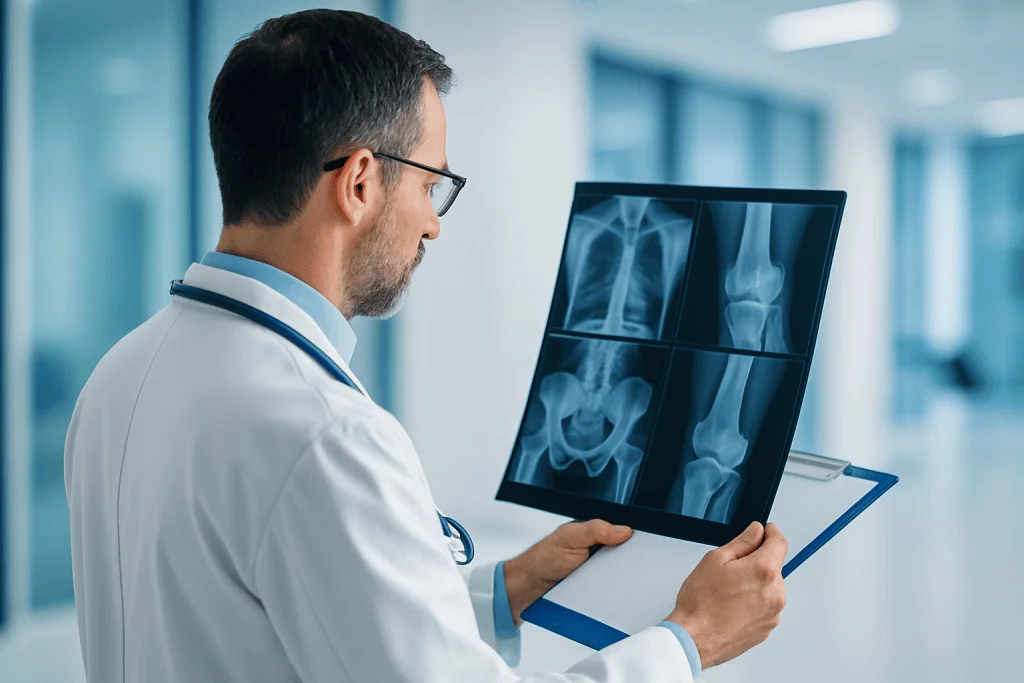
Musculoskeletal radiology is the branch of medical imaging that examines bones, joints, muscles, tendons, ligaments and cartilage. It uses X-ray, ultrasound, CT and MRI to identify fractures, arthritis, sports injuries, spine problems, infection and tumours, and it guides needle procedures such as joint injections and biopsies. Most examinations are outpatient tests with no recovery period; image-guided procedures may need brief observation.
What Is Musculoskeletal Radiology?
Musculoskeletal radiology is the branch of diagnostic imaging that examines bones, joints, muscles, tendons, ligaments, cartilage and the soft tissues around the spine and limbs. Its purpose is practical: to find the structural cause of pain, injury or loss of function, and to guide needle procedures such as joint injections, fluid aspirations and biopsies. It serves anyone whose symptoms involve the moving parts of the body, from a sprained ankle that refuses to settle to a complex spine disorder or a suspected bone tumour.
You will also see the field written as MSK radiology; the abbreviation appears on reports, referral letters and department signage, and it means exactly the same thing. Whatever the label, the core work is imaging of the musculoskeletal system with four main methods, each answering a different kind of question. X-ray is usually the first step for fractures, arthritis, alignment problems and visible bone changes. Ultrasound evaluates tendons, ligaments, muscles, cysts and superficial soft-tissue masses, and it can watch a joint or tendon move in real time. CT produces highly detailed bone images and is valuable in complex fractures, surgical planning and certain spine conditions. MRI gives excellent detail of soft tissues, bone marrow, cartilage, discs, nerves and joints without ionising radiation.
Musculoskeletal radiology is more than taking pictures. It includes selecting the most appropriate examination for the clinical question, tailoring the imaging protocol to your symptoms, interpreting the findings against your history, comparing current images with previous studies and communicating the results to the referring physician in a form that supports a decision. In selected cases it also includes image-guided procedures — injections into joints or tendon sheaths, aspiration of fluid, drainage of a collection, or biopsy of a bone or soft-tissue lesion. These interventions can confirm a diagnosis, relieve symptoms or obtain tissue for pathology.
The goal is always a specific answer. Is there a fracture? Has a ligament torn? Is the arthritis inflammatory or degenerative? Is a lump benign or suspicious? Is there infection? Is the pain coming from a disc, a tendon, a joint surface or changes within the bone marrow? Precise answers shorten the route to the right treatment and reduce the risk of unnecessary procedures. That is the honest value of the specialty: not more scanning, but better-targeted scanning.
What does a musculoskeletal radiologist do?
A musculoskeletal radiologist is a doctor who specialises in interpreting imaging of bones, joints and soft tissues and in performing image-guided procedures on those structures. In daily practice this means reading X-rays, ultrasound studies, CT and MRI of the limbs, pelvis, shoulder girdle and spine; adapting scan protocols so the images actually answer the referring doctor’s question; and discussing findings with orthopaedic surgeons, sports medicine physicians, rheumatologists and oncologists. Many also carry out procedures under imaging guidance — joint and tendon sheath injections, aspiration of swollen joints, drainage of collections and biopsies of suspicious lesions. In complex cases they contribute to multidisciplinary boards, where imaging is weighed alongside pathology, laboratory results and the clinical examination.
How long does it take to become a musculoskeletal radiologist?
In most countries, well over a decade of training in total. The pathway runs through a medical degree, several years of residency or specialty training in general radiology, and then a dedicated subspecialty fellowship in musculoskeletal imaging, typically lasting one to two years. Large academic departments run structured fellowship programmes — UTSW radiology at UT Southwestern in the United States is one well-known example — and learning continues afterwards through case volume, peer review and continuing education. This background matters to you as a patient for a simple reason: subtle findings such as a hairline fracture, an early bone erosion or an unusual marrow signal are easier to recognise, and to weigh correctly, for a reader who looks at these structures every day.
Musculoskeletal Radiology from the Patient’s Perspective
When pain limits walking, a sports injury does not heal as expected, a joint swells or back pain starts to shape your day, you usually want one thing first: a clear explanation. Musculoskeletal radiology is often the pathway to that clarity. It lets your physicians see the structures involved in detail, so treatment decisions rest on precise information rather than guesswork.
Musculoskeletal symptoms are frustrating precisely because the same pain pattern can have several causes. Shoulder pain may come from a tendon tear, bursitis, arthritis, nerve irritation or referred pain from the neck. Knee pain may reflect cartilage damage, ligament injury, early osteoarthritis or inflammation. Back pain may involve discs, joints, muscles, bones or nerves. Good imaging narrows these possibilities and lets the next step be chosen with confidence rather than trial and error.
If you are considering imaging abroad, you probably carry an extra layer of questions. Will the correct test be chosen? Do you need MRI or CT? Is the radiation exposure acceptable? Could your pain be arthritis, a fracture, a spine problem or something more serious? How quickly will results arrive? And if you have already been imaged at home, is a second, more detailed interpretation worthwhile before surgery, injection therapy, rehabilitation or oncology treatment? These are reasonable questions, and this page works through them method by method.
At Acibadem, musculoskeletal radiology sits inside the wider radiology department and within a broader clinical pathway. Radiologists work closely with orthopaedic surgeons, sports medicine physicians, rheumatologists, physical medicine and rehabilitation specialists, neurosurgeons and oncologists when the case requires it. That collaboration matters most for complex injuries, spine disorders, inflammatory joint disease, bone and soft-tissue tumours, and patients who need a coordinated plan while travelling for care.
Who May Need Musculoskeletal Imaging?
Musculoskeletal imaging is considered when you have pain, swelling, weakness, stiffness, reduced range of motion, symptoms after trauma, unexplained bone or joint changes, or a suspected mass in a limb or around a joint. It is also used after surgery or treatment — to monitor healing, evaluate implants, assess disease activity or investigate symptoms that persist when they should not.
Common reasons for referral include persistent joint pain, back or neck pain, pain after a fall or accident, sports injuries, sudden swelling of a joint, difficulty bearing weight, numbness or weakness linked to spine disease, clicking or locking of the knee, shoulder weakness, tendon pain, unexplained bone pain and changes in posture or limb alignment. In children and adolescents, imaging may be requested for sports injuries, growth plate concerns, congenital conditions, scoliosis, limping or suspected infection — always with protocols adapted to a growing skeleton.
Diagnosis rarely starts with a scanner. It starts with a clinical examination and a careful history: how the symptoms began, whether there was trauma, which movements make the pain worse, whether there is fever or weight loss, whether you have an inflammatory disease, and whether you have had previous surgery or cancer. On that basis, the radiology team helps determine which imaging method — if any — is most appropriate for you.
Some patients need only one test: an X-ray for suspected osteoarthritis or a straightforward fracture, for example. Others need a sequence. An X-ray may establish bone alignment, while MRI is added to evaluate ligaments, cartilage or bone marrow. CT is selected when tiny fracture lines or complex anatomy must be seen in fine detail. Ultrasound is chosen when the question involves a tendon, a bursa, a superficial lump or a guided injection. More imaging is not automatically better; the right test is the one that answers the clinical question with the least burden.
International patients often arrive with imaging already performed at home. That prior material is genuinely useful. Comparing old and new studies shows whether a condition is new, stable, improving or progressing, and it can spare you a repeat scan. Bring the actual images — not only the written reports — whenever you can; practical guidance on transferring large DICOM files is available in our guide to sharing large imaging files with your doctor.
Conditions Addressed by Musculoskeletal Radiology
Musculoskeletal radiology supports diagnosis, treatment selection, procedural guidance and follow-up across a wide spectrum, from common injuries to diseases that require multidisciplinary planning. The main groups of conditions are these:
- Fractures and trauma: X-ray and CT identify fractures, dislocations, bone alignment and healing. MRI can reveal occult fractures that are invisible on X-ray, along with the ligament or cartilage injuries that often accompany them. In the wrist, hip and foot, this distinction regularly changes management.
- Sports injuries: MRI and ultrasound evaluate ligament tears, meniscus injuries, tendon tears, muscle strains, cartilage defects and overuse injuries such as stress fractures — including the grading detail that shapes return-to-play decisions.
- Arthritis and joint degeneration: imaging assesses osteoarthritis, inflammatory arthritis, cartilage loss, bone erosions, joint fluid and structural damage, and helps distinguish degenerative wear from active inflammation — a distinction that points towards very different treatments.
- Spine disorders: MRI and CT are used for disc herniation, spinal stenosis, nerve compression, vertebral fractures, deformity, infection and postoperative evaluation.
- Tendon and ligament problems: ultrasound and MRI assess rotator cuff tears, Achilles tendon injuries, tennis elbow, ankle sprains, wrist ligament injuries and tendon inflammation.
- Bone and soft-tissue tumours: MRI, CT, X-ray and image-guided biopsy help characterise a mass, define its extent and support planning with oncology and surgical teams. A painless, slowly enlarging lump still deserves proper characterisation.
- Infection and inflammation: imaging helps detect osteomyelitis, septic arthritis, abscesses, inflammatory muscle conditions and complications after surgery.
- Paediatric musculoskeletal conditions: growth plate injuries, developmental hip conditions, scoliosis, limb deformities, sports injuries and bone pain in children, examined with dose-conscious, age-appropriate protocols.
- Postoperative and implant assessment: X-ray, CT, ultrasound and MRI with metal-adapted protocols evaluate healing, hardware position, complications, recurrent injury or persistent pain after surgery.
The indication determines the strategy. A patient with acute ankle trauma, an elite athlete with a suspected tendon tear, a person with long-standing rheumatoid arthritis and a patient with a soft-tissue mass do not need the same imaging plan. The most valuable examination is the one that answers the clinical question with the least burden and the greatest diagnostic usefulness for that particular patient — sometimes that is a single well-positioned X-ray, sometimes a targeted MRI, sometimes a combination.
How Musculoskeletal Imaging Is Performed
What actually happens depends on the method chosen. The sections below describe each examination in turn, from preparation to the moment the report reaches your treating doctor.
Before the Examination
Your visit begins with a review of your symptoms, referral information and relevant medical history. If you are travelling from abroad, the international patient team helps coordinate appointments, translation support and the transfer of prior medical documents where available. Previous X-rays, MRI and CT scans, ultrasound images, operative notes and pathology results are reviewed when they are relevant to the current problem, because comparison often answers questions a single new scan cannot.
Preparation depends on the examination. X-ray and most ultrasound studies require little or none. For MRI, you will be asked about implanted medical devices, previous surgery, metal fragments, kidney disease, pregnancy, allergies and claustrophobia. Some MRI examinations use contrast material to better evaluate inflammation, tumours, infection or postoperative change; CT may also use contrast in selected cases, particularly when vascular anatomy, infection or a tumour is being assessed. The team explains why contrast is recommended before it is given — our guide to preparing for contrast dye imaging covers what to expect in detail.
You may be asked to remove jewellery, watches, clothing with metal parts or accessories that could interfere with the images, and for some studies you change into a gown. If a joint injection, aspiration or biopsy is planned, you may receive additional instructions about blood-thinning medication, recent infections, fasting or laboratory tests, depending on the procedure and your overall health. Any decision about medication before a procedure belongs to your treating doctor, and the team will confirm the plan with you before anything happens.
During X-Ray Imaging
X-ray is fast and remains the standard first assessment of bones and joints. You are positioned so the area of concern can be imaged from the necessary angles. For certain joint and spine studies you may be asked to stand, because alignment and joint space look different under body weight than when lying down — a standing knee X-ray, for instance, shows cartilage narrowing more honestly than a supine one. The exposure itself takes only a moment, although careful positioning may take several minutes.
Digital X-ray systems produce images that can be reviewed quickly and stored for comparison with future studies. Radiation exposure is kept as low as reasonably achievable while still producing diagnostic-quality images, and protective measures are used where appropriate, particularly for children and sensitive regions. If radiation dose is a concern for you, our guide to radiology and imaging safety for international patients explains how modern departments manage it.
During Ultrasound Imaging
Musculoskeletal ultrasound uses sound waves rather than ionising radiation. Gel is applied to the skin and a handheld probe is moved over the area of concern. Its particular strength is dynamic assessment: the radiologist can watch tendons, muscles and joints during movement, compare the painful side directly with the unaffected side, and guide a needle with precision when an injection or aspiration is needed.
Ultrasound is commonly used for shoulder tendons, Achilles tendon problems, cysts, bursitis, superficial lumps, muscle tears, ligament injuries and certain nerve entrapment conditions. It can also help patients who cannot undergo MRI, although it does not replace MRI for every question, because sound waves cannot see through bone or reach deep structures reliably.
What’s the difference between MRI and MSK ultrasound?
MRI builds a complete, layered picture of both deep and superficial structures, while MSK ultrasound examines superficial soft tissues in real time, during movement. Neither uses ionising radiation, but they answer different questions. MRI shows bone marrow, cartilage, deep ligaments, spinal discs and the interior of joints — structures ultrasound cannot reach — and its result depends mainly on the protocol and equipment. Ultrasound is quicker, allows side-to-side comparison and dynamic testing, and is often preferred for tendon problems and guided injections, but its quality depends heavily on the operator performing it. In practice the two are complementary: a shoulder might be screened with ultrasound and then mapped with MRI before surgery is considered.
During CT Imaging
CT uses X-rays and computer processing to create detailed cross-sectional images. It is particularly helpful for complex fractures, small bone fragments, joint surfaces, spinal bone anatomy, surgical planning and situations where MRI is not possible. CT can also guide biopsies and injections with a high degree of anatomical precision, especially in the spine and pelvis.
During the scan you lie on a table that moves through the scanner; the examination is usually brief. If contrast is required, it is given through a vein. Because CT involves radiation, the protocol is tailored to the clinical need and unnecessary scanning is avoided — the question is always whether the extra detail changes a decision.
During MRI Imaging
An MSK MRI is a magnetic resonance examination tailored to a specific joint, limb or spine region. It uses a strong magnetic field and radiofrequency pulses to create detailed images of soft tissues, bone marrow, cartilage, joints, discs and nerves, and it is often the preferred test for ligament tears, meniscus injuries, tendon injuries, cartilage damage, spine conditions, bone marrow oedema, infection, inflammatory joint disease and tumour evaluation.
You lie on a table that moves into the scanner. You will not feel the magnetic field itself, but you must remain still so the images are sharp. The machine produces loud tapping and knocking sounds; ear protection is provided. Scan time varies with the body part and the clinical question, but many musculoskeletal MRI examinations take approximately 20 to 45 minutes. Contrast studies, complex protocols or multiple body regions take longer.
If you have claustrophobia, pain when lying still or other reasons to find the scan difficult, tell the team in advance. Positioning aids, communication during the scan, careful scheduling and, in selected cases, medication can all help — our guide to sedation for imaging tests lists the questions worth raising beforehand. Safety screening is essential, because some implants and metal fragments are affected by the magnetic field, and the team will go through this with you before you enter the scanner room.
Image-Guided Procedures
Some patients undergo image-guided procedures as part of diagnosis or treatment: joint injections, tendon sheath injections, aspiration of fluid, drainage of a collection, or biopsy of a bone or soft-tissue lesion. Imaging guidance lets the physician see the target and the structures around it, improving accuracy and helping avoid nearby nerves, vessels and organs. A typical procedure follows a predictable sequence:
- Step 1 — Planning: the radiologist reviews your imaging, confirms the target and chooses the guidance method: ultrasound, CT or fluoroscopic X-ray, depending on location and depth.
- Step 2 — Preparation: the skin is cleaned and a local anaesthetic numbs the entry point. For biopsies, sedation may be considered in selected cases.
- Step 3 — Guidance: the needle is advanced under continuous or repeated imaging until it sits precisely at the target.
- Step 4 — Injection, aspiration or sampling: medication is delivered, fluid is withdrawn, or tissue cores are taken and sent to pathology.
- Step 5 — Observation: you are monitored briefly and given instructions about activity, dressing care and what to watch for at the site.
When a tumour is suspected, biopsy planning is especially important and is ideally coordinated with the surgical and oncology teams, because the needle path itself can influence future surgical options. This is one of the clearest examples of why musculoskeletal radiology works best inside a team rather than as an isolated service.
After the Examination
After standard diagnostic imaging, most patients return to normal activities immediately. If contrast was used, you may be observed briefly and encouraged to drink fluids unless your physician has advised otherwise. After an injection, aspiration or biopsy, you may need a short observation period and specific instructions about activity restrictions and dressing care, along with clear guidance on findings such as fever, increasing pain or swelling that should be reported to the team looking after you.
The radiologist then interprets the images and prepares a report for your physician. In complex cases, findings are discussed directly with the treating specialist or reviewed in a multidisciplinary board. Reporting time depends on the complexity of the case and whether prior studies must be compared; how results are delivered, and in what format, is explained in our guide to getting lab test and imaging results during treatment.
Why Acting Early Matters
Musculoskeletal pain is common, and not every ache requires imaging. Evaluation becomes more important when symptoms are persistent, worsening, related to trauma, associated with weakness or numbness, or accompanied by fever, unexplained weight loss or a known cancer history. Early imaging can identify conditions that need prompt treatment and prevent small problems from becoming harder ones.
For injuries, delayed diagnosis of fractures, ligament tears or tendon ruptures can affect healing and long-term function. A missed scaphoid fracture in the wrist is the classic example: small on the day of injury, capable of causing lasting complications if it goes unrecognised. Untreated tendon ruptures may retract and become more difficult to repair over time. In athletes, returning to sport before the injury is properly understood raises the risk of reinjury.
For arthritis and inflammatory joint disease, imaging can detect active inflammation and structural damage. Early diagnosis allows medical treatment and rehabilitation to begin before irreversible joint changes progress. In spine disorders, imaging carries particular weight when symptoms suggest nerve compression, infection, fracture or tumour, because prompt evaluation protects mobility and neurological function.
For bone and soft-tissue masses, timely imaging and a carefully planned biopsy are essential. Most lumps are not cancer, but a mass that is enlarging, deep, firm or unexpectedly large should be characterised properly — and so should a painless one, since the absence of pain says little about what a lesion is. Early, coordinated assessment helps avoid incomplete procedures and supports an organised treatment plan if specialist care turns out to be needed.
Benefits of Musculoskeletal Radiology
The value of the specialty lies in matching the right method to the right question. In concrete terms, that produces the following benefits:
| Benefit | What It Means for You |
|---|---|
| More accurate diagnosis | Imaging can identify fractures, tendon tears, arthritis, inflammation, spine problems, tumours and other causes of pain that examination alone cannot confirm. |
| Personalised treatment planning | Your physician can choose treatment based on the specific structure involved, the severity of the finding and how it relates to your symptoms. |
| Avoidance of unnecessary procedures | Clear imaging may show when surgery is not needed and when rehabilitation, medication, injection therapy or monitoring is the more sensible route. |
| Guidance for precise interventions | Ultrasound, CT or X-ray guidance directs needles to the correct location for injections, aspirations and biopsies. |
| Monitoring over time | Follow-up imaging can show healing, disease progression, treatment response or postoperative change when it is clinically necessary — and only then. |
None of these benefits comes from technology alone. They come from the sequence around it: a clear clinical question, a well-chosen test, a protocol tuned to that question and a report written for the doctor who has to act on it.
What to Expect Afterwards: A Realistic Timeline
What happens next depends on whether you had diagnostic imaging alone or an image-guided procedure such as an injection, aspiration or biopsy. The pattern is broadly as follows:
| Time Period | What Patients Can Expect |
|---|---|
| Day 1 | After X-ray, ultrasound, CT or MRI, most patients resume normal activities immediately. After an injection or biopsy, mild soreness may occur and short observation may be needed. |
| First week | Diagnostic imaging has no recovery period. After a guided procedure, you may be asked to avoid strenuous activity for a short time and to monitor the site for increasing pain, swelling or fever. |
| First month | Your treatment plan begins or is adjusted based on the imaging results — medication, physiotherapy, surgical planning, oncology care or further imaging as required. |
| Longer term | Some conditions need periodic imaging to monitor healing, arthritis progression, tumour response, postoperative recovery or chronic spine and joint disorders. |
Factors That Influence a Good Result
A good result in musculoskeletal radiology begins with choosing the right examination for the right question. The most advanced method is not always the most appropriate first step. A carefully positioned X-ray can be more useful than an MRI for certain alignment and arthritis questions. Ultrasound may be the best test for a dynamic tendon problem. MRI may be essential for cartilage, bone marrow, disc or soft-tissue assessment. CT may give the clearest view of complex bone anatomy. The value lies in matching the technology to the medical need, not in defaulting to the biggest machine.
The quality of the clinical information supplied matters just as much. Radiologists interpret images in context: knowing whether pain began after trauma, whether symptoms are acute or chronic, whether there has been surgery, and whether there is fever, a cancer history or inflammatory disease can change the interpretation of the same picture. For international patients, translated reports, previous imaging and a concise medical history improve continuity and reduce repeated testing.
Image quality is the third pillar. Correct positioning, appropriate protocol selection, motion control and the use of contrast when clinically indicated all contribute to diagnostic accuracy. In MRI, the choice of sequences determines what is visible at all. In CT, slice thickness and reconstruction methods affect how fractures and joint surfaces are judged. In ultrasound, the operator’s experience carries particular weight, because the examination is performed and interpreted in real time — the images that get recorded depend on the questions the examiner thinks to ask.
For image-guided procedures, outcomes depend on accurate targeting, sterile technique, careful patient selection and integration with the overall treatment plan. An injection can provide important diagnostic information even when symptom relief is temporary: if numbing a specific joint abolishes the pain, that joint is very likely the source. A biopsy must be planned thoughtfully, especially when a tumour is possible, because the needle path can influence future surgical management.
Patient factors matter too. Age, bone density, activity level, body habitus, diabetes, smoking, inflammatory disease, medications, previous surgery and the duration of symptoms all affect both the imaging appearance and the plan built on it. A high-performance athlete, an older adult with osteoporosis and a child with a growth plate injury require different interpretation and different clinical pathways, even when their scans superficially resemble one another.
Finally, the usefulness of any scan depends on communication. The report should answer the clinical question clearly, describe the relevant findings and, where appropriate, suggest correlation or further evaluation. In complex cases, a direct conversation between the radiologist and the treating physician aligns the diagnosis with the treatment decision — and that conversation is often where the real value of the examination is realised.
Musculoskeletal Radiology at Acibadem
International patients who come to Acibadem for musculoskeletal imaging are usually looking for more than a scan appointment. They want a reliable diagnostic pathway, access to experienced specialists, efficient coordination and clear communication in a setting familiar with the needs of people travelling for care. Musculoskeletal radiology is treated as part of that broader medical journey, not as a standalone test.
Imaging services are connected with orthopaedic surgery, spine care, sports medicine, rheumatology, oncology, and physical medicine and rehabilitation. This integration matters most when imaging results lead directly to decisions: surgical planning, interventional procedures, medical treatment or a rehabilitation programme. A scan that sits in a folder helps no one; a scan that reaches the right specialist with the right context moves your care forward.
Complex cases can be reviewed through multidisciplinary boards or specialist discussions. This applies particularly to bone and soft-tissue tumours, complicated spine disorders, revision surgery planning, infections, paediatric musculoskeletal conditions and situations where previous treatment has not resolved the symptoms. A radiology report is important on its own, but its interpretation becomes more powerful when weighed alongside clinical examination, laboratory results, pathology and your own goals.
The diagnostic pathways use modern equipment: digital X-ray, high-resolution ultrasound, advanced CT and MRI protocols tailored to specific joints, spine regions and soft-tissue questions. The emphasis is not on performing a scan, but on selecting and conducting the examination in a way that addresses your actual medical question — including metal-adapted MRI protocols after joint replacement and dose-conscious approaches for children.
For patients travelling from Europe, the Middle East, Africa, the United States and elsewhere, coordination shapes the experience as much as the medicine does. Acibadem International supports patients with appointment planning, medical document transfer, language assistance in more than 20 languages, hospital navigation and coordination between departments — which becomes especially useful when imaging, specialist consultation and a treatment plan must fit into a limited travel schedule.
Second opinions are a common part of the work. A patient may have been told they need surgery, may hold conflicting reports, or may not understand why pain persists despite previous treatment. Reviewing existing imaging, and adding targeted studies only where necessary, can clarify the diagnosis. Sometimes the outcome is confirmation of the original plan; sometimes the findings point to a different approach — rehabilitation, injection therapy, rheumatological treatment, oncology evaluation or a different surgical strategy. Both outcomes are useful, and it would be dishonest to promise which one you will get.
Personalised planning is the thread running through all of this. Imaging findings alone do not define a patient: many people have degenerative changes on MRI that are not the true source of their symptoms, while others have subtle findings that explain significant pain or dysfunction. The best plan considers imaging together with physical examination, lifestyle, work demands, athletic goals, medical history and expectations. At Acibadem, radiology is part of that clinical decision-making process rather than an isolated report.
Moving Forward with Clarity
Musculoskeletal radiology can be a turning point in the care of bone, joint, muscle, tendon, ligament and spine conditions. It explains the source of pain, guides treatment, supports safer procedures and helps you avoid unnecessary delays and unnecessary operations alike. For international patients it also offers something structural: a way to obtain a diagnosis, a second interpretation or a treatment plan within one coordinated system rather than across scattered appointments.
Whatever setting you are treated in, a few habits make imaging work better for you. Bring previous images and reports — the files themselves, not just summaries — to any appointment where imaging may be discussed. Be precise about how and when the symptoms started. And treat the scan as a means to a decision, not an end in itself. Questions worth putting to your treating doctor include:
- Which structure do you suspect, and which test shows it best?
- Will the result actually change the treatment plan?
- Is contrast needed, and why?
- Should this study be compared with my previous imaging?
- If the scan is normal, what is the next step?
A well-chosen examination, read in context by a specialist and connected promptly to the clinician who will act on it — that is what musculoskeletal radiology looks like when it works. The technology matters, but the sequence around it is what turns an image into an answer.
Preparation
- Bring previous imaging, reports, referral notes and a list of current medications. Wear comfortable clothing and remove metal items before imaging. Tell the radiology team if you are pregnant, have implants, kidney disease, allergies or may need contrast material.
Aftercare
- Most patients can return to normal activities immediately after diagnostic imaging. If contrast material is used, drinking water may be recommended unless your doctor advises otherwise. Your radiology report is reviewed with the referring physician to guide the next step in care.
Turkey vs UK, Germany & USA
Musculoskeletal radiology can involve different imaging methods depending on the suspected bone, joint, muscle, tendon or ligament condition. Costs and patient experience vary by country, imaging technology, reporting expertise and how care is coordinated.
The comparison below highlights non-price factors that commonly influence the total cost and convenience of musculoskeletal imaging for international patients.
| Factor | Turkey | UK | Germany | USA |
|---|---|---|---|---|
| Cost structure | Private hospital packages may combine imaging, specialist review and coordination for international patients. | Private care is available, while public pathways may depend on referral and eligibility. | Costs are often linked to facility type, imaging modality and specialist reporting. | Billing may vary widely by facility, insurer status, radiologist fees and scan protocol. |
| Hospital and radiology team | International hospitals may offer musculoskeletal radiologists, orthopedic collaboration and modern imaging units. | Access may differ between private imaging centers and hospital-based departments. | University and private hospitals may provide subspecialty radiology and structured diagnostic pathways. | Large academic and private systems may offer advanced imaging and subspecialist reporting. |
| Quality and accreditation | JCI-accredited hospitals and international patient departments can support standardized care processes. | Quality frameworks and professional standards guide radiology practice in both public and private settings. | Radiology departments generally operate within national quality and professional standards. | Accreditation and quality oversight may depend on the hospital, imaging center and state requirements. |
| Waiting and scheduling | Private scheduling for international patients may be arranged efficiently, depending on modality and urgency. | Waiting time can vary between public referral pathways and private appointments. | Availability depends on region, hospital type and requested imaging method. | Scheduling may be fast in some private settings, but authorization and network processes can affect timing. |
| Travel and language logistics | International patient teams may assist with appointments, translation, transfers and report coordination. | Language is straightforward for English speakers, but travel support depends on provider. | Interpreter support may be needed for non-German speakers and varies by hospital. | English-language care is standard, while travel distances and coordination needs can differ by city. |
| What packages may include | Imaging appointment, radiology report, specialist consultation, translation support and care coordination may be grouped. | Private packages may include scan and report, with specialist review billed or arranged separately. | Packages may include imaging and written report, with follow-up depending on the care pathway. | Scan, facility fee, radiologist interpretation and specialist visit may be billed separately depending on provider. |
What affects your final cost
- Type of imaging requested, such as X-ray, ultrasound, CT or MRI.
- Body area examined and whether multiple regions are assessed.
- Use of contrast material, sedation or image-guided procedures.
- Need for urgent scheduling, specialist musculoskeletal reporting or multidisciplinary review.
- Hospital accreditation, imaging technology and radiologist subspecialty experience.
- Travel support, interpreter services, report translation and follow-up consultation needs.
Compare your options
Musculoskeletal radiology includes several clinical options, each suited to different diagnostic questions. Suitability is decided by a specialist based on symptoms, examination findings and prior medical records.
| Option | What it is | Typical use | Key considerations |
|---|---|---|---|
| X-ray | A widely used imaging method for bones and joint alignment. | Fractures, arthritis, deformity, joint space assessment and follow-up after treatment. | Fast and widely available, but soft tissue detail is limited compared with MRI or ultrasound. |
| Ultrasound | Real-time imaging using sound waves to evaluate soft tissues and guide procedures. | Tendon injuries, muscle tears, bursitis, joint fluid, nerve entrapment and guided injections. | Operator expertise is important, and deep structures may be harder to assess. |
| CT | Cross-sectional imaging that shows bone detail with high clarity. | Complex fractures, spine assessment, bone tumors, surgical planning and cases where MRI is not suitable. | Radiation exposure is considered, and contrast may be needed for selected conditions. |
| MRI | Detailed imaging of bones, joints, cartilage, muscles, tendons, ligaments and nerves. | Ligament tears, disc disease, sports injuries, arthritis complications, infection and tumor evaluation. | Scan time, implants, claustrophobia, contrast needs and radiologist expertise can affect planning. |
| Image-guided procedures | Ultrasound, CT or fluoroscopy used to guide needles accurately. | Joint injections, aspirations, biopsies and pain-related diagnostic procedures. | Requires specialist assessment, sterile technique and clear indication based on imaging findings. |
General information only — not medical or financial advice. Final costs depend on the factors above and your individual case; request a free, personalised quote.
Frequently Asked Questions
What affects the cost of musculoskeletal radiology?
The main factors are the imaging method, the body area examined, whether contrast or image guidance is needed, the hospital setting, the radiologist’s subspecialty review and any added consultation, translation or care coordination services.
How can I get a personalised quote?
You can request a free consultation and share your symptoms, referral notes, previous imaging and medical history. The clinical team can then advise which scan may be appropriate and prepare a personalised estimate.
Is MRI always more appropriate than X-ray or ultrasound?
No. Each modality answers different clinical questions. X-ray may be best for bone alignment, ultrasound may be useful for tendons and guided procedures, CT may clarify complex bone problems and MRI may show soft tissues in detail. A specialist decides suitability.
Can imaging and specialist consultation be arranged together?
In many international patient pathways, imaging can be coordinated with an orthopedic, rheumatology, neurosurgery or oncology review when clinically relevant. This may improve convenience and help determine the next step in care.
Are reports available in English?
International hospitals commonly provide support for English communication, and report translation may be arranged depending on the service package. You should confirm language needs before travel.
Is this information medical or financial advice?
No. It is general educational information. The appropriate imaging test and final cost depend on specialist assessment, medical indication and the services included in your personalised quote.
Medically reviewed by the Acıbadem International Medical Board — August 31, 2026
See our medical review board →
Update history
- PublishedJune 8, 2026
- Medical review approvedAugust 31, 2026
- Last content updateAugust 31, 2026
Trusted care for international patients
Doctors Performing This Treatment

Prof. Dr. Metin Türkmen
Orthopedic Surgery & Traumatology
Prof. Dr. Cihangir Tetik
Orthopedic Surgery & Traumatology
Prof. Dr. Harzem Özger
Orthopedic Surgery & Traumatology
Prof. Dr. Ahmet Alanay
Orthopedic Surgery & Traumatology
Prof. Dr. Mustafa Karahan
Orthopedic Surgery & Traumatology
Prof. Dr. Barış Kocaoğlu
Orthopedic Surgery & Traumatology
Prof. Dr. Mustafa Seyhan
Orthopedic Surgery & Traumatology
Prof. Dr. Ata Can Atalar
Orthopedic Surgery & Traumatology
Prof. Dr. Fatih Dikici
Orthopedic Surgery & Traumatology
Prof. Dr. Levent Eralp
Orthopedic Surgery & Traumatology
Prof. Dr. İbrahim Tuncay
Orthopedic Surgery & Traumatology
Prof. Dr. İbrahim Kaya
Orthopedic Surgery & Traumatology
Prof. Dr. Alper Kaya
Orthopedic Surgery & Traumatology
Prof. Dr. Korhan Özkan
Orthopedic Surgery & Traumatology
Prof. Dr. Metin Uzun
Orthopedic Surgery & Traumatology
Prof. Dr. Burak Akan
Orthopedic Surgery & Traumatology
Prof. Dr. Kerem Bilsel
Orthopedic Surgery & Traumatology
Prof. Dr. Göksel Dikmen
Orthopedic Surgery & Traumatology
Prof. Dr. Kerim Sarıyılmaz
Orthopedic Surgery & Traumatology
Prof. Dr. Aziz Kaya Alturfan
Orthopedic Surgery & Traumatology
Prof. Dr. Hüseyin Bayram
Orthopedic Surgery & Traumatology
Prof. Dr. Mehmet Serdar Binnet
Orthopedic Surgery & Traumatology
Prof. Dr. Mahir Gülşen
Orthopedic Surgery & Traumatology
Prof. Dr. Mustafa Herdem
Orthopedic Surgery & TraumatologyMedical Units
Available at These Hospitals