Craniocervical Junction Treatment: How It Works, Results and What to Expect

The craniocervical junction is the connection between the skull, first two cervical vertebrae and nearby nerves, spinal cord and blood vessels. Treatment depends on the underlying condition, symptoms, neurological examination and carefully interpreted imaging.
Key Takeaways
- The craniocervical junction is the connection between the skull, first two cervical vertebrae and nearby nerves, spinal cord and blood vessels.
- Treatment depends on the underlying condition, symptoms, neurological examination and carefully interpreted imaging.
- Conservative care may help some people, but structural instability with neurological deterioration may require surgical stabilization.
- Occipitocervical fusion aims to stabilize the skull-to-neck connection and protect neural structures, but it reduces some neck movement.
- Recovery after surgery is gradual and includes pain management, wound care, activity restrictions and rehabilitation.
- New weakness, walking difficulty, swallowing or breathing problems, or changes in bladder or bowel control require urgent medical assessment.
Craniocervical junction treatment is tailored to the cause and severity of instability or compression where the skull meets the upper cervical spine. It may include monitoring, targeted rehabilitation and pain care, while surgery is considered when there is progressive neurological compromise, significant instability or persistent disabling symptoms.
Overview: How craniocervical junction treatment works
Craniocervical junction treatment addresses problems at the point where the base of the skull joins the upper neck. This region includes the occiput, atlas (C1) and axis (C2), and it protects the lower brainstem, upper spinal cord and important nerves. Treatment is designed to relieve symptoms where possible, prevent further neurological injury and improve stability when the bones and supporting ligaments are not providing adequate support.
The right plan depends on why the junction is affected. Causes can include congenital bone differences, inflammatory disease, injury, infection, tumors, degenerative changes or connective-tissue disorders. Some people have symptoms that can be managed without surgery; others need a specialist assessment because compression or instability may threaten neurological function.
Care is usually coordinated by neurosurgery or spine surgery alongside neurology, neuroradiology, rehabilitation and pain-management professionals. A treatment plan should be based on the whole clinical picture, rather than on an imaging finding alone.
Symptoms, causes and candidacy for treatment
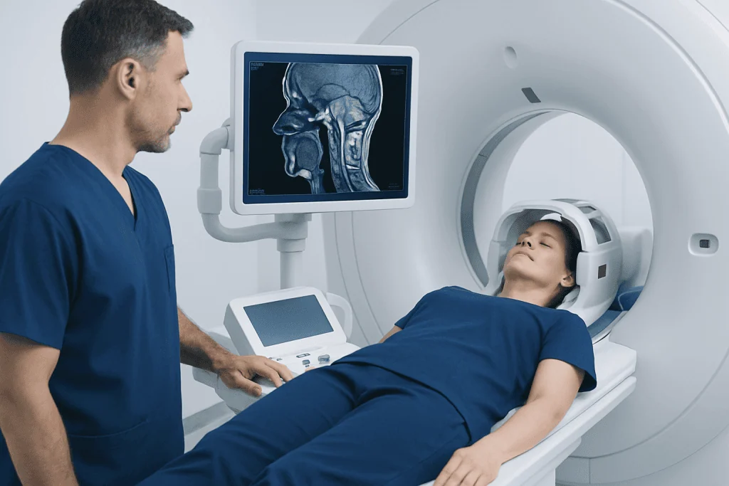
Symptoms vary widely because the craniocervical junction is close to the brainstem and spinal cord. A person may experience upper-neck or occipital headache, neck pain, a feeling of head heaviness, reduced balance, dizziness, numbness, weakness, clumsiness or changes in walking. Some conditions can also be associated with swallowing difficulty, voice changes, visual symptoms or sleep-related breathing concerns.
Not every headache, neck pain or abnormal scan indicates craniocervical instability. Clinicians consider the pattern and progression of symptoms, medical history, neurological findings and whether imaging shows a meaningful structural problem. Other disorders of the upper spine and nervous system may also need consideration during assessment.
People may be candidates for surgery when there is clearly demonstrated instability, compression of the spinal cord or brainstem, progressive neurological deficits, deformity, or symptoms that remain substantially limiting despite appropriate non-surgical care. The decision is individualized. Surgery is generally not recommended solely for nonspecific symptoms without supportive clinical and radiological evidence.
How painful is craniocervical instability? Pain can range from mild intermittent upper-neck discomfort to severe, persistent pain at the base of the skull. It may worsen with movement, maintaining an upright posture or activities that strain the neck. Pain severity does not always match the degree of instability on imaging, so a medical assessment is important for identifying the cause and choosing safe care.
Diagnosis and imaging: Will an MRI show craniocervical instability?
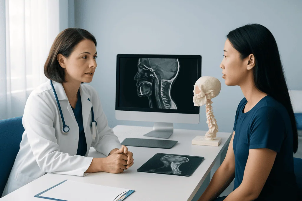
Diagnosis begins with a careful history and neurological examination. The clinician may assess strength, reflexes, sensation, coordination, gait, neck movement and signs that the brainstem or spinal cord could be affected. Previous injuries, inflammatory conditions, congenital diagnoses and connective-tissue disorders are also relevant.
Will an MRI show craniocervical instability? MRI can show the brainstem, spinal cord, ligaments, discs, soft tissues and signs of neural compression. It is very useful when doctors need to assess associated changes such as spinal cord compression, fluid-flow issues or soft-tissue abnormalities. However, standard MRI is usually performed lying down and may not fully demonstrate movement-related instability.
Depending on the suspected problem, imaging may also include CT to define bone anatomy and carefully selected flexion-extension X-rays or other dynamic studies to examine alignment during movement. Measurements on scans require expert interpretation and must be correlated with symptoms and examination findings. In selected complex cases, a multidisciplinary review can help clarify whether a finding is clinically important.
Step by step: Non-surgical care and craniocervical instability surgery
When there is no urgent compression or progressive neurological deficit, treatment may begin with education, activity modification, symptom-directed medication prescribed by a clinician, and rehabilitation. Physical therapy should be individualized and gentle, focusing on posture, movement control and functional goals. Forceful neck manipulation or unsupervised high-velocity techniques should be avoided when instability is suspected.
If surgery is needed, the most common stabilizing procedure is occipitocervical fusion. The surgeon joins the skull base to selected upper cervical vertebrae using specialized screws, rods and bone graft material. This creates a stable construct that allows the bones to fuse over time. In some cases, decompression is performed as well to create more space for compressed neural structures.
Before surgery, the team reviews imaging, general health, medications, anesthesia considerations and the planned fusion levels. During the operation, the patient receives general anesthesia and is positioned carefully to protect the neck. The surgeon makes an incision at the back of the head and neck, places fixation under image guidance or navigation as appropriate, prepares the bone surfaces and applies graft material. The wound is then closed, and the patient is monitored closely after anesthesia.
The purpose of occipitocervical fusion is stabilization, not restoration of normal joint motion. Because the upper cervical spine contributes substantially to head rotation and nodding, patients should expect some permanent restriction in neck movement after fusion. The surgical team discusses this trade-off in detail before treatment.
Benefits, risks and expected results
Potential benefits of surgery include improved stability, protection of the brainstem and spinal cord, prevention of further structural deterioration, and reduction of symptoms related to compression or abnormal motion. Individual results vary according to the cause of the condition, duration and severity of neurological symptoms, other health conditions and whether there is irreversible nerve or spinal cord injury before surgery.
What is the success rate of craniocervical instability surgery? There is no single reliable success rate that applies to every person. Craniocervical instability is uncommon and includes several different causes, while studies use different definitions of success, surgical methods and follow-up periods. In appropriately selected patients, stabilization can achieve solid fusion and may improve symptoms related to proven instability or compression, but it cannot guarantee relief of every symptom.
Risks include bleeding, infection, blood clots, wound problems, leakage of spinal fluid, injury to nerves or the spinal cord, problems with fixation or bone healing, persistent pain, and the possible need for additional surgery. Fusion also limits movement permanently and can place additional stress on nearby spinal segments over time. The surgeon explains personal risks after reviewing the individual anatomy and health profile.
What happens if craniocervical instability is left untreated? The outlook depends on its cause and severity. Some stable or mild findings may be monitored without progression, while significant untreated instability or compression can lead to worsening pain, weakness, sensory changes, impaired coordination, difficulty walking or other neurological problems. Progressive symptoms should not be ignored, because timely specialist review helps determine whether observation remains safe.
Recovery timeline and self-care after treatment
After surgery, patients are monitored in hospital for pain control, neurological checks and early recovery. The length of stay varies with the procedure, the person’s overall health and whether decompression or treatment of another condition was also required. A neck brace may be used in selected cases, based on the surgeon’s instructions.
During the first weeks, fatigue, incision discomfort and reduced neck mobility are common. Patients are usually asked to avoid heavy lifting, strenuous activity, driving until cleared, and movements that place excessive strain on the neck. Walking and gradual activity are commonly encouraged when safe. Wound-care instructions, medication guidance and warning signs should be followed closely.
Follow-up appointments and imaging help the surgical team monitor alignment, hardware and bone fusion. Rehabilitation is introduced at a pace suited to healing and individual needs. Return to work and regular activities may take weeks to months, especially for jobs requiring physical exertion or sustained neck movement.
Acibadem International’s multidisciplinary specialists and JCI-accredited hospitals assess and treat complex craniocervical junction conditions for international patients, with care plans guided by detailed clinical and imaging evaluation.
When to seek medical care
Arrange a medical appointment for persistent or worsening pain at the base of the skull or upper neck, especially when it is accompanied by numbness, weakness, poor balance, hand clumsiness, dizziness or a change in walking. A clinician can determine whether symptoms are related to the craniocervical junction or another, more common cause of neck or neurological symptoms.
Seek urgent medical care for new or rapidly worsening weakness, difficulty walking, loss of coordination, trouble swallowing, breathing difficulty, sudden severe neck pain after trauma, or new changes in bladder or bowel control. These symptoms can have several causes, but prompt assessment is important when neurological involvement is possible.
People with a known diagnosis should attend recommended follow-up visits and contact their clinical team if symptoms change. Avoid self-diagnosis based on online measurements or scan reports alone; expert review is essential before considering a collar, intensive exercise program or surgery.
Frequently asked questions
What is craniocervical junction treatment?
Craniocervical junction treatment refers to care for conditions affecting the connection between the skull and upper cervical spine. Depending on the cause, it may involve monitoring, rehabilitation, symptom management, treatment of an underlying disease or surgery to decompress and stabilize the area.
What is the success rate of craniocervical instability surgery?
A single success rate is not available because causes of instability, surgical techniques and definitions of improvement differ between patients and studies. When surgery is recommended for well-documented instability or neural compression, it can provide stabilization and may improve related symptoms, but outcomes cannot be guaranteed.
What happens if craniocervical instability is left untreated?
Some mild or stable cases can be observed under specialist guidance. However, significant instability or compression may worsen over time and can cause increasing pain, balance problems, weakness, sensory symptoms or other neurological difficulties. Progressive symptoms require prompt medical review.
How painful is craniocervical instability?
Pain varies substantially. Some people have mild upper-neck or occipital discomfort, while others have persistent pain that becomes worse with head movement, upright posture or exertion. A clinician should evaluate persistent pain because many other conditions can produce similar symptoms.
Will an MRI show craniocervical instability?
MRI is valuable for examining the brainstem, spinal cord, ligaments and soft tissues, and for detecting compression. Because routine MRI is usually done while lying down, it may not show all movement-related instability. CT and selected dynamic X-rays may be needed, depending on the clinical question.
How long does recovery take after occipitocervical fusion?
Early recovery commonly takes several weeks, while bone fusion and return to fuller activity may take months. Timing varies according to the extent of surgery, healing, overall health, work demands and the need for rehabilitation. The surgical team provides individualized activity restrictions and follow-up plans.
References
- National Institute of Neurological Disorders and Stroke
- American Association of Neurological Surgeons
- American College of Radiology
- National Institute of Arthritis and Musculoskeletal and Skin Diseases
This article is for general information only and is not a substitute for professional medical advice. Please consult a qualified doctor about your individual situation.
Joint and spine care in Turkey — expert assessment & treatment
JCI-accredited · board-certified surgeons · reply within 24h
Add us as a Preferred Source to see more of our trusted health content across Google Search, AI Overviews and Discover.