CT Scan Kidney Cancer: Preparation, Procedure and Results

CT imaging uses X-rays and computer processing to produce detailed cross-sectional images of the kidneys and nearby organs. Contrast dye is often used to show how a kidney mass takes up blood, which helps distinguish suspicious lesions from some noncancerous findings.
Key Takeaways
- CT imaging uses X-rays and computer processing to produce detailed cross-sectional images of the kidneys and nearby organs.
- Contrast dye is often used to show how a kidney mass takes up blood, which helps distinguish suspicious lesions from some noncancerous findings.
- A CT scan may help determine the size, location and extent of a kidney tumor, but some cases require further imaging, monitoring or biopsy.
- Patients should tell the imaging team about pregnancy, kidney disease, diabetes medicines, allergies and previous contrast reactions before the scan.
- Most people can return to usual activities shortly after a kidney CT scan, unless their clinician gives different instructions.
A CT scan for kidney cancer is a detailed imaging test used to identify a kidney mass, assess its features and look for possible spread beyond the kidney. It cannot always confirm cancer by itself, but it provides essential information for diagnosis, staging and treatment planning.
CT Scan for Kidney Cancer: What It Shows
A CT scan for kidney cancer is a noninvasive imaging examination that creates detailed pictures of the kidneys, abdomen and pelvis. It is commonly used when a kidney mass is found on ultrasound or another scan, or when symptoms and test results suggest a possible kidney problem. The scan can help clinicians assess whether a mass has features that may be consistent with cancer and whether it involves nearby structures.
CT stands for computed tomography. During the test, an X-ray scanner rotates around the body while a computer combines multiple images into thin, detailed slices. A contrast agent, usually injected into a vein, is often used because it shows blood vessels and kidney tissues more clearly. The timing of images after contrast is especially useful for evaluating a renal mass.
For people with confirmed or strongly suspected kidney cancer, CT can support staging. This means evaluating the tumor’s size and position, checking nearby lymph nodes and looking for signs that disease may have extended into veins, surrounding tissue or other organs. CT findings are considered alongside medical history, blood and urine tests, and sometimes other imaging or tissue testing.
How a Kidney CT Scan Works and Who May Need One
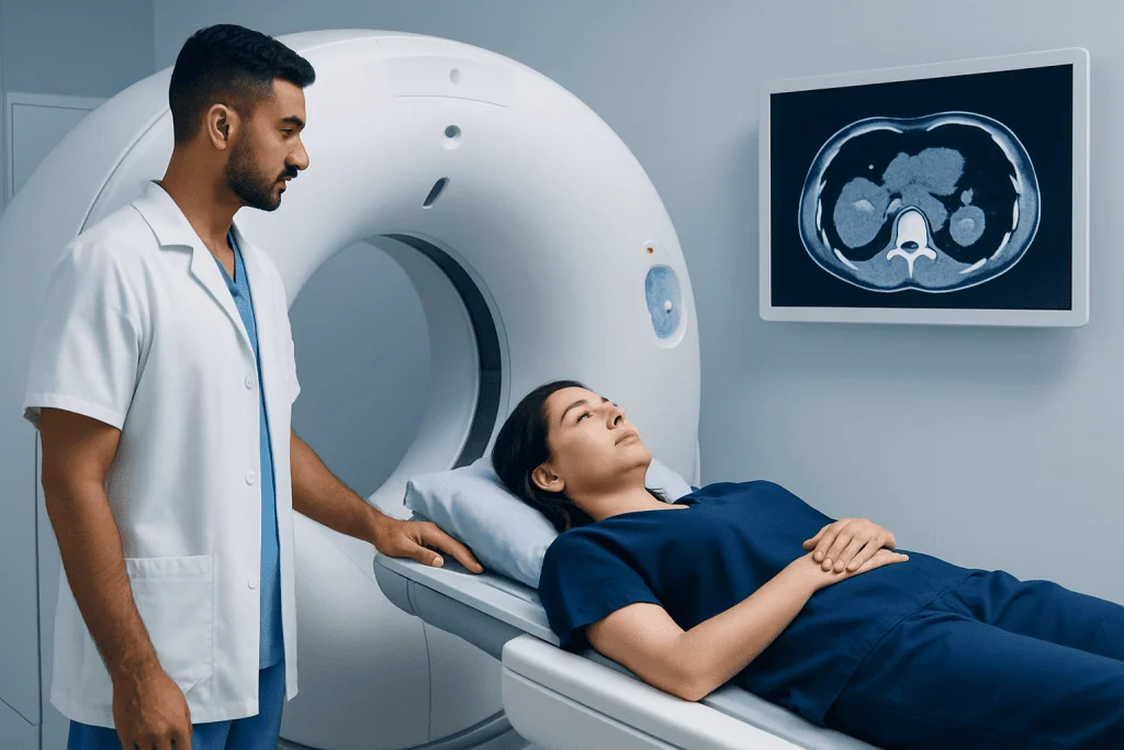
A kidney CT scan may be recommended after blood in the urine, persistent flank discomfort, an unexplained kidney finding or an abnormal ultrasound. It may also be used to investigate injuries, kidney stones, infections, cysts or structural differences. Not every kidney lesion is cancer; simple cysts, benign growths and inflammatory changes can sometimes appear as abnormalities on imaging.
The examination is particularly helpful when clinicians need a clear map of a suspected tumor before discussing treatment. It can show whether a mass is limited to the kidney, close to important blood vessels or affecting the collecting system that drains urine. These details can guide decisions about observation, further testing or procedures such as kidney cancer treatment.
CT may not be the preferred examination for every person. Ultrasound or MRI may be considered when radiation should be minimized, when contrast cannot be used safely, or when CT findings need clarification. The imaging specialist and referring clinician select the most suitable test based on the individual’s symptoms, kidney function, medical history and prior imaging.
What Do I Need to Do Before a Kidney CT Scan?

Preparation instructions vary depending on whether contrast will be used and the reason for the examination. The radiology department may ask the patient not to eat for several hours beforehand, especially when intravenous contrast is planned. Clear fluids may be permitted, but patients should follow the specific instructions provided by their healthcare team rather than making changes on their own.
Before the appointment, patients should provide a complete medical history. This includes kidney disease, reduced kidney function, diabetes, thyroid disorders, asthma, allergies, prior reactions to contrast dye and current medicines or supplements. A blood test to check kidney function may be arranged before contrast is given, particularly for people with known kidney concerns or other risk factors.
Patients should bring previous scan reports or image discs if these are available. Comparing new images with earlier ultrasound, CT or MRI studies can help determine whether a kidney lesion is stable or changing. It is also important to tell the team about pregnancy or the possibility of pregnancy, as imaging plans may need to be adjusted.
- Wear comfortable clothing and remove metal items if requested.
- Ask in advance whether fasting is required and how long it should last.
- Bring a current medication list, including over-the-counter products.
- Arrange language assistance or a support person if this would make the visit easier.
What Should You Not Do Before a CT Scan?
Patients should not ignore fasting or medication instructions given by the imaging department. If contrast is planned, eating immediately before a scan may be discouraged in some settings. However, patients should not stop prescribed medicines, including diabetes medicines, unless their own clinician or radiology team has specifically advised them to do so.
It is also important not to withhold information about allergies, previous contrast reactions or kidney problems. A prior mild reaction does not necessarily mean contrast cannot be used, but the team may need to take precautions, consider alternatives or adjust the plan. Similarly, dehydration can make it harder to place an intravenous line and may be a concern for some people with impaired kidney function, so patients should ask whether they should drink fluids before the test.
Patients should avoid arriving late if possible, as there may be safety screening, paperwork and intravenous line placement before the scan. They should not assume that all CT scans require the same preparation; abdominal CT with contrast, CT for kidney stones and CT of another body area can have different instructions.
Step by Step: What Happens During and After the Scan
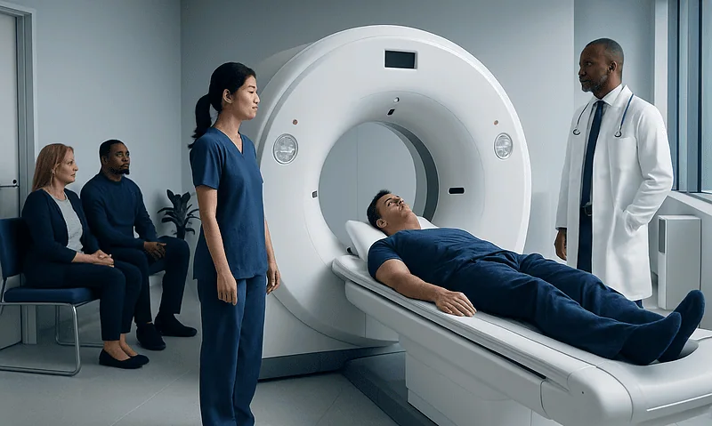
On arrival, the patient is usually asked to confirm their identity, symptoms, medical history and contrast safety information. If contrast is required, a small intravenous cannula is placed in an arm or hand. The patient then lies on a padded table that moves through the doughnut-shaped CT scanner. The scanner is open at both ends and does not enclose the body like some MRI machines.
During the scan, the radiographer may ask the patient to hold their breath for a few seconds. If contrast is injected, a temporary warm sensation, metallic taste or feeling of needing to urinate can occur. These sensations are common and generally pass quickly. The image-taking portion is usually brief, although preparation and multiple imaging phases can make the overall visit longer.
Afterward, most people can leave promptly and resume normal activities. Drinking fluids may be advised after contrast unless fluid intake is restricted for another medical reason. The care team will provide instructions if there are any special considerations. Delayed allergic-type reactions are uncommon, but patients should seek medical advice if they develop a widespread rash, swelling, breathing difficulty or feel unwell after leaving.
The images are reviewed by a radiologist, who prepares a report for the referring clinician. Results may be available the same day or over the following days, depending on local processes and whether comparison with prior studies is needed.
Can You Tell if a Kidney Tumor Is Cancerous from a CT Scan?
CT can often provide strong clues about whether a kidney tumor is likely to be cancerous, especially when a solid mass enhances after contrast or has certain growth patterns. It can also identify simple kidney cysts, which commonly have a benign appearance and may not need treatment. However, CT cannot determine with complete certainty whether every kidney mass is cancerous.
Some lesions have indeterminate features, meaning that the images do not provide a definite answer. In these cases, clinicians may recommend MRI, repeat imaging after a period of monitoring, a biopsy or referral to a urologist. A biopsy involves taking a small tissue sample for examination in a laboratory, but it is not necessary for every kidney mass.
CT is also valuable even after a diagnosis is established. It helps show the tumor’s relationship to the kidney, adrenal gland, lymph nodes and blood vessels, and may help evaluate the chest or other areas when clinically appropriate. This information supports individualized care for kidney cancer and helps the clinical team discuss suitable options.
What Is the Next Step After a Kidney CT Scan?
The next step depends on what the scan shows. If the finding appears to be a simple cyst or another clearly noncancerous condition, the clinician may recommend reassurance or periodic follow-up. If a mass looks suspicious, the patient may be referred to a urologist, oncologist or multidisciplinary team for further assessment and discussion.
Additional evaluation can include blood and urine tests, MRI, chest imaging, follow-up CT or, in selected cases, a kidney biopsy. If cancer is confirmed or considered likely, the team will discuss the tumor’s size, location, stage, overall health and kidney function. Options may include active surveillance for selected small masses, surgery, ablation or systemic therapies in more advanced situations.
For localized tumors, a kidney-sparing approach may be possible in some people, while others may need removal of all or part of the kidney. Decisions should be made with a specialist team and tailored to the individual. Acibadem International’s multidisciplinary specialists and JCI-accredited hospitals support diagnosis and treatment planning for international patients with kidney conditions.
Benefits, Risks and When to Seek Medical Care
The main benefit of CT is that it provides fast, detailed information that can clarify a kidney finding and help plan further care. It is widely available and can assess the kidneys as well as nearby organs and blood vessels in one examination. The information may reduce uncertainty and help avoid unnecessary procedures when a lesion has clearly benign imaging features.
CT uses ionizing radiation. The dose is managed according to the clinical purpose, and clinicians generally request CT when the expected benefit outweighs the potential risk. Contrast can rarely cause allergic reactions and may require additional caution in people with significantly reduced kidney function. The imaging team reviews these issues before the examination and may recommend an alternative test if needed.
When to seek medical care: a person should arrange medical assessment for visible blood in the urine, ongoing flank or back pain, an unexplained abdominal lump, persistent fever with urinary symptoms, or unintended weight loss. These symptoms can have many causes and do not automatically mean cancer, but they deserve timely evaluation. Urgent medical care is appropriate for severe pain, inability to pass urine, heavy bleeding in the urine, fainting, breathing difficulty or signs of a serious allergic reaction after contrast.
Frequently asked questions
How long does a CT scan for kidney cancer take?
The actual image acquisition often takes only a few minutes. The full appointment may take longer because of registration, safety checks, intravenous contrast placement and preparation for different imaging phases. The radiology department can provide a more specific estimate before the visit.
Does a kidney CT scan hurt?
The scan itself is painless. The patient must lie still on a table, and there may be a brief needle stick if contrast is injected. Contrast can cause a short-lived warm feeling or metallic taste, which is usually normal.
Will a CT scan show whether kidney cancer has spread?
A CT scan can evaluate nearby tissues, lymph nodes, major blood vessels and, depending on the scan area, other organs for possible spread. Additional tests may still be needed because no single scan can answer every staging question. The clinician interprets the scan together with other clinical findings.
Can a CT scan detect small kidney tumors?
CT can detect many small kidney masses, particularly when contrast-enhanced images are obtained. Very small findings may still be difficult to characterize fully, and follow-up imaging may be recommended. Detection does not by itself establish whether a lesion is cancerous.
Is contrast dye always needed for a kidney CT scan?
No. A non-contrast CT may be used for certain questions, such as evaluating kidney stones. Contrast-enhanced CT is commonly useful when assessing a possible kidney tumor because it helps show blood flow and tissue characteristics.
When will I receive my kidney CT scan results?
Timing varies by healthcare setting and the urgency of the scan. A radiologist usually reviews the images and sends a report to the clinician who ordered the test. That clinician then explains the findings and any next steps.
References
- National Cancer Institute
- American Cancer Society
- Radiological Society of North America
- American College of Radiology
- European Association of Urology
This article is for general information only and is not a substitute for professional medical advice. Please consult a qualified doctor about your individual situation.
Kidney Cancer in Turkey — costs, top hospitals & a free quote
JCI-accredited · board-certified surgeons · reply within 24h
Add us as a Preferred Source to see more of our trusted health content across Google Search, AI Overviews and Discover.

Medical Oncology Department
Medical treatment of cancer with chemotherapy, immunotherapy and targeted therapies under a multidisciplinary tumor board.
60 specialists in this unit