Mass on Brain MRI: Preparation, Procedure and Results

A brain MRI can detect an abnormal area, but a “mass” is a descriptive finding rather than a final diagnosis. Possible explanations include benign tumors, malignant tumors, cysts, inflammation, infection, vascular changes, or bleeding.
Key Takeaways
- A brain MRI can detect an abnormal area, but a “mass” is a descriptive finding rather than a final diagnosis.
- Possible explanations include benign tumors, malignant tumors, cysts, inflammation, infection, vascular changes, or bleeding.
- MRI is painless and does not use ionizing radiation, although contrast material may be recommended for clearer detail.
- The urgency of follow-up depends on the MRI appearance, symptoms, and whether there is pressure or swelling in the brain.
- A biopsy or surgery may sometimes be needed because MRI alone cannot always determine the exact tissue type or tumor grade.
A mass on brain MRI is an area that appears different from surrounding brain tissue. It does not by itself confirm cancer; a radiologist and neurology or neurosurgery team use its features, symptoms, and sometimes further tests to identify the cause and guide care.
Overview: What a Mass on Brain MRI Means
A mass on brain MRI means that the scan has identified an area with a different appearance from normal brain tissue. The word “mass” describes what is seen on imaging, such as a lump, lesion, swelling, or space-occupying area. It is not, on its own, a diagnosis of cancer.
Brain MRI uses a strong magnetic field and radio waves to produce detailed images of the brain. It can show the size, location, borders, fluid content, blood products, and effects of an abnormality on nearby structures. A radiologist interprets these features and sends a report to the clinician who ordered the test.
Some findings require prompt assessment, while others are small, slow-growing, or unrelated to the symptoms that led to the scan. The appropriate next step depends on the MRI findings, a person’s medical history, neurological examination, and any symptoms such as headaches, seizures, weakness, or changes in vision.
How Brain MRI Works and Who May Need It
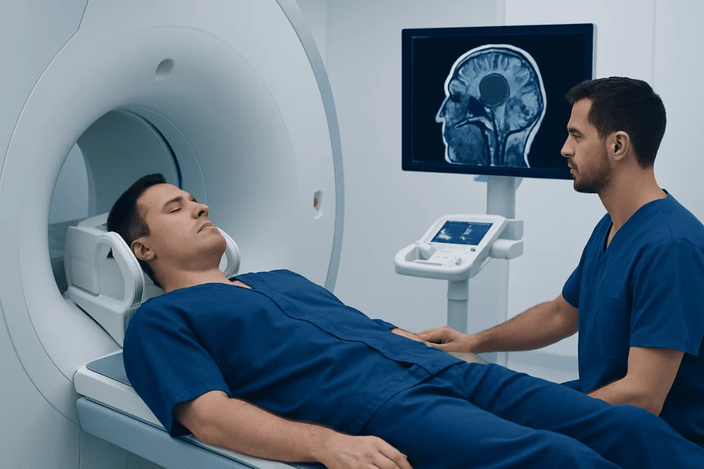
MRI does not use X-rays. During the examination, the patient lies still on a moving table that enters a scanner. The machine takes images in several sequences, each designed to highlight different characteristics of brain tissue, fluid, blood flow, or inflammation.
A contrast agent called gadolinium may be injected through a vein when additional detail is needed. Contrast can help show whether a lesion has an altered blood-brain barrier or an increased blood supply, which may help narrow the possible causes. It is commonly used when investigating a suspected tumor, infection, inflammation, or certain vascular abnormalities.
Brain MRI may be recommended for persistent or unusual headaches, seizures, unexplained neurological symptoms, hormonal changes related to the pituitary gland, hearing or vision changes, follow-up of a known lesion, or assessment after head injury. It is also used to evaluate conditions such as brain tumors and other disorders affecting the brain and nervous system.
Most people can have an MRI safely. However, the imaging team needs to know about implanted medical devices, metal fragments, prior surgery, pregnancy, kidney disease, allergies, and severe claustrophobia before the appointment. Some devices are MRI-compatible only under specific conditions.
Preparing for a Brain MRI and What to Expect Step by Step
Preparation is usually straightforward. The imaging center may ask the patient to arrive early to complete a safety questionnaire and change into a gown. Jewelry, watches, hairpins, hearing aids, removable dental devices, bank cards, and other metal-containing items must be removed before entering the scan room.
If contrast is planned, the clinical team may review kidney function and previous reactions to contrast agents. People who are anxious in enclosed spaces should discuss this in advance, as practical measures or prescribed sedation may sometimes be arranged. When sedation is used, the person generally needs someone to take them home and should follow the center’s fasting instructions.
During the scan, a head coil is placed around the head to improve image quality. The patient can communicate with the MRI staff through an intercom and is usually offered ear protection because the scanner makes loud tapping and knocking sounds. Remaining still is important, and the scan commonly takes around 30 to 60 minutes, depending on whether contrast and specialized sequences are needed.
After the scan, most people can return to usual activities immediately. If contrast was given, normal hydration is generally encouraged unless a clinician has advised fluid restriction. If sedation was used, driving, alcohol, operating machinery, and important decisions should be avoided until the effects have fully worn off.
What Not to Do Before an MRI of the Brain?
Before a brain MRI, a person should not bring metal objects or electronic items into the scanner room. This includes jewelry, piercings that cannot be confirmed as safe, watches, keys, coins, phones, credit cards, hair accessories, and clothing with metallic fasteners. Certain cosmetics may contain metallic particles, so the imaging center may advise arriving without makeup, particularly eye makeup.
It is also important not to withhold information about implants or previous injuries. The MRI team should be told about pacemakers, neurostimulators, cochlear implants, aneurysm clips, insulin pumps, medication patches, artificial joints, surgical clips, retained shrapnel, or possible metal exposure from work or an injury. Never assume an implant is safe without confirmation.
Unless sedation is planned, food and drink restrictions are often unnecessary for a standard brain MRI. However, instructions vary when contrast, anesthesia, or sedation is involved. Patients should follow the instructions provided by the imaging center and continue regular medicines unless their clinician advises otherwise.
What Does a Mass on an MRI of the Brain Indicate?
A mass on an MRI of the brain can indicate many different conditions. It may represent a tumor, but it can also be a cyst, an abscess caused by infection, an area of inflammation, a blood vessel abnormality, a collection of blood, or a change related to a previous stroke or injury. In some situations, a mass-like appearance is caused by swelling rather than a discrete growth.
Common characteristics considered on MRI include location, size, shape, margins, whether the area contains fluid or blood, whether it enhances after contrast, and whether it causes swelling or shifts nearby brain structures. These details help clinicians build a focused list of possible diagnoses, but they may not provide certainty.
Some masses are found incidentally during imaging performed for another reason. Even an incidental finding deserves appropriate review, but it is not automatically dangerous or cancerous. Depending on the appearance, a specialist may recommend comparison with older scans, repeat MRI surveillance, additional imaging, blood tests, or a consultation with neurology, oncology, or neurosurgery.
When an abnormality appears likely to require intervention, a team may discuss brain tumor surgery or other treatments based on the lesion’s location, suspected type, growth pattern, and effect on neurological function.
Can You Tell If a Brain Tumor Is Malignant From an MRI?
MRI can provide important clues about whether a brain tumor may be more or less aggressive, but it cannot always determine with certainty whether a tumor is malignant. Features such as rapid growth on serial scans, irregular borders, extensive surrounding swelling, tissue breakdown, and particular contrast-enhancement patterns can raise concern. However, benign tumors can sometimes have complex appearances, and malignant tumors can occasionally look less typical.
The only way to confirm the exact tumor type and grade in many cases is to examine tissue under a microscope. This may be obtained through a biopsy or during surgery when treatment is appropriate. Pathology testing may also include molecular analysis, which can help classify the tumor and inform treatment planning.
Advanced MRI methods, such as perfusion imaging, diffusion imaging, spectroscopy, and functional MRI, can add useful information. Additional tests may include CT, body imaging, laboratory testing, or neurological assessment, depending on the situation. Decisions are ideally made by a multidisciplinary team that includes neuroradiology, neurology, neurosurgery, pathology, and oncology specialists.
Treatment may involve monitoring, surgery, radiation therapy, systemic medicines, or a combination of approaches. The recommended plan is individualized and should balance diagnostic certainty, symptom relief, preservation of brain function, and the person’s overall health.
How Quickly Do You Get Results From a Brain MRI?
Brain MRI images are available immediately after the scan, but the formal interpretation takes time. A radiologist reviews the images carefully and prepares a report for the clinician who requested the examination. In many routine settings, results are communicated within a few days, although the timing varies by hospital, scan urgency, and the need for comparison with previous imaging.
When the scan is ordered urgently, such as for sudden neurological symptoms or concern about bleeding, stroke, or significant pressure in the brain, the images are usually reviewed much more quickly. The treating team may discuss preliminary findings on the same day.
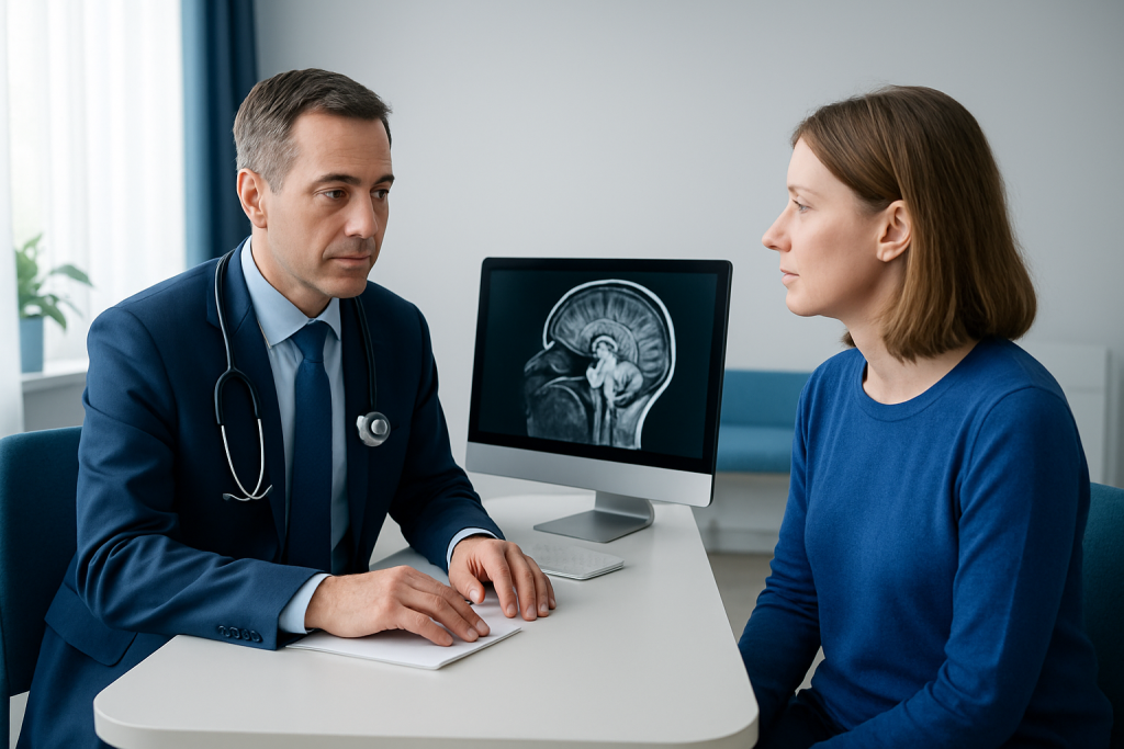
A report may use terms that sound worrying without giving a final diagnosis. Patients should review the findings with the referring clinician, who can explain what the result means in the context of symptoms and medical history. Keeping copies of MRI images and reports can also be helpful if specialist review or future comparison is needed.
For international patients, Acibadem International’s multidisciplinary specialists at JCI-accredited hospitals can coordinate diagnostic evaluation and treatment planning when a brain MRI identifies an abnormal finding.
When to Seek Medical Care
A person should seek urgent medical care for sudden weakness or numbness on one side of the body, difficulty speaking, a new seizure, sudden severe headache, fainting, severe confusion, loss of vision, or rapidly worsening balance problems. These symptoms can have several causes and require prompt clinical assessment rather than waiting for a routine imaging appointment.
Medical review should also be arranged promptly for persistent or progressively worsening headaches, repeated vomiting without a clear cause, new personality or memory changes, gradual loss of coordination, persistent vision changes, or headaches that are different from a usual pattern. These symptoms do not necessarily indicate a brain mass, but they warrant professional evaluation.
After a mass on brain MRI is reported, patients should ask who will explain the result, whether specialist referral is needed, and what warning symptoms should prompt urgent help. If a follow-up scan is recommended, attending it on schedule is important because interval changes can guide decisions about observation or treatment.
Frequently asked questions
Is a mass on brain MRI always a tumor?
No. A mass-like finding can be caused by a tumor, but it may also reflect a cyst, infection, inflammation, bleeding, a vascular abnormality, or tissue changes after injury or stroke. The MRI pattern and clinical assessment help determine which causes are most likely.
Does a brain MRI with contrast mean cancer is suspected?
No. Contrast is used for many reasons because it can show blood flow and changes in the blood-brain barrier more clearly. It may be helpful when evaluating tumors, but also infections, inflammation, vascular conditions, and some post-treatment changes.
Can a brain mass be benign?
Yes. Some brain masses are benign, meaning they do not spread to distant organs and may grow slowly. Even benign lesions can need monitoring or treatment if they press on important brain structures or cause symptoms.
Will a brain MRI show whether a mass is growing?
A single MRI shows the current appearance and size of an abnormality. Growth is best assessed by comparing it with earlier imaging or with a follow-up MRI performed at an interval recommended by the treating clinician.
What happens after a suspicious brain MRI result?
The referring clinician usually reviews the report and may arrange consultation with a neurologist, neurosurgeon, or oncologist. Next steps can include further MRI sequences, repeat imaging, other tests, observation, or tissue sampling when a definitive diagnosis is needed.
Can I drive after a brain MRI?
Most people can drive after a standard brain MRI, including one performed with contrast, if they feel well. Driving should be avoided if sedation or anti-anxiety medication was used, and the imaging team’s discharge instructions should be followed.
References
- American College of Radiology
- National Cancer Institute
- National Institute of Neurological Disorders and Stroke
- Radiological Society of North America
- World Health Organization
This article is for general information only and is not a substitute for professional medical advice. Please consult a qualified doctor about your individual situation.
Oncology care in Turkey — second opinion and treatment plan
JCI-accredited · board-certified surgeons · reply within 24h
Add us as a Preferred Source to see more of our trusted health content across Google Search, AI Overviews and Discover.