

CT Scan: Uses, Radiation Dose, Contrast, and Result Timing
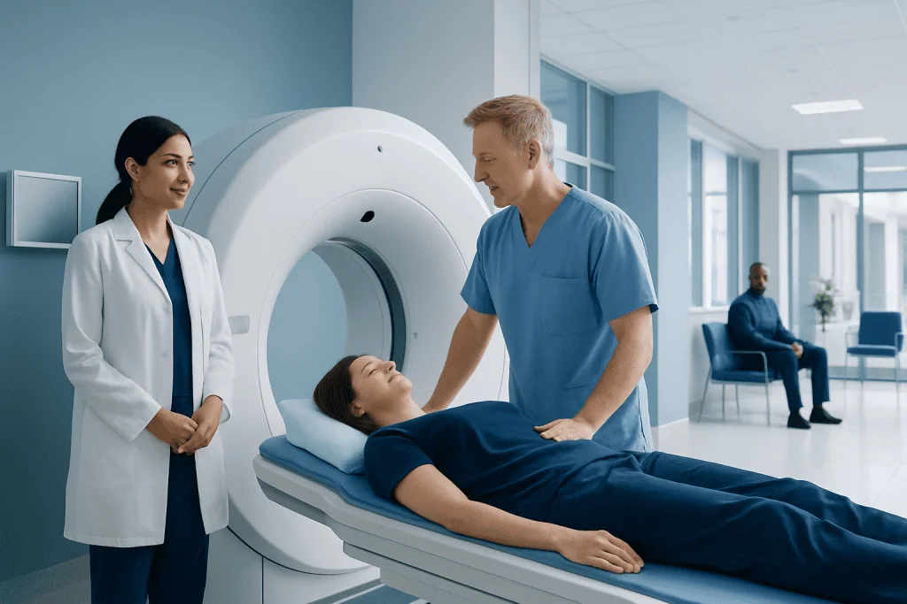
A CT scan uses X-rays and computer processing to create detailed cross-sectional images of the body. CT is often chosen when doctors need quick, precise information about bones, organs, blood vessels, or internal bleeding.
Key Takeaways
- A CT scan uses X-rays and computer processing to create detailed cross-sectional images of the body.
- CT is often chosen when doctors need quick, precise information about bones, organs, blood vessels, or internal bleeding.
- Radiation exposure is kept as low as reasonably possible, and the expected benefit usually outweighs the small potential risk when the test is medically needed.
- Contrast material may be given by vein, by mouth, or by rectum to highlight blood vessels, organs, bowel, or inflammation.
- Result timing varies from urgent same-day reporting to routine reports within a few days, depending on the clinical situation and facility workflow.
A CT scan is a fast, detailed imaging test that helps doctors diagnose injuries, infections, cancers, blood vessel problems, and many other conditions. Understanding radiation dose, contrast material, preparation, and result timing can help patients feel more confident before the examination.
Overview
A CT scan, also called computed tomography or a CAT scan, is a medical imaging test that uses X-rays and advanced computer processing to create detailed pictures of the inside of the body. Unlike a standard X-ray, which produces a flat image, CT creates thin cross-sectional slices that can be viewed individually or reconstructed into three-dimensional images.
CT scans are used in many areas of medicine because they are fast, widely available, and highly detailed. They can show bones, lungs, abdominal organs, blood vessels, the brain, and soft tissues. In emergency situations, CT may help doctors quickly identify internal bleeding, stroke, severe infection, or traumatic injuries.
The test is usually painless and often takes only a few minutes of actual scanning time. Depending on the body area and the reason for the scan, contrast material may be used to make certain structures easier to see. A radiologist, a doctor specially trained in medical imaging, reviews the images and sends a report to the referring doctor.
Common Uses of a CT Scan
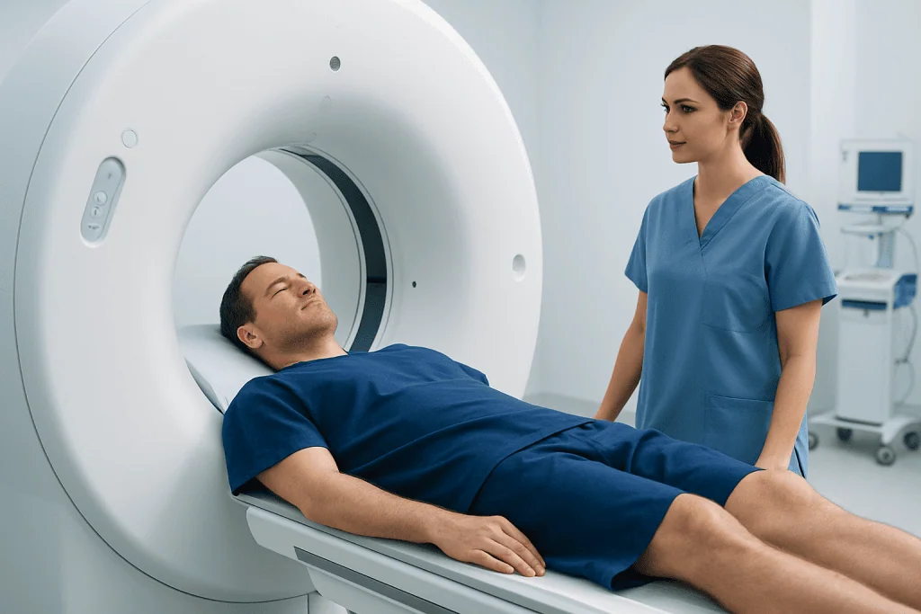
Doctors request CT scans for many reasons, usually when more detailed information is needed than a physical examination, blood test, ultrasound, or standard X-ray can provide. CT is particularly useful for evaluating complex anatomy and conditions that require quick diagnosis.
Common uses include assessing head injuries, suspected stroke or bleeding in the brain, chest pain, lung disease, abdominal pain, kidney stones, appendicitis, bowel problems, fractures, spinal injuries, and suspected tumors. CT can also guide certain procedures, such as biopsies or drainage of fluid collections.
Examples of situations where CT may be recommended include:
- Trauma after a car accident, fall, or sports injury
- Severe or unexplained abdominal or pelvic pain
- Shortness of breath or suspected blood clot in the lungs
- Monitoring cancer treatment response or detecting recurrence
- Planning surgery, radiotherapy, or interventional procedures
- Evaluating blood vessels with CT angiography
The decision to perform a CT scan depends on the patient’s symptoms, medical history, examination findings, and the clinical question that needs to be answered. In some cases, another test such as ultrasound or MRI may be more appropriate.
Radiation Dose and Safety
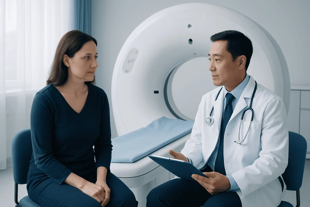
A CT scan uses ionizing radiation, which is the same type of energy used in standard X-rays. Because CT produces many images from different angles, the radiation dose is usually higher than a single plain X-ray, but the dose varies widely depending on the body part, patient size, scan technique, and whether multiple phases are needed.
Modern CT scanners and imaging protocols are designed to use the lowest radiation dose that can still produce useful diagnostic images. This principle is often described as keeping exposure as low as reasonably achievable. Radiology teams adjust scan settings, limit the scanned area, and avoid repeated scans unless they are clinically necessary.
For most patients, when a CT scan is appropriately recommended, the expected diagnostic benefit is greater than the small potential risk associated with radiation. However, patients should tell their doctor if they are pregnant or may be pregnant, because special consideration is needed. Children and young adults may also require tailored protocols because they are more sensitive to radiation over a lifetime.
Patients should not refuse an important CT scan solely because of radiation concerns, but it is reasonable to ask why the scan is needed, whether previous imaging can answer the question, and whether a non-radiation test such as ultrasound or MRI is suitable. These questions help ensure that imaging is both safe and clinically useful.
Contrast Material: Why It Is Used and What to Expect
Contrast material is sometimes used during a CT scan to make blood vessels, organs, inflammation, tumors, or the digestive tract more visible. The most common CT contrast is iodine-based and is given through a vein. Some abdominal or pelvic scans may also use oral contrast, and less commonly rectal contrast, to outline the bowel.
Intravenous contrast may cause a warm feeling, a metallic taste, or the sensation of needing to urinate; these effects usually pass quickly. Oral contrast may require the patient to drink liquid before the scan and may add preparation time. The radiology team will explain which type is needed and why.
Most people receive CT contrast safely. Before contrast is given, patients are typically asked about previous contrast reactions, allergies, asthma, kidney disease, diabetes medications, thyroid disease, pregnancy, and breastfeeding. A blood test to check kidney function may be needed for some patients, especially those with kidney problems or certain risk factors.
Serious allergic-type reactions to iodinated contrast are uncommon, but the imaging team is trained to recognize and treat them. Patients should report itching, hives, breathing difficulty, throat tightness, dizziness, or swelling immediately. Mild nausea or warmth alone is usually not dangerous, but staff should still be informed if any symptom feels concerning.
How to Prepare and What Happens During the Scan
Preparation depends on the type of CT scan. Some scans require no special preparation, while others may require fasting for several hours, drinking oral contrast, or having blood tests before intravenous contrast. Patients should follow the instructions from the imaging center and bring previous imaging reports if available.
Before the scan, patients may be asked to remove jewelry, eyeglasses, hearing aids, removable dental work, or clothing with metal parts. A hospital gown may be provided. If IV contrast is needed, a small cannula is placed in a vein, usually in the arm or hand.
During the scan, the patient lies on a narrow table that moves through a large, doughnut-shaped scanner. The machine does not usually enclose the whole body for long, and most people tolerate it well. The technologist may ask the patient to hold their breath briefly, especially for chest or abdominal imaging, because movement can blur the images.
The scanning itself is often very quick, sometimes less than a minute, although the full appointment can take longer because of registration, preparation, contrast administration, and observation afterward. Patients should remain still and follow breathing instructions carefully to help produce clear images and avoid repeat scanning.
CT Scan Results and Timing
After the scan, the images are reviewed by a radiologist. The radiologist interprets the findings in the context of the clinical question and prepares a written report for the referring doctor. In many cases, the referring doctor discusses the results with the patient and explains what they mean for diagnosis, treatment, or follow-up.
Result timing varies. In emergency departments or urgent situations, a preliminary or final interpretation may be available quickly, sometimes within the same visit. For routine outpatient CT scans, reports may take from the same day to a few business days, depending on the complexity of the study, comparison with previous scans, and local workflow.
Some findings require correlation with symptoms, blood tests, physical examination, or additional imaging. A CT scan can show important abnormalities, but it does not always provide a complete diagnosis by itself. Occasionally, the report may recommend follow-up imaging, MRI, ultrasound, biopsy, or specialist consultation.
If patients have access to the radiology report before speaking with their doctor, unfamiliar terms can feel confusing. It is best to avoid drawing conclusions from isolated phrases. The referring physician can explain whether a finding is significant, incidental, stable, or requires further evaluation.
Risks, Limitations, and Aftercare
CT is a safe and valuable test when used appropriately, but like all medical procedures it has limitations. Radiation exposure is considered before the scan is ordered, and contrast material may not be suitable for every patient. The radiology team balances image quality, diagnostic need, and safety precautions for each case.
Possible issues after CT with intravenous contrast include mild nausea, a temporary rash, or discomfort at the injection site. Rarely, contrast can leak outside the vein into surrounding tissue, causing swelling or pain. Patients should tell staff immediately if they feel burning, tightness, or increasing pain during injection.
After most CT scans, patients can return to normal activities right away. If contrast was used, drinking fluids may be recommended unless the patient has been told to restrict fluids for another medical reason. People who received sedatives, which is uncommon for standard CT but may be used in selected cases, should follow discharge instructions and avoid driving until cleared.
CT also has diagnostic limitations. Very small abnormalities may be difficult to detect, and some conditions are better evaluated with MRI, ultrasound, endoscopy, or laboratory testing. If symptoms continue despite a normal CT scan, patients should follow up with their doctor rather than assuming no further care is needed.
When to Speak With a Doctor
Patients should speak with their doctor before a CT scan if they are pregnant, may be pregnant, have had a previous reaction to iodinated contrast, have kidney disease, are on dialysis, have severe allergies or asthma, or take medications that may affect kidney function. This does not always mean the scan cannot be done, but it helps the team choose the safest approach.
After the scan, patients should seek medical advice if they develop symptoms such as a spreading rash, swelling of the face or throat, breathing difficulty, faintness, persistent vomiting, fever, or increasing pain and swelling at the IV site. These symptoms are not typical, but they should be assessed promptly.
Patients should also contact their referring doctor if they have not received results within the expected timeframe, if symptoms worsen, or if they do not understand the next steps after the report. Imaging is one part of care, and results are most helpful when reviewed together with the patient’s full clinical picture.
For international patients, Acibadem International’s multidisciplinary specialists and JCI-accredited hospitals provide diagnostic imaging services, including CT, as part of coordinated medical evaluation and treatment planning. Patients should always discuss the appropriateness of any scan with a qualified healthcare professional.
Frequently asked questions
Is a CT scan painful?
The scan itself is painless. Some patients feel brief discomfort from lying still or from the IV cannula if contrast is used. Intravenous contrast can cause a temporary warm sensation or metallic taste, which usually passes quickly.
How long does a CT scan take?
The actual scanning time is often only a few minutes or less. The full visit may take longer because of registration, changing clothes, placing an IV, drinking contrast, or waiting after contrast administration. Complex scans or emergency workflows can also affect timing.
Does every CT scan require contrast?
No. Some CT scans are done without contrast, especially when looking for kidney stones, certain lung findings, bleeding, or bone injuries. Contrast is used when it helps answer the medical question more clearly, such as evaluating blood vessels, tumors, infection, or abdominal organs.
Is CT radiation dangerous?
CT uses ionizing radiation, so it is ordered only when the expected benefit is meaningful. Modern CT protocols aim to minimize dose while preserving image quality. For a medically necessary scan, the benefit of accurate diagnosis usually outweighs the small potential risk.
Can a pregnant patient have a CT scan?
Pregnancy should always be reported before a CT scan. In some situations, CT may still be necessary, especially in emergencies, but the medical team will consider the body area, urgency, and possible alternatives such as ultrasound or MRI. Protective and dose-reduction measures may be used when appropriate.
When will CT scan results be available?
Urgent CT results may be available quickly, sometimes during the same emergency visit. Routine outpatient reports may take from the same day to a few business days. The referring doctor is usually the best person to explain the findings and next steps.
Can a CT scan detect cancer?
CT can detect many tumors and is often used for cancer diagnosis, staging, treatment planning, and follow-up. However, it cannot detect every cancer, and some findings require further testing such as MRI, PET-CT, endoscopy, blood tests, or biopsy. The interpretation depends on the scan type and the patient’s clinical situation.
References
- American College of Radiology
- Radiological Society of North America
- U.S. Food and Drug Administration
- International Atomic Energy Agency
- European Society of Radiology
This article is for general information only and is not a substitute for professional medical advice. Please consult a qualified doctor about your individual situation.
Explore treatments in Turkey — costs, top hospitals & a free quote
JCI-accredited · board-certified surgeons · reply within 24h
Add us as a Preferred Source to see more of our trusted health content across Google Search, AI Overviews and Discover.

More from the Health Library



Related Specialists

Prof. Dr. Ercan Karaarslan
Radiology
Prof. Dr. Bora Peynircioğlu
Interventional Radiology
Prof. Dr. Koray Güven
Interventional Radiology
Assoc. Prof. Dr. Deniz Can Aliş
Radiology