

Dental X-Rays: Safety, Types, and What Dentists Look For
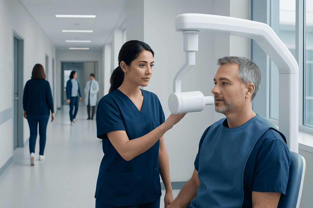
Dental X-rays help detect tooth decay, bone loss, infections, impacted teeth, and other conditions that may not be visible during a dental exam. The type and frequency of X-rays depend on age, oral health, symptoms, dental history, and treatment needs.
Key Takeaways
- Dental X-rays help detect tooth decay, bone loss, infections, impacted teeth, and other conditions that may not be visible during a dental exam.
- The type and frequency of X-rays depend on age, oral health, symptoms, dental history, and treatment needs.
- Modern digital dental X-rays use low levels of radiation, and dentists follow safety principles to keep exposure as low as reasonably achievable.
- Children, pregnant patients, and people with medical concerns can usually have dental X-rays when needed, with appropriate precautions.
- Patients should tell their dentist about pregnancy, recent X-rays, medical conditions, and any concerns before imaging.
Dental X-rays, also called dental radiographs, help dentists see areas of the teeth, gums, jaw, and bone that are not visible during a routine examination. When used appropriately, they are a safe and valuable tool for diagnosing problems early and planning dental treatment.
Overview
Dental X-rays are images that allow dentists to examine parts of the mouth that cannot be seen clearly with the eyes alone. They can show tooth roots, the spaces between teeth, the jawbone, developing teeth, and areas under existing fillings, crowns, or bridges. This makes them an important part of preventive care as well as diagnosis and treatment planning.
Many dental problems begin silently. Early tooth decay between teeth, small infections near the root, bone loss from gum disease, and impacted wisdom teeth may not cause pain at first. X-rays can help identify these changes before they become more complex to treat.
Dental imaging is not the same for every patient. A dentist decides whether X-rays are needed based on symptoms, examination findings, age, oral health risk, previous dental records, and the type of treatment being considered. The goal is to use the right image at the right time, while avoiding unnecessary exposure.
Common Types of Dental X-Rays
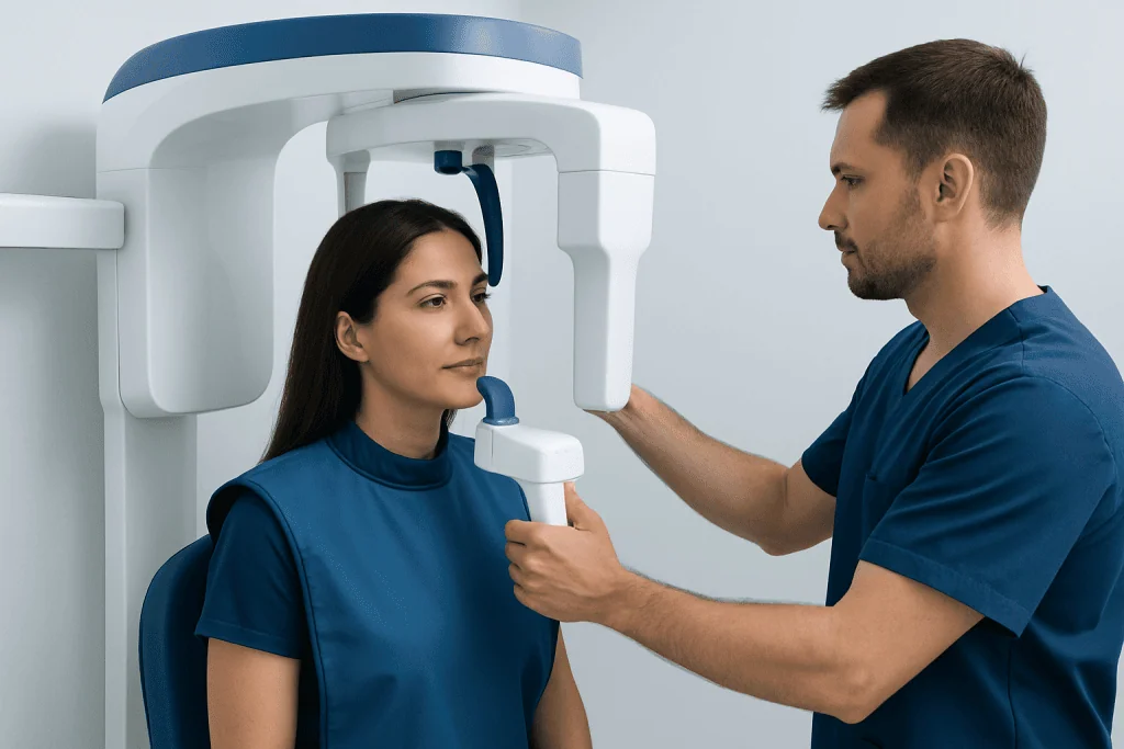
Different types of dental X-rays provide different views. Intraoral X-rays are taken with a small sensor or film inside the mouth, while extraoral X-rays are taken with the sensor outside the mouth. The dentist selects the type that best answers the clinical question.
Bitewing X-rays show the upper and lower back teeth together when the patient bites down. They are especially useful for detecting cavities between teeth and checking the height of the bone that supports the teeth. Periapical X-rays show the whole tooth from crown to root tip and are often used to evaluate tooth pain, infection, root shape, or previous root canal treatment.
Panoramic X-rays create a broad view of the entire mouth, including all teeth, jaws, temporomandibular joints, sinuses, and developing or impacted teeth. They are commonly used for wisdom teeth, orthodontic assessment, implant planning, and overall jaw evaluation. Occlusal X-rays show a larger area of the upper or lower jaw and may be used to locate extra teeth, jaw lesions, or developing teeth in children.
In some situations, dentists may use cone beam computed tomography, often called CBCT. This is a three-dimensional dental scan that provides detailed information about bone, nerves, tooth roots, and jaw structures. It is typically reserved for cases where 3D information is important, such as complex implant planning, impacted teeth, certain root canal problems, or jaw surgery assessment.
What Dentists Look For on Dental X-Rays

Dental X-rays help dentists look beyond the visible tooth surface. One of the most common uses is detecting tooth decay, especially cavities between teeth or under existing restorations. These areas can be difficult to evaluate during a visual exam alone, even with careful probing.
Dentists also assess the bone around teeth. Bone levels can indicate whether gum disease has caused bone loss, whether teeth are stable, or whether there are changes around implants. X-rays may also show tartar below the gumline, root shape, and signs of trauma such as fractures or root damage.
Another important use is identifying infection or inflammation. A dark area near the tip of a tooth root may suggest an abscess or chronic infection, although the dentist will interpret this together with symptoms and clinical tests. X-rays can also help evaluate cysts, certain jaw lesions, sinus-related findings, and abnormalities that may require further assessment.
For children and teenagers, dental X-rays can help monitor tooth development. Dentists may check whether permanent teeth are forming normally, whether teeth are missing or extra, and whether orthodontic referral is needed. In adults, X-rays are often used before extractions, root canal treatment, crowns, bridges, dentures, implants, and other dental procedures.
Safety and Radiation Exposure
Modern dental X-rays use low levels of radiation, particularly when digital sensors are used. Dentists are trained to follow radiation safety principles, including taking images only when they are clinically justified and choosing the smallest area and lowest exposure that provides a useful diagnostic image.
Radiation exposure from dental imaging is generally low, but it is still important to avoid unnecessary X-rays. Dental teams may use protective measures such as lead aprons and thyroid collars when appropriate, especially for children and people who may benefit from added shielding. Equipment is also maintained and checked to ensure images are produced efficiently and safely.
Patients can help by telling their dentist about recent dental X-rays taken elsewhere. If previous images are available and still relevant, they may reduce the need for repeat imaging. Patients should also mention pregnancy, medical conditions, previous radiation therapy to the head or neck, or any concerns about radiation before the appointment.
For most people, the benefit of a needed dental X-ray outweighs the small radiation risk. Finding an infection, untreated decay, or bone loss early can prevent pain, tooth loss, and more extensive treatment later. The decision should always be individualized and explained clearly by the dental professional.
Dental X-Rays for Children, Pregnancy, and Special Situations
Children may need dental X-rays because their mouths are growing and changing quickly. X-rays can show whether permanent teeth are developing correctly, whether cavities are forming between teeth, and whether orthodontic problems are emerging. However, children are more sensitive to radiation than adults, so dentists use child-appropriate settings and take images only when needed.
During pregnancy, dental care remains important, because untreated dental infections or gum disease can affect overall health and comfort. Dental X-rays can usually be taken during pregnancy when they are necessary for diagnosis or urgent treatment, using appropriate shielding and modern techniques. Elective imaging that is not needed immediately may be postponed until after pregnancy, depending on the situation.
Patients with braces, dental implants, complex restorations, or a history of gum disease may need X-rays at different intervals from patients with low cavity risk and stable oral health. People with unexplained tooth pain, swelling, injury, or suspected infection may need imaging even if their last routine X-rays were recent.
There is no single schedule that fits everyone. A patient who has frequent cavities, dry mouth, gum disease, or many restorations may need monitoring more often than someone with excellent oral health and low risk. The dentist should explain why an image is recommended and how it will guide care.
What to Expect During a Dental X-Ray
Dental X-rays are usually quick and painless. For intraoral images, the dental professional places a small sensor or film inside the mouth and asks the patient to bite gently or hold still for a few seconds. Some people may feel mild pressure from the sensor, especially if they have a small mouth, a strong gag reflex, or tender gums.
For a panoramic X-ray or CBCT scan, the patient usually stands or sits while the machine rotates around the head. The dental team will position the chin, forehead, or bite guide to keep the head steady. It is important to remain still during the scan so the image is clear and does not need to be repeated.
After the images are taken, the dentist reviews them together with the clinical examination. The images may be displayed on a screen, making it easier to point out cavities, bone levels, impacted teeth, or other findings. Patients are encouraged to ask what the images show and why any recommended treatment is needed.
How Often Are Dental X-Rays Needed?
The timing of dental X-rays depends on risk and need. A new patient may need images so the dentist can understand current oral health, previous dental work, bone levels, and hidden problems. If the patient brings recent high-quality X-rays, the dentist may be able to use those instead of taking new ones.
For routine monitoring, dentists consider several factors, including cavity risk, gum health, symptoms, age, diet, oral hygiene, dry mouth, medical conditions, and previous dental treatment. Some patients may need bitewing X-rays at shorter intervals, while others may safely go longer between images. Panoramic X-rays and CBCT scans are not usually taken at every checkup; they are used when a broader or three-dimensional view is clinically helpful.
Guidelines from professional dental organizations emphasize individualized decisions rather than fixed schedules for everyone. This approach helps dentists detect disease early while keeping radiation exposure as low as reasonably achievable. If a patient is unsure why an X-ray is being suggested, it is appropriate to ask for an explanation.
When to See a Dentist
A dental examination is recommended if a person has tooth pain, sensitivity, swelling, bleeding gums, a broken tooth, jaw discomfort, or a bad taste that does not go away. X-rays may be needed to determine whether the problem is caused by decay, infection, gum disease, trauma, or another condition. Prompt assessment can often make treatment simpler and more comfortable.
Regular dental visits are also important even when there are no symptoms. Many oral health problems develop gradually and may be found only during a professional exam or with the help of X-rays. Preventive visits allow dentists to monitor changes over time and recommend care before problems progress.
Patients who are planning orthodontic treatment, dental implants, wisdom tooth removal, root canal treatment, or major restorative work should expect imaging as part of the evaluation. In complex cases, a dentist may coordinate care with oral surgeons, orthodontists, periodontists, endodontists, or radiology specialists.
Acibadem International’s multidisciplinary dental and medical specialists, within JCI-accredited hospitals, diagnose and treat dental and jaw conditions for international patients. Patients should always discuss their personal imaging needs, benefits, and alternatives with a qualified dentist.
Frequently asked questions
Are dental X-rays safe?
Dental X-rays are considered safe when they are used appropriately and only when clinically needed. Modern digital equipment uses low radiation levels, and dental teams follow safety practices to minimize exposure. The dentist should explain why an X-ray is recommended and how it will help with diagnosis or treatment.
Do dental X-rays hurt?
Dental X-rays do not hurt, but the sensor placed inside the mouth may feel slightly uncomfortable for a short time. Patients with a small mouth, sensitive gums, or a gag reflex can tell the dental team so positioning can be adjusted. The image itself usually takes only a few seconds.
Can dental X-rays show cavities?
Yes, dental X-rays can show many cavities, especially those between teeth or under existing fillings where a dentist cannot easily see. Very early enamel changes may not always be visible on an X-ray, so dentists also rely on visual examination and risk assessment. X-rays are one part of a complete dental evaluation.
Are dental X-rays safe during pregnancy?
Dental X-rays can be taken during pregnancy when they are necessary for diagnosis or urgent dental care, using appropriate precautions. Untreated dental infections should not be ignored during pregnancy. Patients should always tell the dental team if they are pregnant or may be pregnant so the need and timing can be reviewed.
How often should a person have dental X-rays?
There is no single schedule for everyone. The frequency depends on age, oral health, cavity risk, gum health, symptoms, and previous dental treatment. A dentist may recommend more frequent imaging for high-risk patients and less frequent imaging for those with stable, low-risk oral health.
What is the difference between a panoramic X-ray and a bitewing X-ray?
A bitewing X-ray shows the upper and lower back teeth in detail and is often used to find cavities between teeth and check bone levels. A panoramic X-ray shows a broad view of the entire mouth and jaws, including wisdom teeth, jaw joints, and developing teeth. Each image type is chosen for a different diagnostic purpose.
References
- American Dental Association
- U.S. Food and Drug Administration
- International Atomic Energy Agency
- European Commission Radiation Protection Guidelines
- American Academy of Pediatric Dentistry
This article is for general information only and is not a substitute for professional medical advice. Please consult a qualified doctor about your individual situation.
Explore treatments in Turkey — costs, top hospitals & a free quote
JCI-accredited · board-certified surgeons · reply within 24h
Add us as a Preferred Source to see more of our trusted health content across Google Search, AI Overviews and Discover.

Dental & Oral Health
Full-scope dental care from implants and aesthetics to oral and maxillofacial surgery.
158 specialists in this unitRelated Treatments
More from the Health Library



Related Specialists

Assoc. Prof. Dr. Ferit Bayram
Oral & Dental Health
Dt. Alara Naz Kenir
Oral & Dental Health
Dt. Gonca Katmer
Oral & Dental HealthDt. Doğa Baysal
Oral & Dental Health