Diagnostic Mammogram vs Screening Mammogram: Key Differences Explained
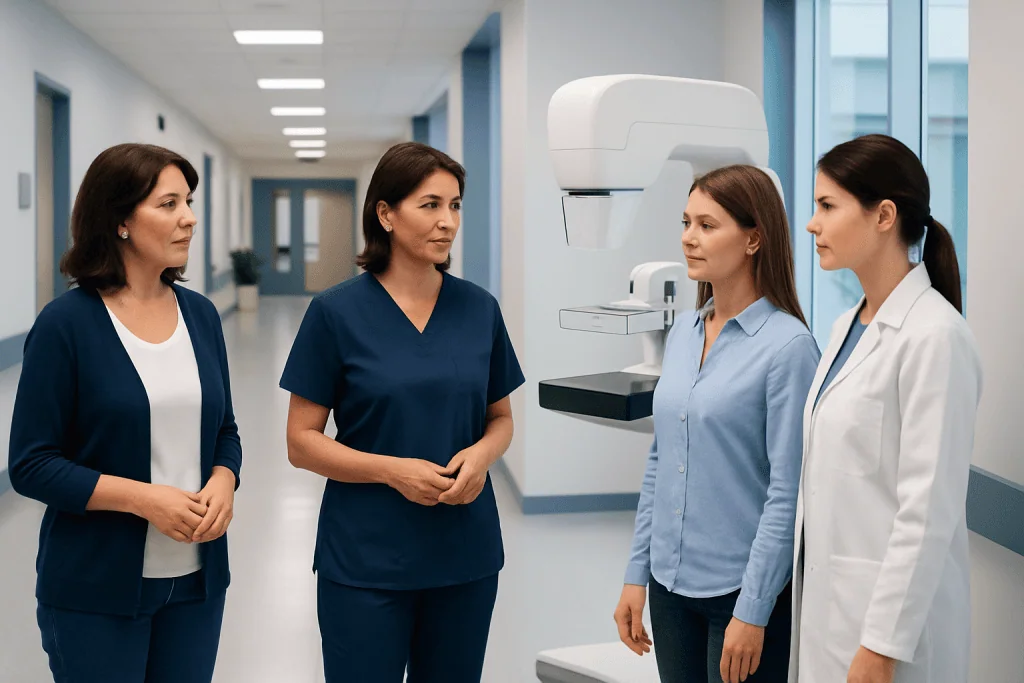
Screening mammograms are routine tests for people without breast symptoms. Diagnostic mammograms are used to assess a specific concern, such as a lump, pain, nipple discharge, or an abnormal screening result.
Key Takeaways
- Screening mammograms are routine tests for people without breast symptoms.
- Diagnostic mammograms are used to assess a specific concern, such as a lump, pain, nipple discharge, or an abnormal screening result.
- A diagnostic mammogram usually includes additional views and may take longer than a screening exam.
- Needing a diagnostic mammogram does not automatically mean cancer; many findings are benign.
- Follow-up may include breast ultrasound, MRI, short-term imaging review, or biopsy depending on the results.
A screening mammogram is a routine test used to look for early breast changes in people without symptoms. A diagnostic mammogram is a more detailed exam used when there is a breast symptom, an abnormal screening result, or a need for closer evaluation.
Overview: What Is the Difference?
The main difference in diagnostic mammogram vs screening mammogram is the reason the test is done. A screening mammogram is a routine breast X-ray for people who do not have symptoms. Its goal is to find early breast changes, sometimes before they can be felt. A diagnostic mammogram is performed when there is a specific concern that needs closer attention.
That concern may be a new lump, breast pain in one area, nipple discharge, skin thickening, or a change seen on a previous screening mammogram. In these cases, the radiology team takes extra images and focuses on the area of concern. The aim is not simply to screen, but to evaluate and clarify what is happening.
Both tests use low-dose X-rays and are performed using the same basic mammography equipment. However, the process, number of images, and how the radiologist interprets the exam can differ. Understanding these differences can help patients know what to expect and reduce unnecessary worry.
What Happens During a Screening Mammogram?
A screening mammogram is designed for routine breast cancer screening. It is usually offered based on age, family history, personal risk factors, and local screening guidelines. During the exam, each breast is positioned and gently compressed for a few seconds so clear images can be taken.
In many cases, standard screening includes two images of each breast taken from different angles. The visit is often relatively brief. After the images are reviewed, results are usually sent later rather than discussed immediately, although timing can vary by center.
The purpose of screening is to look for small changes that may not cause any symptoms. These can include tiny calcium deposits, areas of asymmetry, or masses that need comparison with past images. Screening mammography is an important part of preventive breast care, but it is not meant to explain every symptom or answer every question in a single visit.
What Happens During a Diagnostic Mammogram?
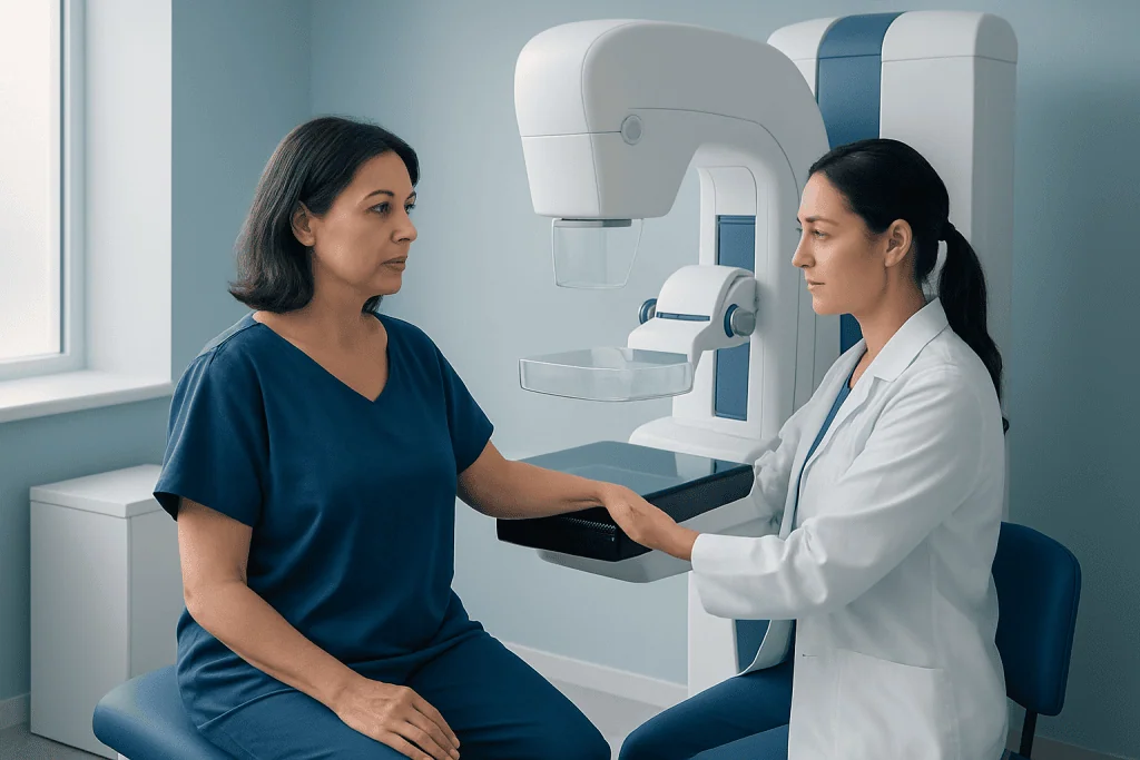
A diagnostic mammogram is more focused and tailored to the patient’s individual concern. It may be recommended after an abnormal screening mammogram, or because a person has developed symptoms such as a palpable lump, one-sided nipple discharge, skin dimpling, or persistent focal breast pain. The radiologist may request extra views to examine one area more closely.
These additional images can include magnification views, spot compression views, or images from different angles. Because the exam is problem-focused, it often takes longer than a screening mammogram. In many centers, the radiologist reviews the images during the visit and may decide whether more imaging is needed right away.
Follow-up tests may include breast ultrasound to better assess a lump or dense tissue, or breast MRI in selected cases when more detail is needed. If imaging still leaves uncertainty, the next step may be tissue sampling with a biopsy to determine whether a finding is benign or needs treatment.
Why Someone May Need One or the Other
A screening mammogram is typically recommended for people who feel well and have no new breast symptoms. It is part of routine preventive care and is used to detect possible problems at an early stage. The exact timing and frequency depend on age, breast density, family history, genetic factors, prior radiation exposure, and national or regional guidelines.
A diagnostic mammogram is recommended when there is a reason to investigate further. Common reasons include:
- An abnormality found on a screening mammogram
- A new breast lump or thickening
- One-sided or bloody nipple discharge
- Changes in breast skin, shape, or nipple position
- Follow-up of a previous finding that needs short-term recheck
- Evaluation after treatment for a known breast condition
Sometimes a patient starts with screening and is then asked to return for diagnostic imaging. This can feel stressful, but callback imaging is common and often leads to a non-cancer explanation. Diagnostic testing helps the radiologist decide whether a finding is normal tissue overlap, a harmless cyst, or something that requires closer management.
How Results Are Interpreted
Mammogram results are commonly reported using a standardized system called BI-RADS, which helps describe what was seen and what follow-up is recommended. A result may be negative or benign, meaning no suspicious finding is present. In other cases, the report may say that additional imaging is needed, that short-term follow-up is advised, or that biopsy should be considered.
Screening mammograms may identify an area that simply needs a second look. This does not mean cancer has been found. Overlapping tissue, benign calcifications, cysts, or dense breast tissue can create findings that need clarification on diagnostic images. A diagnostic mammogram gives the radiologist more detailed views to sort out these possibilities.
If a suspicious finding remains after diagnostic evaluation, the doctor may discuss whether it could be related to a condition such as breast cancer or another breast disorder. Even then, imaging alone may not provide a final diagnosis. When necessary, biopsy is the test that confirms what the tissue actually is.
Benefits, Limitations, and Breast Density
Both screening and diagnostic mammograms are valuable tools in breast imaging. Screening can find early abnormalities before symptoms develop, which may improve treatment planning if cancer is present. Diagnostic mammography can provide more precise assessment of an area of concern and guide the next steps in care.
At the same time, mammography has limitations. Not every cancer is visible on a mammogram, especially in people with dense breast tissue. Dense breasts can make interpretation more difficult because both dense tissue and some abnormalities appear white on the image. This is one reason extra imaging may sometimes be recommended.
Mammograms can also lead to false-positive results, where an abnormality is suspected but later found to be benign. While this can cause anxiety, it is part of careful breast evaluation. In selected cases, additional tools such as ultrasound, MRI, or image-guided procedures help create a clearer and more complete picture.
Preparing for the Exam and What to Expect Afterward
Preparation for both types of mammograms is similar. Patients are often advised not to use deodorant, powders, lotions, or creams on the chest or underarms on the day of the exam, as these can appear on images. Wearing a two-piece outfit can make changing easier. It is also helpful to bring prior mammograms or ensure the imaging center can access them for comparison.
Some people prefer to schedule the test when their breasts are less tender, such as avoiding the days just before a menstrual period. During the exam, compression may feel uncomfortable, but it is brief and helps produce clear images with the lowest practical radiation dose. Patients should tell the technologist if they are pregnant, breastfeeding, have implants, or have difficulty standing still.
After a screening mammogram, results are often communicated within days. After a diagnostic mammogram, the radiologist may discuss preliminary findings sooner, depending on the center. If more tests are needed, the care team explains the reason and the next step. Near the end of the diagnostic pathway, patients seeking coordinated international care may also choose centers such as Acibadem International, where multidisciplinary specialists in JCI-accredited hospitals evaluate breast concerns for international patients.
When to See a Doctor
A person should speak with a doctor promptly if they notice a new breast lump, persistent focal pain, nipple discharge, skin dimpling, redness that does not improve, swelling, or a change in the shape of the breast or nipple. Most breast changes are not cancer, but they still deserve proper assessment. Early evaluation helps identify whether the cause is benign or needs treatment.
It is also important to follow through on callback imaging after a screening mammogram. Delaying a diagnostic exam can prolong uncertainty and postpone care if treatment is needed. Even when the chance of cancer is low, completing the recommended workup is the safest course.
Anyone with a strong family history of breast or ovarian cancer, known genetic risk, or previous high-risk breast findings should ask a doctor about the most appropriate screening plan. Some individuals may need a different schedule or additional imaging beyond standard mammography. Personalized advice from a qualified clinician is the best way to decide which test is right and when it should be done.
Frequently asked questions
Is a diagnostic mammogram the same as a screening mammogram?
No. A screening mammogram is a routine test for people without symptoms, while a diagnostic mammogram is used to investigate a specific breast concern or an abnormal screening result. A diagnostic exam usually includes additional, targeted images.
Does being called back after a screening mammogram mean cancer?
Not necessarily. Many callback findings are caused by overlapping normal tissue, cysts, benign calcifications, or technical factors that require clearer images. A diagnostic mammogram helps determine whether the finding is harmless or needs further testing.
Which mammogram takes longer?
A diagnostic mammogram usually takes longer than a screening mammogram. That is because the radiologist may request extra views during the visit and may review the images in real time to decide whether more evaluation is needed.
Can a diagnostic mammogram detect cancer?
A diagnostic mammogram can identify suspicious changes that may suggest cancer, but it does not confirm the diagnosis by itself. If imaging remains concerning, a biopsy may be recommended to examine the tissue directly.
Do both tests use radiation?
Yes. Both screening and diagnostic mammograms use low-dose X-rays to create images of the breast. The amount is kept as low as reasonably possible while still producing clear images for accurate interpretation.
What if I have dense breasts?
Dense breast tissue can make mammograms harder to interpret because dense tissue can hide some abnormalities. Depending on the situation, a doctor may recommend additional imaging such as ultrasound or MRI along with mammography.
This article is for general information only and is not a substitute for professional medical advice. Please consult a qualified doctor about your individual situation.
Explore treatments in Turkey — costs, top hospitals & a free quote
JCI-accredited · board-certified surgeons · reply within 24h
Add us as a Preferred Source to see more of our trusted health content across Google Search, AI Overviews and Discover.

More from the Health Library

When Do You Need Urgent Imaging for Headache, Abdominal Pain, or Injury?

Cerebral Angiography Risks and Recovery: What Patients Should Expect

Coronary CT Angiography: Imaging Heart Arteries Without a Catheter
How to Prepare for a Diagnostic Mammogram: Clothing, Timing, and Questions to Ask

What Is a Lab Test Panel and Why Are Tests Grouped Together?