Elevated Right Hemidiaphragm Treatment: How It Works, Results and What to Expect

An elevated right hemidiaphragm is an imaging finding, not a diagnosis by itself. Many people have no symptoms and do not require a procedure.
Key Takeaways
- An elevated right hemidiaphragm is an imaging finding, not a diagnosis by itself.
- Many people have no symptoms and do not require a procedure.
- Tests help distinguish diaphragm weakness from lung, abdominal or liver-related causes of elevation.
- Diaphragm plication can improve lung expansion and breathlessness in carefully selected people with persistent symptoms.
- Recovery and outlook vary according to the cause, overall lung health and whether nerve function can recover.
Elevated right hemidiaphragm treatment is guided by the underlying cause, symptoms and effect on breathing. Some people only need observation or treatment of a related condition, while persistent, disabling diaphragm weakness may be treated with surgery called diaphragm plication.
Overview: elevated right hemidiaphragm treatment
Elevated right hemidiaphragm treatment depends on why the right side of the diaphragm sits higher than expected and whether it is affecting breathing or daily life. Treatment may range from monitoring and managing an underlying lung, nerve or abdominal condition to breathing rehabilitation; for selected people with lasting symptoms due to diaphragm weakness, diaphragm plication surgery may be considered.
The diaphragm is the main muscle used for breathing. It separates the chest from the abdomen and normally moves downward as a person breathes in. The right side naturally rests slightly higher than the left because the liver lies beneath it, but a marked or new elevation on a chest X-ray or scan may need further assessment.
An elevated hemidiaphragm can occur when the diaphragm is weak or paralyzed, when tissue below it pushes upward, or when changes in the lung reduce its volume. The finding is sometimes discovered incidentally during imaging performed for another reason. The aim of care is to identify the cause, assess breathing function and choose the least invasive approach likely to help.
Is an elevated right hemidiaphragm serious?
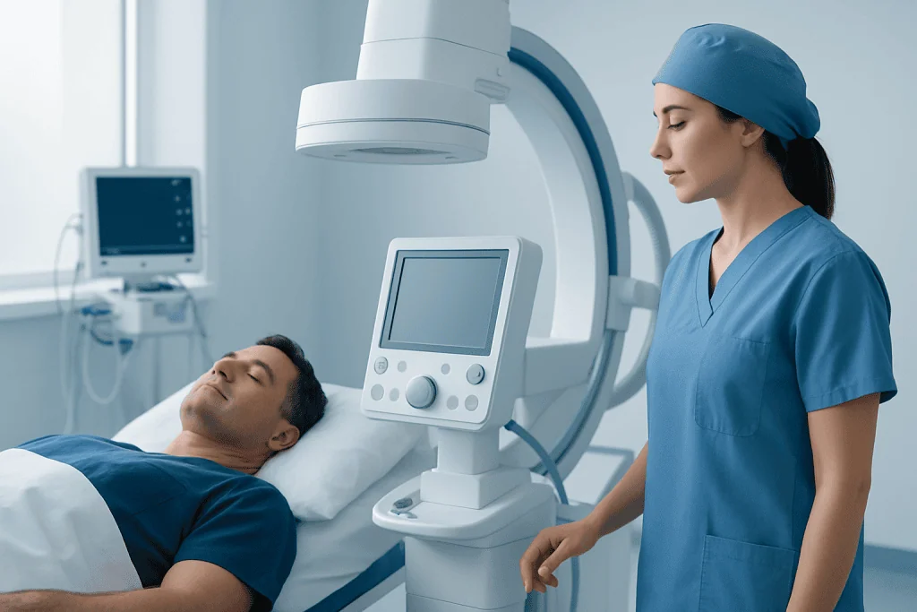
An elevated right hemidiaphragm is not always serious. Some people have no noticeable symptoms, stable imaging findings and normal or near-normal breathing tests. In these situations, a clinician may recommend observation and follow-up rather than active treatment.
However, it can be important when it is new, progressively worsening or associated with shortness of breath, reduced exercise tolerance, repeated chest infections, sleep-related breathing symptoms, chest discomfort or low oxygen levels. It also needs prompt evaluation when there are signs of an underlying problem involving the lungs, the phrenic nerve that controls the diaphragm, the spinal cord, or organs in the abdomen.
The level of concern is determined by the whole clinical picture rather than an X-ray appearance alone. A respiratory specialist, thoracic surgeon, neurologist or other relevant clinician may work together to clarify the cause and decide whether treatment is needed.
Causes, symptoms and candidacy for treatment

Possible causes include phrenic nerve injury or dysfunction, prior chest, heart, neck or upper abdominal surgery, trauma, viral or inflammatory nerve conditions, and some neurological disorders. Lung collapse or scarring can pull the diaphragm upward. Abdominal causes, such as enlargement beneath the diaphragm, may push it upward; on the right, the liver is often considered during assessment. In some cases, no definite cause is found.
Symptoms vary widely. A person may notice breathlessness during activity, difficulty lying flat, fatigue, reduced stamina, cough, disturbed sleep or recurrent respiratory infections. Symptoms may be more noticeable in people who already have asthma, chronic obstructive pulmonary disease, obesity, heart disease or weakness of the other side of the diaphragm.
People are usually considered for an intervention such as plication when imaging and functional testing support diaphragm weakness or paralysis, symptoms remain meaningful despite appropriate nonsurgical care, and the condition has been stable long enough to judge likely recovery. Surgery is not automatically appropriate for an elevated diaphragm caused by a temporary illness or a treatable abdominal or lung problem. Individual assessment is essential.
- Breathlessness that limits usual activities
- Persistent symptoms with documented diaphragm dysfunction
- Reduced lung function related to diaphragm position or movement
- A stable condition after a suitable period of observation, when spontaneous recovery appears unlikely
How do you fix an elevated hemidiaphragm?
There is no single treatment for every elevated hemidiaphragm. When an underlying condition is responsible, treatment focuses first on that cause. For example, care may involve managing a lung disorder, addressing an abdominal process, treating infection or inflammation when present, or reviewing whether a prior injury or surgery could have affected the phrenic nerve.
For mild or incidental elevation, clinicians may recommend monitoring with follow-up imaging, breathing tests and symptom review. Pulmonary rehabilitation, breathing exercises, physical activity adapted to the person’s abilities, weight management where appropriate, and treatment of coexisting lung or heart conditions can help reduce breathlessness and improve conditioning. These measures do not necessarily restore nerve function, but they can improve day-to-day function.
When one side of the diaphragm is persistently weak or paralyzed and causes disabling symptoms, diaphragm plication may be an option. During this operation, the surgeon tightens and flattens the loose, elevated diaphragm with sutures. This reduces its paradoxical upward movement and can create more room for the lung to expand. The procedure improves mechanics but does not directly repair a damaged nerve.
How diaphragm plication works: steps, benefits and risks
Before surgery, the care team reviews imaging, lung function, oxygen levels when needed, medical history and fitness for anesthesia. Tests may include chest X-ray, CT scanning, ultrasound of diaphragm movement, fluoroscopy in selected cases, and pulmonary function testing. The team also checks for causes that would be better treated in another way before recommending plication.
Plication is performed under general anesthesia. Depending on anatomy, surgical expertise and the individual situation, the surgeon may use an open chest incision, minimally invasive thoracoscopic surgery or a robotic-assisted approach. The elevated diaphragm is gently pulled taut and folded or tightened with permanent sutures. The surgical approach and expected hospital stay should be discussed with the treating team.
The potential benefits are improved lung expansion, less breathlessness and better activity tolerance for appropriately selected patients. As with any operation, risks include bleeding, infection, pain, blood clots, anesthesia-related complications, injury to nearby structures, fluid or air around the lung, and an incomplete improvement in symptoms. Existing heart or lung disease can also influence surgical risk and expected benefit.
Results are usually assessed through symptoms, activity tolerance and repeat breathing tests or imaging over time. Improvement may be meaningful but is not identical for everyone, particularly when breathlessness has more than one cause.
How long does it take for the diaphragm to heal?
Healing time depends on the reason the diaphragm is elevated. If the problem is related to a temporary phrenic nerve irritation or injury, nerve recovery may take months and occasionally longer. This is one reason clinicians may observe some cases before recommending surgery, provided symptoms are manageable and there is no urgent underlying cause.
After diaphragm plication, early recovery begins in the hospital and continues at home over several weeks. Pain control, gradual walking, breathing exercises and avoiding activities that strain the chest or abdomen are commonly part of recovery. The exact restrictions and timeline depend on the surgical technique, general health and the surgeon’s instructions.
Many people notice changes in breathing gradually as postoperative discomfort settles and activity levels increase. Full functional recovery may take several months. Follow-up appointments allow the care team to review wound healing, breathing symptoms, lung function and any need for pulmonary rehabilitation.
What is the prognosis for an elevated hemidiaphragm?
The prognosis for an elevated hemidiaphragm is often favorable, especially when a person has no symptoms or the underlying cause can be treated. Some cases remain stable without affecting everyday life. When diaphragm weakness follows a reversible nerve problem, movement may improve over time, although the pace and extent of recovery cannot be predicted precisely.
For people with long-standing unilateral paralysis and significant breathlessness, plication can improve respiratory mechanics and quality of life when careful evaluation confirms that the diaphragm is a major contributor to symptoms. It is important to have realistic expectations: surgery stabilizes the diaphragm position but may not eliminate breathlessness caused by other lung, heart, sleep or weight-related factors.
Prognosis is influenced by age, overall fitness, lung reserve, the health of the opposite diaphragm, the cause of nerve dysfunction and the presence of other medical conditions. Regular follow-up helps ensure that new symptoms or changes in function are addressed appropriately.
When to seek medical care
Medical assessment is advisable for new or unexplained shortness of breath, reduced exercise capacity, persistent cough, difficulty breathing when lying down, repeated chest infections or an elevated diaphragm newly reported on imaging. A clinician can determine whether the finding is likely incidental or whether additional tests are needed.
Urgent medical care is appropriate for severe or rapidly worsening breathing difficulty, chest pain, bluish lips or face, fainting, confusion, or symptoms occurring after major trauma or recent surgery. These symptoms can have several causes and should not be assumed to be due only to the diaphragm.
Acibadem International’s multidisciplinary specialists and JCI-accredited hospitals assess and treat diaphragm-related conditions for international patients, coordinating respiratory, thoracic surgical, imaging and rehabilitation care when appropriate.
Frequently asked questions
What does an elevated right hemidiaphragm mean?
It means the right half of the diaphragm appears higher than expected on a chest X-ray or other scan. Because the liver is beneath the right diaphragm, it normally sits a little higher than the left, but a more marked elevation can be related to diaphragm weakness, lung changes or conditions below the diaphragm.
Can an elevated right hemidiaphragm cause shortness of breath?
Yes, particularly if the diaphragm is weak or paralyzed and cannot move normally during breathing. Some people have no symptoms, while others notice breathlessness with exertion, when lying flat or when another lung or heart condition is also present.
Can an elevated hemidiaphragm go back to normal?
It can improve when the cause is temporary or reversible, such as some forms of phrenic nerve irritation or a treatable lung or abdominal condition. If there is permanent nerve damage, the elevation may persist, although symptoms can still be managed.
Is diaphragm plication a cure for diaphragm paralysis?
Diaphragm plication does not repair the phrenic nerve or restore normal nerve signaling. It stabilizes and flattens the elevated diaphragm to improve lung expansion and reduce symptoms in selected people with persistent unilateral dysfunction.
How is diaphragm weakness diagnosed?
Diagnosis usually combines symptoms, physical examination, chest imaging and pulmonary function tests. Ultrasound or other dynamic tests may be used to evaluate diaphragm movement, while CT or other studies can help identify causes in the chest or abdomen.
Can breathing exercises lower an elevated diaphragm?
Breathing exercises generally do not physically lower a persistently elevated diaphragm caused by paralysis. However, pulmonary rehabilitation and guided breathing strategies may improve conditioning, breathing efficiency and confidence with activity.
References
- American Thoracic Society
- European Respiratory Society
- National Heart, Lung, and Blood Institute
- Society of Thoracic Surgeons
This article is for general information only and is not a substitute for professional medical advice. Please consult a qualified doctor about your individual situation.
Explore treatments in Turkey — costs, top hospitals & a free quote
JCI-accredited · board-certified surgeons · reply within 24h
Add us as a Preferred Source to see more of our trusted health content across Google Search, AI Overviews and Discover.