Endobronchial Ultrasound Biopsy: What to Expect Before and After
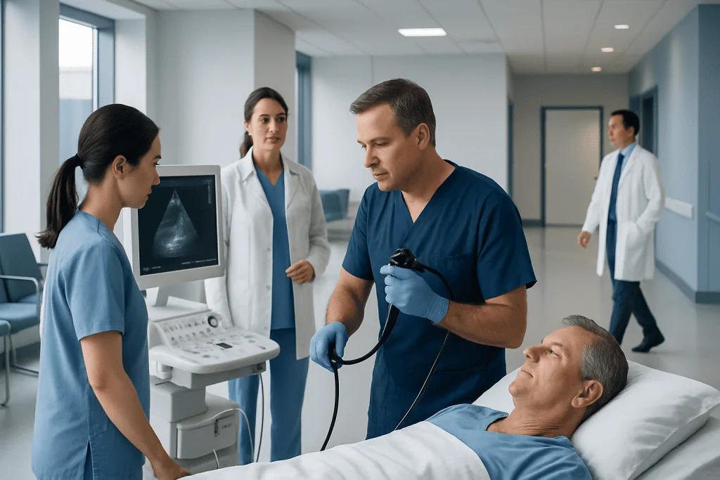
Endobronchial ultrasound biopsy, often called EBUS, combines bronchoscopy and ultrasound to guide tissue sampling. It is commonly used to evaluate enlarged lymph nodes, lung abnormalities, infections, inflammation, and suspected cancer.
Key Takeaways
- Endobronchial ultrasound biopsy, often called EBUS, combines bronchoscopy and ultrasound to guide tissue sampling.
- It is commonly used to evaluate enlarged lymph nodes, lung abnormalities, infections, inflammation, and suspected cancer.
- Most people go home the same day after sedation or anesthesia and recover quickly.
- Mild sore throat, cough, or small amounts of blood-tinged mucus can happen briefly after the test.
- Results from tissue analysis help doctors confirm a diagnosis and plan the next steps in care.
Endobronchial ultrasound biopsy is a minimally invasive procedure used to collect tissue samples from lymph nodes or areas near the lungs and airways. It helps doctors diagnose conditions and guide treatment while avoiding more invasive surgery in many cases.
Overview of Endobronchial Ultrasound Biopsy
Endobronchial ultrasound biopsy is a procedure that allows doctors to look inside the airways and obtain tissue samples using real-time ultrasound guidance. It is often called EBUS biopsy. During the procedure, a thin flexible tube with a camera and ultrasound probe is passed through the mouth into the windpipe and bronchial tubes. The ultrasound helps locate lymph nodes or other structures next to the airways, and a fine needle is used to collect samples.
This technique is especially useful because many important lymph nodes and tissues in the chest sit close to the airways. In the past, reaching some of these areas often required a more invasive surgical procedure. With EBUS, doctors can often obtain the needed tissue through the natural airway route, which can mean less discomfort, a shorter recovery, and fewer risks for the patient.
Endobronchial ultrasound biopsy is commonly used when imaging tests such as chest X-ray, CT, or PET scan show enlarged lymph nodes or suspicious areas. It may help diagnose infection, inflammation, lung cancer, or the spread of cancer from another part of the body. In many cases, it is an important step in deciding the most appropriate treatment plan.
Why Doctors Recommend It
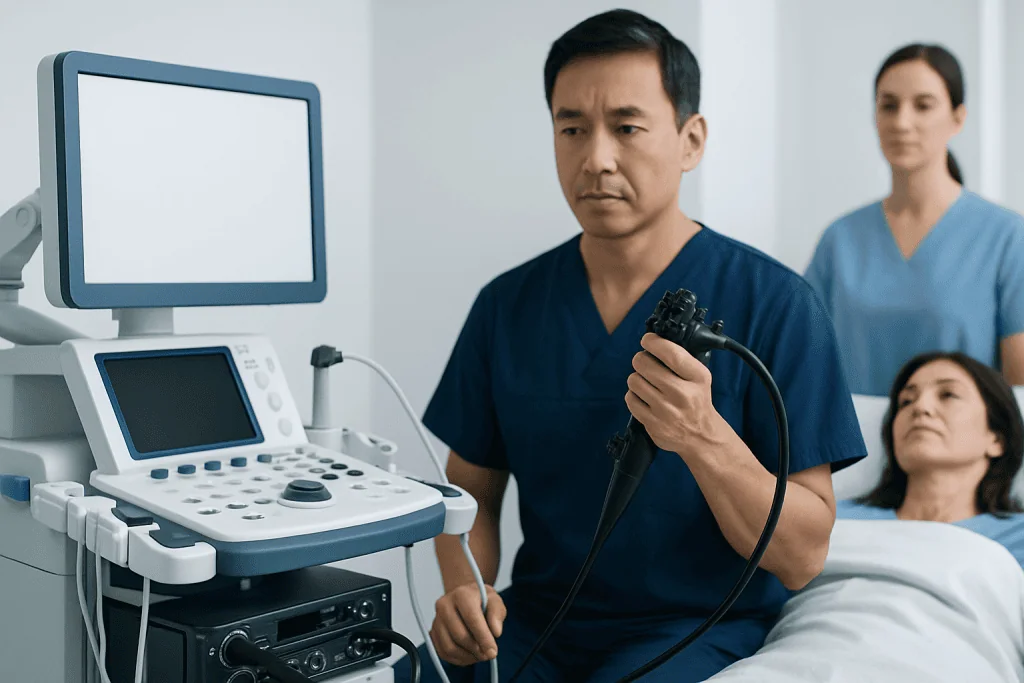
Doctors recommend endobronchial ultrasound biopsy when they need a tissue diagnosis from lymph nodes or structures in the middle of the chest, also called the mediastinum, or from areas near the large airways. Imaging can show that something is abnormal, but only a tissue sample can often confirm exactly what is causing the change.
The procedure may be used for several reasons. It can help determine whether enlarged lymph nodes are due to infection, inflammatory conditions such as sarcoidosis, or cancer. It is also frequently used to stage lung cancer by checking whether cancer cells have spread to nearby lymph nodes. This information is important because treatment can differ greatly depending on the stage.
In some people, EBUS is used after an abnormal scan or persistent symptoms such as unexplained cough, weight loss, or coughing up blood. In others, it helps clarify findings found during follow-up of a known condition. When tissue is needed, EBUS may be preferred over surgery because it is less invasive while still providing valuable diagnostic information.
What to Expect Before the Procedure

Preparation usually begins with a medical review. The care team asks about current symptoms, allergies, medicines, previous reactions to anesthesia, and any heart or lung conditions. Blood-thinning medicines may need special instructions, because they can increase the risk of bleeding during a biopsy. Patients should only stop or adjust these medicines if their doctor advises it.
Most people are asked not to eat or drink for several hours before the test. The exact timing depends on the hospital and the type of sedation or anesthesia planned. The doctor may also request blood tests, breathing assessment, or imaging review before the procedure. It is usually important to arrange for someone to drive the patient home, as sedative medicines can affect alertness for the rest of the day.
Before the biopsy starts, the team explains the process and asks for consent. A small intravenous line is placed for medications. The throat may be numbed with a local anesthetic spray, and sedation or general anesthesia may be given depending on the person, the hospital protocol, and how extensive the sampling needs to be. Many patients are understandably nervous, but the procedure is routinely performed and closely monitored throughout.
How the Biopsy Is Performed
During endobronchial ultrasound biopsy, the doctor inserts the bronchoscope through the mouth and gently advances it into the airways. The camera provides a direct view inside the breathing passages, while the ultrasound probe shows structures just outside the airway walls. This combined view helps the doctor target the correct area with precision.
Once the lymph node or area of interest is identified, a very fine needle is passed through the bronchoscope and through the airway wall to collect small tissue or cell samples. Several passes may be made from the same site or from more than one lymph node to improve the chance of an accurate diagnosis. The samples are then sent to a laboratory for detailed examination.
The procedure often takes less than an hour, although timing can vary. Throughout the test, the patient’s oxygen level, heart rate, and blood pressure are monitored. In some centers, EBUS may be performed as part of a broader bronchoscopy evaluation, and the team may combine visual inspection of the airways with ultrasound-guided sampling to gather the most useful information in one session.
What to Expect After the Procedure
After the biopsy, the patient is moved to a recovery area for observation until the effects of sedation or anesthesia begin to wear off. The throat may feel numb for a short time, so eating and drinking are usually delayed until it is safe to swallow normally. Mild throat discomfort, hoarseness, cough, or tiredness can happen for a day or so and usually improve on their own.
Some people notice a small amount of blood-tinged sputum after the procedure. This can be expected for a short time, especially after a biopsy, but it should not be heavy or persistent. Most patients go home the same day with instructions about rest, medicines, diet, and warning signs to watch for.
Biopsy results are not usually available immediately because the samples need to be examined in the laboratory. Depending on the tests required, results may take a few days or longer. Once available, they can guide the next steps, which may include follow-up imaging, medication, referral to a specialist, or further care such as lung cancer treatment if a cancer diagnosis is confirmed.
Benefits, Risks, and Limitations
The main benefit of endobronchial ultrasound biopsy is that it can provide important diagnostic information without the need for open surgery in many cases. It allows doctors to sample lymph nodes and nearby chest structures accurately, often with a shorter recovery time and less discomfort than more invasive approaches. It is especially valuable for diagnosing and staging lung-related conditions.
Like any medical procedure, EBUS does have risks, although serious complications are uncommon. Possible risks include bleeding, infection, temporary low oxygen levels, sore throat, fever, or reactions to sedative or anesthetic medicines. In rare cases, breathing problems or other complications may require additional treatment or observation.
There are also some limitations. A biopsy may occasionally collect too little tissue, or the sample may not fully explain the abnormal finding. If that happens, doctors may recommend repeating the procedure or using another test such as mediastinoscopy to obtain a larger sample. The best approach depends on the location of the abnormality and the question the medical team is trying to answer.
Recovery, Self-care, and When to Seek Medical Advice
Recovery after endobronchial ultrasound biopsy is usually straightforward. Most people are advised to rest for the remainder of the day and avoid driving, alcohol, or important decisions until the effects of sedation have fully worn off. Drinking fluids and returning gradually to normal eating can help once swallowing feels comfortable again.
It is sensible to follow all discharge instructions carefully, especially regarding regular medicines. If blood thinners were paused before the test, the doctor will explain when to restart them. Patients should also keep their follow-up appointment so the team can review biopsy results and explain what they mean in clear terms. Sometimes the results are reassuring; other times they help identify a condition that can then be treated more precisely.
Medical advice should be sought promptly if there is worsening shortness of breath, chest pain, heavy bleeding, fever that does not settle, difficulty swallowing, or severe weakness after the procedure. While these problems are not common, they deserve medical attention. For patients seeking coordinated care, Acibadem International’s multidisciplinary specialists and JCI-accredited hospitals diagnose and treat chest and airway conditions for international patients, including further evaluation when biopsy results need additional follow-up.
Frequently asked questions
Is endobronchial ultrasound biopsy painful?
Most patients do not feel pain during the procedure because sedation or anesthesia is used, and the throat is often numbed. Afterward, mild sore throat, hoarseness, or cough can occur, but these symptoms are usually temporary and manageable.
How long does an EBUS biopsy take?
The procedure itself often takes less than an hour, although preparation and recovery add more time to the hospital visit. The total stay may be several hours, especially if sedation or anesthesia is used.
Why is a biopsy needed if a scan already showed a problem?
Scans can show that an area looks abnormal, but they usually cannot confirm the exact cause. A biopsy provides cells or tissue that a pathologist can examine to determine whether the problem is due to infection, inflammation, cancer, or another condition.
When will the biopsy results be ready?
Results are often available within a few days, but timing can vary depending on the type of analysis needed. Some samples need additional laboratory tests, which may take longer before the doctor can give a final answer.
Can endobronchial ultrasound biopsy diagnose lung cancer?
Yes, it can help diagnose lung cancer and is also commonly used to check whether cancer has spread to nearby lymph nodes. This information is important for staging the disease and planning treatment.
What symptoms after the procedure should not be ignored?
Patients should contact a doctor if they develop heavy bleeding, worsening shortness of breath, chest pain, persistent fever, or severe weakness. These symptoms are not expected and may need prompt medical assessment.
References
- American Thoracic Society
- European Respiratory Society
- National Cancer Institute
- National Health Service
- American College of Chest Physicians
This article is for general information only and is not a substitute for professional medical advice. Please consult a qualified doctor about your individual situation.
Ultrasound in Turkey — costs, top hospitals & a free quote
JCI-accredited · board-certified surgeons · reply within 24h
Add us as a Preferred Source to see more of our trusted health content across Google Search, AI Overviews and Discover.

Check-up & Preventive Medicine
Comprehensive health screenings designed for early detection and prevention.
3 specialists in this unit