Endovascular Treatment for Brain AVM: Options, Risks, and Treatment Goals

A brain AVM is an abnormal tangle of blood vessels connecting arteries and veins without the usual capillaries. Endovascular treatment usually involves embolization, which blocks part or all of the AVM through a catheter.
Key Takeaways
- A brain AVM is an abnormal tangle of blood vessels connecting arteries and veins without the usual capillaries.
- Endovascular treatment usually involves embolization, which blocks part or all of the AVM through a catheter.
- Treatment goals vary and may include lowering bleeding risk, reducing symptoms, or preparing for surgery or radiosurgery.
- Not every AVM needs the same approach; decisions depend on size, location, bleeding history, and the person’s overall health.
- Possible risks include stroke, bleeding, vessel injury, and incomplete closure of the AVM.
- Care is best planned by a multidisciplinary team with expertise in interventional neurology, neurosurgery, and radiation treatment.
Endovascular treatment for brain AVM is a minimally invasive procedure that blocks abnormal blood vessels from inside the bloodstream. It may be used on its own in selected cases, but more often it helps make surgery or radiosurgery safer and more effective.
Overview of Brain AVM and Endovascular Treatment
A brain arteriovenous malformation, or AVM, is a tangle of abnormal blood vessels in which arteries connect directly to veins. In normal circulation, capillaries slow blood flow and help exchange oxygen and nutrients. In an AVM, that step is missing, so blood can move quickly through fragile vessels. This may increase the risk of bleeding and can also affect nearby brain tissue.
Endovascular treatment for brain AVM is a minimally invasive procedure performed from inside the blood vessels. A specialist guides a thin catheter, usually through an artery in the wrist or groin, toward the vessels feeding the AVM. Then a blocking material, often called an embolic agent, is delivered to reduce or stop blood flow in the abnormal vessels. This approach is commonly referred to as brain AVM embolization.
Embolization may be used for different reasons. In some people, it is part of a staged treatment plan before open surgery or radiosurgery. In selected situations, it may be used to treat a specific weak point within the AVM, such as an associated aneurysm, or to reduce symptoms related to high blood flow. Complete cure with embolization alone is possible in some cases, but it is not the expected outcome for every AVM.
Because every AVM is different, treatment planning is individualized. Doctors consider the AVM’s size, shape, location, the vessels feeding it, whether bleeding has already occurred, and the person’s symptoms and general health. A multidisciplinary team often reviews these details together to recommend the safest and most appropriate option.
Symptoms and Why Treatment May Be Considered
Some brain AVMs cause no symptoms and are found by chance during imaging for another reason. Others may cause headaches, seizures, weakness, numbness, vision changes, balance problems, or trouble with speech, depending on where the AVM is located. In some cases, the first sign is bleeding in or around the brain, which can lead to sudden severe headache or neurological symptoms.
Treatment may be considered for several reasons. One goal may be to lower the chance of future bleeding, especially if the AVM has already ruptured. Another goal may be to reduce blood flow to relieve symptoms, protect nearby brain tissue, or address a specific high-risk feature seen on imaging. When an AVM is large or complex, embolization may help simplify it before another treatment.
Not every AVM needs immediate intervention. For some unruptured AVMs, the risks of treatment may outweigh the likely benefit, particularly if the AVM is in a highly sensitive area of the brain. In those situations, careful observation with follow-up imaging and symptom management may be the best approach.
People often hear the term brain AVM during evaluation, but the practical question is whether treatment would improve safety or quality of life overall. That decision is made by balancing the natural risks of the AVM against the short- and long-term risks of treatment.
How Endovascular Embolization Works

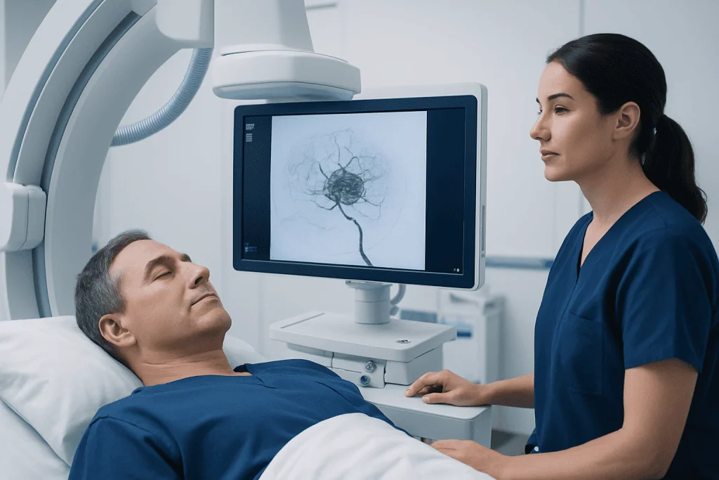
Brain AVM embolization is usually performed in a specialized angiography suite under imaging guidance. During the procedure, the doctor threads a microcatheter into the arteries that feed the AVM. Through this catheter, an embolic material is injected to block abnormal vessels. Different materials may be used depending on the AVM’s structure and the treatment goal.
The aim is not always to close the entire AVM in one session. For larger or more complex AVMs, treatment may be staged over more than one procedure to improve safety. Embolization can reduce blood flow, shrink the nidus of the AVM, close dangerous weak spots, or make later surgery less bloody and easier to perform.
Before the procedure, patients usually have detailed imaging such as MRI, CT, and cerebral angiography. Angiography is especially important because it shows the exact anatomy of the feeding arteries, draining veins, and any associated aneurysms. This information helps the team decide whether embolization is appropriate and what result is realistically achievable.
After embolization, some people go on to brain surgery or Gamma Knife radiosurgery as part of a combined plan. Others may only need monitoring if the treatment goal was symptom control or reduction of a specific bleeding risk rather than complete cure.
Treatment Goals and Who May Benefit
The treatment goals for a brain AVM are not the same for every person. In general, doctors may aim to completely eliminate the AVM, reduce the risk of bleeding, treat a related aneurysm, improve seizure control in selected patients, or prepare the AVM for another treatment. A clear treatment goal is important because it shapes both the procedure and the expected outcome.
People who may benefit from endovascular treatment include those with AVMs that have bled, those with high-risk vascular features, and those whose AVM can be made safer for surgery or radiosurgery through embolization. It may also be useful when there is a deep feeder vessel that is difficult to reach during open surgery. In carefully selected cases with favorable anatomy, embolization alone may offer durable closure.
However, not all AVMs are suitable for endovascular treatment. If the feeding arteries are too small or tortuous, or if the AVM is supplied by vessels that also nourish critical areas of the brain, the risk may be too high. Likewise, some AVMs are better treated with observation, microsurgery, or focused radiation depending on their location and overall risk profile.
Multidisciplinary review is especially helpful in complex cases. Specialists in interventional neurology, neurosurgery, neurology, and radiation oncology can compare the relative benefits of embolization, surgery, focused radiosurgery, or observation and then tailor care to the individual.
Risks, Limitations, and Recovery
As with any procedure involving the brain and blood vessels, endovascular treatment for brain AVM has risks. Possible complications include stroke, bleeding during or after the procedure, blockage of a normal artery, vessel injury, swelling in the brain, seizures, infection, and reactions related to contrast dye or anesthesia. The exact risk varies depending on the AVM’s anatomy, the treatment goal, and the person’s overall health.
Another important limitation is that embolization may not completely cure the AVM. Even when blood flow is significantly reduced, some abnormal vessels may remain open or reopen over time. For this reason, follow-up imaging is essential, and some patients need additional treatment sessions or another therapy such as surgery or radiosurgery.
Recovery is often shorter than with open surgery, but it still requires observation. Patients are usually monitored in the hospital so the team can watch for neurological changes, headache, blood pressure issues, or bleeding. Temporary fatigue, mild groin or wrist discomfort, or headache can occur after the procedure.
Many people can gradually return to usual activities based on their doctor’s advice, but recovery depends on whether the AVM had bled beforehand and whether other treatments are planned. It is important to attend follow-up appointments and report new symptoms promptly, especially sudden severe headache, weakness, confusion, or seizure activity.
Diagnosis, Planning, and Follow-Up
Accurate diagnosis is the foundation of safe AVM treatment. Initial tests often include CT or MRI scans to look at the brain and identify bleeding or structural changes. The most detailed test is cerebral angiography, which maps the blood vessels in real time and remains the key study for confirming the anatomy of an AVM.
During planning, the team looks at several factors: the AVM’s size, whether it is superficial or deep, the number and type of feeding arteries, the pattern of venous drainage, and whether there are associated aneurysms. Doctors also consider age, symptoms, prior bleeding, seizure history, and any other medical conditions that could affect safety.
Follow-up is an important part of treatment. After embolization, repeat angiography or other imaging may be needed to confirm how much of the AVM has been closed and whether blood flow has changed in a meaningful way. If the person proceeds to surgery or radiosurgery, follow-up also helps coordinate timing and assess the result.
Long-term monitoring may continue even after successful treatment, especially in younger patients or in cases where complete cure was uncertain. If symptoms such as seizures are present, neurologists may also help manage medicines and recovery needs during this period.
Questions to Ask and When to Seek Medical Care
When discussing treatment, patients may find it helpful to ask what the main goal of embolization is in their case. They can also ask whether the AVM is expected to be cured, reduced, or prepared for another treatment, what the short-term and long-term risks are, and what kind of follow-up imaging will be required. Understanding these points can make decision-making clearer and less overwhelming.
Urgent medical care is needed if symptoms suggest bleeding in the brain. Warning signs include a sudden severe headache, weakness on one side, difficulty speaking, confusion, seizures, fainting, or sudden vision changes. These symptoms should not be ignored, whether or not a person has already been diagnosed with an AVM.
Even without emergency symptoms, medical review is important for new seizures, persistent headaches, progressive neurological symptoms, or concerns after a recent procedure. People who have been told they have an AVM but are unsure about the best next step may benefit from consultation at a center with experience in arteriovenous malformations and advanced imaging.
For international patients seeking specialist evaluation, Acibadem International offers multidisciplinary assessment and treatment through JCI-accredited hospitals, with teams experienced in interventional neurology, neurosurgery, and radiosurgical care.
Frequently asked questions
Is endovascular treatment for brain AVM a surgery?
It is considered a minimally invasive procedure rather than open brain surgery. The doctor reaches the AVM through blood vessels using a catheter, usually inserted at the wrist or groin. Even so, it is still a specialized treatment that carries important risks and requires expert planning.
Can embolization completely cure a brain AVM?
Sometimes, but not always. In selected AVMs with favorable anatomy, embolization alone may fully close the malformation. More commonly, it is used to reduce blood flow or make another treatment, such as surgery or radiosurgery, safer and more effective.
What are the main risks of brain AVM embolization?
The main risks include stroke, bleeding, vessel injury, brain swelling, and incomplete treatment of the AVM. The risk level depends on the AVM’s size, location, blood supply, and the treatment goal. A specialist can explain how these risks apply to an individual case.
How long does recovery take after endovascular treatment?
Recovery varies from person to person. Many patients spend a short period in the hospital for monitoring and then gradually return to normal activities, depending on symptoms and whether other treatments are planned. Recovery may take longer if the AVM has already bled or if there are neurological symptoms.
Will a person still need surgery after embolization?
Possibly. Embolization is often one step in a broader treatment plan and may be followed by surgery or radiosurgery. Whether more treatment is needed depends on how much of the AVM remains and what the original treatment goal was.
How do doctors decide whether to treat an unruptured AVM?
Doctors weigh the natural risk of the AVM against the risks of treatment. They consider factors such as prior bleeding, symptoms, location in the brain, size, vessel anatomy, and the person’s age and overall health. In some cases, careful observation is safer than intervention.
References
- World Health Organization
- National Institute of Neurological Disorders and Stroke
- American Stroke Association
- Society of NeuroInterventional Surgery
- Radiological Society of North America
This article is for general information only and is not a substitute for professional medical advice. Please consult a qualified doctor about your individual situation.
Explore treatments in Turkey — costs, top hospitals & a free quote
JCI-accredited · board-certified surgeons · reply within 24h
Add us as a Preferred Source to see more of our trusted health content across Google Search, AI Overviews and Discover.