Ependymoma on MRI: Preparation, Procedure and Results
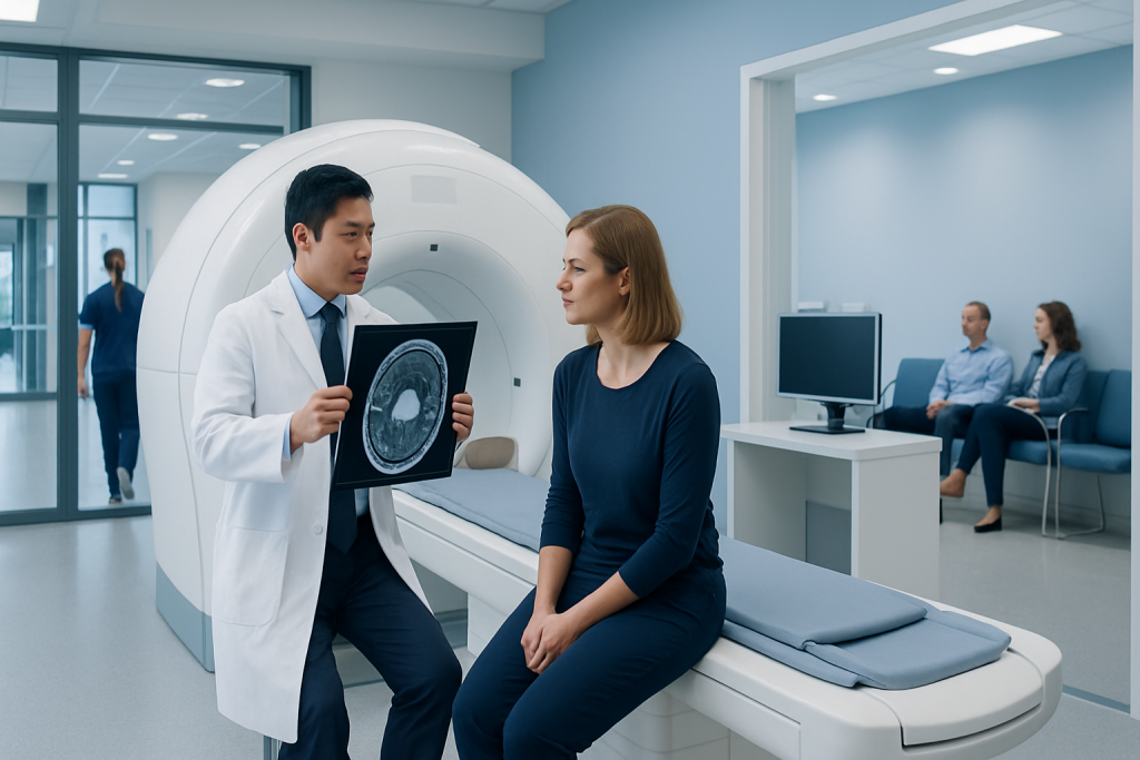
MRI is the main imaging test used to evaluate a suspected ependymoma in the brain or spinal cord. Contrast-enhanced MRI helps clinicians assess tumor boundaries, internal features and possible spread.
Key Takeaways
- MRI is the main imaging test used to evaluate a suspected ependymoma in the brain or spinal cord.
- Contrast-enhanced MRI helps clinicians assess tumor boundaries, internal features and possible spread.
- An MRI scan is painless and uses no ionizing radiation, but some people need support for claustrophobia or contrast safety assessment.
- Ependymoma may be benign-appearing or malignant in behavior; pathology testing is needed for a definitive diagnosis.
- Surgery is often the first treatment when it can be performed safely, sometimes followed by radiotherapy.
- Follow-up MRI is important because ependymoma can recur, sometimes years after initial treatment.
Ependymoma on MRI is assessed using detailed magnetic images of the brain or spine to identify a suspected tumor, describe its features and guide next steps. MRI can strongly support diagnosis and treatment planning, but tissue examination after biopsy or surgery is usually needed to confirm the tumor type and grade.
Overview: What ependymoma on MRI can show
Ependymoma on MRI refers to the appearance of an ependymoma—a tumor arising from ependymal cells that line fluid-filled spaces in the brain and the central canal of the spinal cord—on magnetic resonance imaging. MRI provides high-detail images of soft tissues and is the preferred scan for evaluating a suspected tumor in the brain or spine. It can show where the lesion is located, its size, its relationship to nearby structures and whether there are features that require prompt specialist assessment.
Ependymomas occur in both children and adults, although their usual locations differ by age. In children, they more often arise in the brain, especially around the fourth ventricle in the back of the brain. In adults, they are commonly found in the spinal cord. MRI findings can suggest ependymoma, but they cannot confirm it with certainty. A neuropathologist examines tumor tissue obtained during surgery or biopsy to establish the exact diagnosis and tumor classification.
The scan also plays an essential role beyond diagnosis. It helps neurosurgeons plan the safest approach, helps oncologists decide whether further treatment may be appropriate and provides a baseline for later follow-up scans. A multidisciplinary assessment is especially important because treatment decisions depend on the tumor location, symptoms, age, surgical options and pathology results.
How MRI works and who may need it
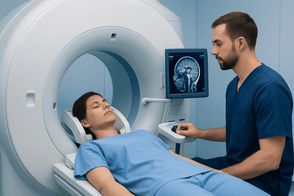
MRI uses a strong magnetic field and radiofrequency waves to create detailed images. Unlike X-rays and CT scans, it does not use ionizing radiation. For suspected ependymoma, imaging commonly includes MRI of the brain or spinal cord with and without intravenous contrast. The contrast agent, usually gadolinium-based, can make areas with altered blood-brain barrier or increased blood supply more visible.
A doctor may request MRI when a person has symptoms that could be caused by pressure on the brain, obstruction of cerebrospinal fluid flow or involvement of the spinal cord. Symptoms vary by location and may include persistent or worsening headache, nausea, balance changes, double vision, weakness, sensory changes, back or neck pain, or changes in bladder or bowel function. These symptoms are common in many conditions and do not necessarily indicate a tumor.
MRI may not be suitable without special precautions for people with some implanted medical devices, retained metal fragments or severe kidney impairment when contrast is being considered. Many modern implants are MRI-compatible, but the imaging team must verify the device details. Pregnancy, previous contrast reactions and kidney disease should also be discussed before the appointment.
- MRI may be used to investigate a new neurological symptom pattern.
- It is used to assess a lesion found on another imaging test.
- It helps plan surgery and radiotherapy when an ependymoma is suspected or confirmed.
- It supports long-term surveillance after treatment.
Preparing for an ependymoma MRI
Most people can eat, drink and take regular medicines as usual before a standard MRI. The imaging center will provide specific instructions, particularly if sedation is planned or contrast will be used. It is important to tell the team about kidney problems, allergies, pregnancy, implanted devices, previous surgeries and any possibility of metal in the body from work or injury.
Before entering the scan room, the person removes metal items such as jewelry, watches, hearing aids, removable dental devices, belts and clothing with metallic parts. The radiographer may ask screening questions more than once because MRI magnets are powerful and safety checks are essential. A hospital gown may be provided.
Some people feel uneasy in enclosed spaces or find it difficult to remain still because of pain, age or anxiety. The care team can discuss practical measures such as music, communication through an intercom, a support person where permitted, or prescribed medication. Children and some adults may need sedation or anesthesia so that the images are clear and the experience is as comfortable as possible.
The procedure: step by step and what to expect
During the scan, the person lies on a padded table that moves slowly into the MRI scanner. For a brain MRI, a lightweight head coil is placed around the head; for a spine MRI, a coil is positioned under or around the relevant area. Cushions and straps may be used to support comfort and reduce movement. The radiographer remains in contact throughout the examination and can see and hear the patient.
The scanner makes rhythmic knocking, tapping or humming sounds while images are acquired. Earplugs or headphones are provided. It is important to stay as still as possible, as even small movements can blur images and may mean that a sequence needs to be repeated. The scan itself is painless, although the table can feel firm and lying still may be tiring.
If contrast is needed, a small intravenous cannula is placed in the arm or hand. Images are usually taken before and after contrast administration. The entire appointment often takes between 30 and 60 minutes, though timing varies according to the area being scanned, the number of sequences needed and whether both brain and spine imaging are required.
In some cases, clinicians recommend MRI of the entire neuraxis—the brain and the full spine—because ependymoma cells can occasionally spread through cerebrospinal fluid pathways. This is not needed for every person, but it may be considered depending on the initial tumor location, pathology and treatment stage.
What are the MRI findings of ependymomas?
Ependymomas can have varied MRI appearances, partly because they occur in different parts of the central nervous system. In the brain, an ependymoma may appear as a mass near a ventricle, often the fourth ventricle. It can extend through normal openings of the ventricle into nearby spaces. In the spinal cord, it commonly appears as a centrally located, well-defined mass that expands the spinal cord over one or more spinal levels.
On MRI, the tumor is often different in signal from normal brain or spinal cord tissue on T1- and T2-weighted images. It may enhance after contrast administration, but the enhancement can be uneven. Larger tumors may contain cyst-like areas, calcification or prior bleeding, which can create a mixed internal appearance. Associated swelling, fluid buildup in the brain or a fluid cavity within the spinal cord may also be visible.
These patterns are useful clues, but no individual MRI feature proves that a tumor is an ependymoma. Other tumors and noncancerous conditions can sometimes look similar. Radiologists interpret the scan together with the person’s age, symptoms, location of the lesion and other imaging characteristics. Confirmation generally requires pathological examination of tissue.
Results, diagnosis and the meaning of cancerous
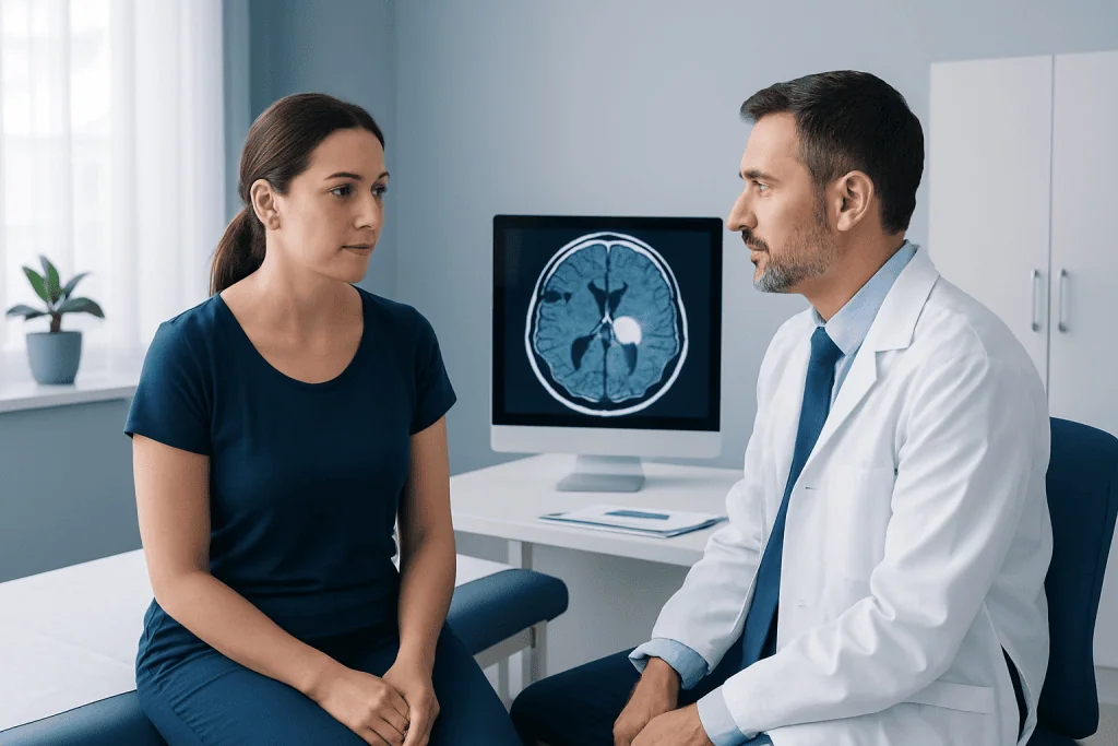
A radiologist reviews the MRI images and sends a report to the referring doctor. The report generally describes the lesion’s location, dimensions, enhancement pattern, effects on surrounding tissue and whether there are signs of blocked cerebrospinal fluid flow or spinal cord compression. If the scan raises concern for a tumor, referral to neurology, neurosurgery, neuro-oncology or a specialized tumor team may follow.
Is ependymoma cancerous? Ependymoma is a central nervous system tumor that can vary in biological behavior. Some tumors grow relatively slowly, while others are more likely to grow, recur or spread within the central nervous system. Rather than relying only on the word “cancer,” specialists use the tumor’s pathological classification, molecular features, location and extent of surgical removal to estimate outlook and recommend treatment.
Imaging alone cannot determine the full biological behavior of an ependymoma. Following surgical removal or biopsy, pathology testing identifies the tumor type and may include molecular testing that provides more precise information. This detailed assessment supports individualized treatment planning and follow-up.
For related information about tumors affecting the nervous system, readers can explore brain tumor information if available through their care pathway.
Treatment options, recovery and follow-up imaging
Is it possible to remove an ependymoma? In many cases, surgery is the main initial treatment and the goal is to remove as much tumor as can be done safely. Whether complete removal is possible depends on the tumor’s position and how closely it involves important brain, spinal cord or nerve structures. The surgical team balances tumor removal with protecting functions such as movement, sensation, speech, swallowing and balance.
Radiotherapy may be recommended after surgery, especially when there is remaining tumor, when pathology suggests a higher risk of recurrence or when the clinical team believes it will improve local tumor control. Chemotherapy may be used in selected situations, particularly in children or recurrent disease, but its role varies. The appropriate plan is made by a multidisciplinary team that includes neurosurgeons, neuro-oncologists, radiation oncologists, neuroradiologists and rehabilitation specialists.
Recovery after an MRI is immediate for most people. If contrast was used, normal activities can usually resume the same day unless the doctor advises otherwise. Recovery after surgery is more variable and may involve a hospital stay, symptom management and rehabilitation for balance, strength, coordination or other neurological needs. Follow-up MRI is scheduled at intervals determined by the treatment team.
Does ependymoma always come back? No. Some people remain free of tumor progression after treatment, particularly when the tumor can be fully removed and treated appropriately. However, ependymoma can recur, and recurrence may occur locally or, less commonly, elsewhere along the brain and spinal cord pathways. Regular follow-up imaging is therefore important even when a person feels well.
Acibadem International’s multidisciplinary specialists in JCI-accredited hospitals diagnose and treat central nervous system tumors for international patients, coordinating imaging, pathology, surgery and follow-up according to individual clinical needs.
When to seek medical care
A person should arrange medical assessment for persistent, progressive or unexplained neurological symptoms, especially if they interfere with daily activities. Examples include a new pattern of severe or worsening headaches, repeated vomiting without a clear cause, changes in balance, vision changes, weakness, numbness, seizures, persistent neck or back pain, or new bladder or bowel changes.
Urgent medical attention is needed for sudden severe headache, a first seizure, rapidly worsening weakness, loss of consciousness, severe confusion or new loss of bladder or bowel control with weakness or numbness. These symptoms can have several possible causes, but prompt assessment is important.
People who have been treated for ependymoma should contact their care team if new neurological symptoms develop or if they have concerns between planned follow-up appointments. Keeping scheduled MRI appointments helps clinicians identify changes early and discuss appropriate next steps calmly and clearly.
Frequently asked questions
Can MRI diagnose ependymoma by itself?
MRI can identify features that make ependymoma a possibility and shows the tumor's location and extent in detail. However, MRI cannot usually confirm the diagnosis alone. Examination of tumor tissue by a neuropathologist is generally needed for confirmation and classification.
Does an ependymoma always enhance with MRI contrast?
Many ependymomas show contrast enhancement, but the degree and pattern can vary. Enhancement may be uneven, particularly when a tumor has cystic areas, calcification or bleeding. A lack of a typical enhancement pattern does not by itself rule in or rule out ependymoma.
How long does an MRI for suspected ependymoma take?
A brain or spine MRI commonly takes around 30 to 60 minutes, though the time varies by protocol. Imaging of both the brain and entire spine may take longer. The radiology team can provide a more specific estimate before the appointment.
Is MRI contrast safe?
Gadolinium-based contrast agents are widely used and are generally well tolerated when clinically appropriate. The team checks kidney function, previous reactions and other health factors before contrast is given. Serious reactions are uncommon, but patients should report any prior contrast reaction or kidney condition.
Why might the whole spine be scanned after an ependymoma diagnosis?
Ependymoma can occasionally spread through cerebrospinal fluid pathways within the brain and spinal canal. MRI of the full spine can help check for additional areas requiring attention. Whether this imaging is needed depends on the individual tumor type, location and clinical plan.
How often is MRI needed after ependymoma treatment?
The follow-up schedule varies according to age, tumor location, pathology results, treatment received and previous scan findings. Scans are often more frequent in the earlier period after treatment and may continue long term. The treating specialist will set a surveillance plan tailored to the individual.
References
- World Health Organization
- National Cancer Institute
- American Cancer Society
- European Association of Neuro-Oncology
- RadiologyInfo.org
This article is for general information only and is not a substitute for professional medical advice. Please consult a qualified doctor about your individual situation.
Joint and spine care in Turkey — expert assessment & treatment
JCI-accredited · board-certified surgeons · reply within 24h
Add us as a Preferred Source to see more of our trusted health content across Google Search, AI Overviews and Discover.