Ewing Sarcoma Xray: Preparation, Procedure and Results
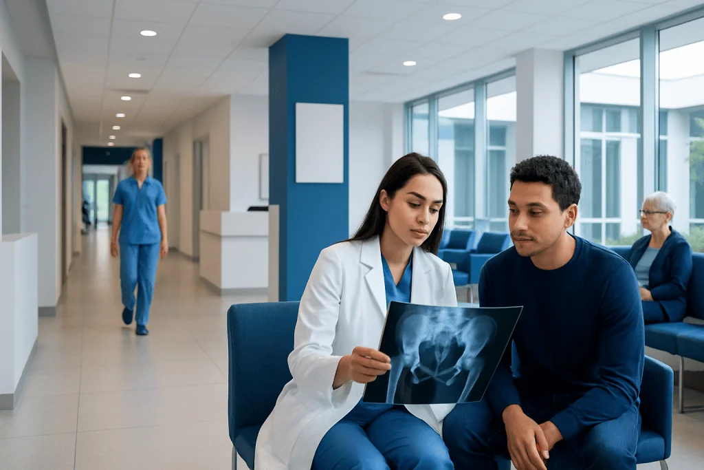
An X-ray is a quick, painless first test that can show bone destruction, new bone formation, or a soft-tissue mass. Normal blood tests do not rule out Ewing sarcoma.
Key Takeaways
- An X-ray is a quick, painless first test that can show bone destruction, new bone formation, or a soft-tissue mass.
- Normal blood tests do not rule out Ewing sarcoma.
- MRI, CT, bone or PET imaging, and biopsy help confirm the diagnosis and determine whether the cancer has spread.
- Persistent bone pain, swelling, or a growing lump should be assessed promptly, especially in children, teenagers, and young adults.
- Ewing sarcoma is a serious but treatable cancer that requires care from an experienced multidisciplinary sarcoma team.
An Ewing sarcoma Xray is often the first imaging test used when persistent bone pain, swelling, or an unexplained lump raises concern. It can identify abnormal bone patterns, but it cannot diagnose Ewing sarcoma on its own; further imaging and a biopsy are usually needed.
Ewing Sarcoma Xray: What It Shows and Why It Is Used
An Ewing sarcoma Xray is commonly the first imaging examination when a clinician is investigating ongoing bone pain, swelling, tenderness, or a lump near a bone. The test uses a small amount of radiation to create images of dense structures, including bone. It is fast, widely available, and can help show whether symptoms may be related to an injury, infection, a noncancerous condition, or a possible bone tumor.
On an X-ray, Ewing sarcoma may appear as an area where bone has been damaged or replaced by abnormal tissue. Radiologists may see a destructive pattern within the bone, changes in the outer bone layer, new bone formation around the affected area, or a nearby soft-tissue swelling. These findings can raise suspicion, but they are not specific enough to confirm the condition.
Ewing sarcoma is an uncommon cancer that usually begins in bone, although it can also develop in soft tissue. It occurs most often in children, adolescents, and young adults. The pelvis, thigh bone, shin bone, ribs, chest wall, and upper arm are among the areas that may be affected. A suspicious X-ray should lead to timely assessment by a specialist team familiar with Ewing sarcoma.
How the X-ray Works, Who May Need One, and How to Prepare
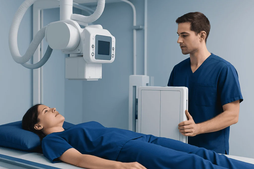
X-rays pass through the body in different amounts depending on the tissue being examined. Bone absorbs more radiation than surrounding soft tissue, creating the contrast that allows radiologists to assess its shape, density, and structure. Images are usually taken from two or more angles so that the area can be evaluated in three dimensions.
A clinician may request an X-ray for persistent pain in a bone or limb that does not improve as expected, pain that occurs at rest or at night, unexplained swelling, a firm lump, reduced movement, or a fracture after little or no trauma. Fever, tiredness, or weight loss can occur in some people, although these symptoms have many possible causes and are not diagnostic of sarcoma.
Preparation is usually minimal. The person may need to remove jewellery, watches, belts, or clothing items with metal near the area being imaged. It is important to tell the radiology team about a possible pregnancy, previous surgery or implants, and any difficulty holding a position. No fasting, injections, contrast dye, or sedation is usually required for a standard bone X-ray.
What Happens During an Ewing Sarcoma Xray and Afterward
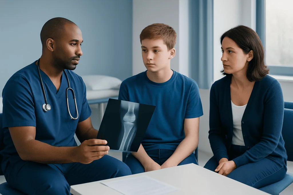
During the procedure, a radiographer positions the body part on an X-ray table or in front of an upright detector. The person may be asked to sit, stand, or lie down and to hold still for a few seconds while each image is taken. Gentle repositioning may be needed to obtain views from different directions.
The examination itself often takes only a few minutes, although the full appointment may take longer if several body areas or additional views are needed. The test does not hurt. However, positioning may be uncomfortable when the area is painful or swollen, and the person should let the radiographer know so that positioning can be adapted safely where possible.
There is no recovery period after a routine X-ray. People can generally return to school, work, eating, drinking, and normal activities immediately. A radiologist reviews the images and sends a report to the requesting clinician. If the X-ray is concerning, the next steps may be arranged urgently rather than waiting for symptoms to change.
The benefits of X-ray include speed, accessibility, and its ability to identify important bone abnormalities. Radiation exposure from a diagnostic X-ray is low, and the clinical benefit usually outweighs this small risk when imaging is medically indicated. Its main limitation is that it cannot reliably define the full extent of a tumor or tell exactly what type of tumor is present.
What Happens If the X-ray Is Abnormal?
An abnormal X-ray does not automatically mean cancer. Infection, healing injuries, benign bone lesions, and other cancers can sometimes create similar appearances. For this reason, clinicians interpret the images alongside symptoms, examination findings, medical history, and further tests.
MRI is often the most useful next scan for assessing the bone marrow and nearby muscles, nerves, blood vessels, and joints. CT may be used to examine the chest or evaluate certain bones in more detail. PET-CT, bone scanning, or other staging tests may be considered to check whether disease has spread. These investigations also help the team plan treatment safely.
A biopsy is needed to confirm Ewing sarcoma. A specialist removes a small sample of tissue, usually using an image-guided needle, for examination by a pathologist. Molecular testing can identify characteristic genetic changes in tumor cells. Biopsy planning is important: it should be coordinated by the sarcoma team because the route used can affect later surgical options.
Once results are available, care is planned by specialists in pediatric or medical oncology, orthopedic oncology, radiology, pathology, radiation oncology, surgery, nursing, rehabilitation, and supportive care. Treatment may include systemic chemotherapy, surgery, and sometimes radiotherapy, depending on the tumor location, response to treatment, and stage.
What Is the 2 Week Rule for Sarcoma?
The “2 week rule” generally refers to urgent referral pathways used in some healthcare systems. It aims for a person with symptoms that could suggest cancer, including possible sarcoma, to be assessed by an appropriate specialist within about two weeks. It is a referral target, not a test that diagnoses sarcoma and not a rule that symptoms must have been present for exactly two weeks.
Features that may justify urgent assessment include a lump that is growing, larger than about 5 cm, deep beneath the skin, painful, or returning after removal. Persistent unexplained bone pain, particularly pain at night or pain with swelling, also deserves prompt medical review. Children and teenagers should not be expected to “wait it out” when symptoms are persistent or worsening.
Urgent referral does not mean that cancer is likely. Most lumps and bone pains are caused by noncancerous conditions. The purpose is to ensure that the small number of people who need specialist imaging and evaluation receive it without unnecessary delay.
Would Ewing Sarcoma Show Up in Blood Work?
Ewing sarcoma cannot be diagnosed or ruled out with routine blood work. Blood tests may be normal, especially early in the disease. This is why persistent symptoms should be evaluated based on the whole clinical picture rather than reassurance from normal laboratory results alone.
Doctors may still order blood tests to assess general health and look for signs such as anemia, inflammation, or changes in organ function. Some results can be abnormal in Ewing sarcoma, but none is specific enough to confirm the diagnosis. Tests are also useful before treatment and for monitoring how the body is coping with therapy.
Imaging and biopsy remain the key diagnostic steps. If an X-ray or symptoms raise concern, referral to a specialist sarcoma service is appropriate even when blood test results are unremarkable.
How Fast Does Ewing Sarcoma Progress, and How Serious Is It?
Ewing sarcoma can grow and spread more quickly than many benign bone conditions, which is why timely evaluation is important. However, the rate of progression varies from person to person. Symptoms may develop over weeks or months, and the exact behavior of an individual tumor cannot be predicted from an X-ray alone.
Ewing sarcoma is serious because it is a malignant cancer and can spread, most often to the lungs, other bones, or bone marrow. At the same time, it is treatable. Outcomes depend on several factors, including whether the cancer has spread, where it began, how it responds to chemotherapy, and whether local control with surgery and/or radiation can be achieved.
Modern treatment is carefully coordinated and usually takes place over many months. Supportive care is also central, including pain management, nutrition, emotional support, fertility discussions when appropriate, rehabilitation, and follow-up imaging. Families are encouraged to ask the care team to explain the purpose and timing of every investigation and treatment step.
When to Seek Medical Care
Medical advice should be sought promptly for bone pain that persists, worsens, wakes a person at night, or is not clearly explained by an injury. An assessment is also important for unexplained swelling, a growing lump, limping, reduced use of an arm or leg, repeated fractures, or pain accompanied by fever, marked fatigue, or unintentional weight loss.
Emergency care may be needed for severe sudden pain after a suspected fracture, inability to bear weight, new weakness or numbness, breathing difficulty, or symptoms of spinal cord compression such as new bowel or bladder changes. These symptoms are not always caused by sarcoma, but they require urgent evaluation.
After a suspicious scan, it is best not to delay specialist review or attempt treatment for a presumed injury without follow-up. Acibadem International’s multidisciplinary specialists at JCI-accredited hospitals can assess and treat Ewing sarcoma for international patients, with diagnostic and treatment planning tailored to the individual clinical situation.
Frequently asked questions
Can an X-ray diagnose Ewing sarcoma?
No. An X-ray can show bone changes that may be suspicious for Ewing sarcoma, but it cannot confirm the diagnosis. MRI and other staging scans help define the abnormality, while a biopsy is required to identify the tumor type.
What does Ewing sarcoma look like on an X-ray?
It may appear as an area of bone destruction, an abnormal pattern of new bone formation, thinning or disruption of the outer bone layer, or a soft-tissue mass. These features can also occur in other conditions, including infection and other bone tumors. A specialist radiologist interprets the X-ray in context.
Is an Ewing sarcoma X-ray painful?
The X-ray itself is painless and does not involve needles or contrast material. Holding the affected body part in position can be uncomfortable if there is pain or swelling. The radiographer can often adjust positioning to reduce discomfort.
Would Ewing sarcoma show up in blood work?
Routine blood tests may be normal and cannot exclude Ewing sarcoma. Some people have nonspecific changes, such as anemia or markers of inflammation, but these do not diagnose the cancer. Imaging and biopsy are essential when there is clinical concern.
How fast does Ewing sarcoma progress?
Ewing sarcoma can be fast-growing, but progression varies between individuals. Symptoms may become noticeable over weeks or months, and the tumor may spread if untreated. Prompt assessment is important, but only specialist testing can determine the extent and behavior of a particular tumor.
How serious is Ewing sarcoma?
Ewing sarcoma is a serious cancer because it can spread to other parts of the body. It is nevertheless treatable with coordinated chemotherapy and local treatment such as surgery and, in some cases, radiation therapy. The care team can explain the individual outlook after staging and response assessment.
References
- National Cancer Institute
- American Cancer Society
- European Society for Medical Oncology
- NHS England
- Children's Oncology Group
This article is for general information only and is not a substitute for professional medical advice. Please consult a qualified doctor about your individual situation.
Oncology care in Turkey — second opinion and treatment plan
JCI-accredited · board-certified surgeons · reply within 24h
Add us as a Preferred Source to see more of our trusted health content across Google Search, AI Overviews and Discover.