Excision for Melanoma: Procedure, Recovery and Results

Wide local excision removes melanoma plus a safety margin of surrounding skin. The required margin depends mainly on the melanoma’s thickness and location.
Key Takeaways
- Wide local excision removes melanoma plus a safety margin of surrounding skin.
- The required margin depends mainly on the melanoma’s thickness and location.
- Most procedures are performed with local anesthetic, although some patients need more complex reconstruction or general anesthesia.
- Initial wound healing commonly takes about 1 to 3 weeks, while scar maturation takes months.
- Early-stage melanoma can often be cured with complete excision, but regular skin follow-up remains important.
Excision for melanoma is surgery to remove a melanoma along with a planned border of healthy-looking skin. It is the main treatment for most localized melanomas, and the laboratory analysis of the removed tissue helps confirm whether the cancer has been fully removed and whether additional care is needed.
Overview: what excision for melanoma involves
Excision for melanoma is a surgical procedure that removes a confirmed melanoma and a measured margin of nearby skin that may contain microscopic cancer cells. In many cases, the original biopsy has already removed the visible lesion. A second procedure, called a wide local excision, is then performed to remove additional skin around the biopsy site and reduce the likelihood that melanoma cells remain locally.
The amount of normal-appearing skin removed is based on the thickness of the melanoma, measured in millimeters by a pathologist. This is why the pathology report from the initial biopsy is central to surgical planning. The aim is complete removal while preserving as much healthy tissue and function as possible.
For melanoma that appears confined to the skin, surgery is often the most important treatment. Some people with thicker melanomas may also be offered sentinel lymph node biopsy, an operation that checks the first lymph nodes most likely to receive melanoma cells. This helps with staging and decisions about follow-up or additional treatment.
Who may need melanoma excision and how it is planned
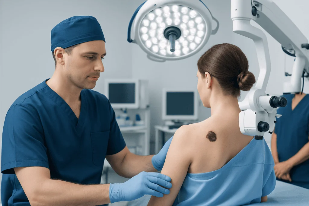
Nearly everyone with an invasive melanoma or melanoma in situ needs surgical removal, unless an individual health issue makes surgery unsuitable. Dermatologists, surgical oncologists, plastic surgeons and pathologists may work together when a melanoma is on the face, ear, scalp, hand, foot, or another area where closure is more complex.
Before surgery, the clinical team reviews the biopsy report, including melanoma type, thickness, ulceration and whether the biopsy edges contain melanoma. They also consider the lesion’s location, medical history, medicines that affect bleeding, allergy history and the person’s ability to care for the wound after surgery.
Margins are individualized according to established clinical guidance. Melanoma in situ usually requires a narrower margin than invasive melanoma, while thicker melanomas generally require a wider margin. A larger margin does not automatically mean a better outcome; the surgical plan balances cancer control with wound closure, appearance and function.
People with melanoma may also benefit from education about melanoma, skin surveillance and sun protection. If lymph node assessment or systemic treatment is appropriate, the care team explains why and how each option may contribute to overall care.
How the procedure is performed
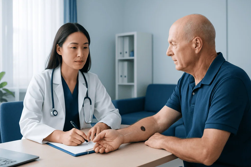
Most straightforward melanoma excisions are outpatient procedures. The clinician marks the planned margin around the biopsy scar or melanoma site, cleans the skin and injects local anesthetic. Local anesthetic numbs the area but allows the person to remain awake; gentle pressure or pulling may be felt, but sharp pain should not occur.
The surgeon removes an oval-shaped area of skin and tissue beneath it. The specimen is labeled and sent to a pathology laboratory to assess the surgical margins. Depending on the size and location of the wound, the skin may be closed directly with stitches, allowed to heal gradually, or repaired using a skin graft or flap.
When sentinel lymph node biopsy is recommended, it is often performed at the same time as the wide local excision. A tracer helps identify the relevant lymph node or nodes, which are removed through a separate small incision and examined for melanoma cells. This procedure is mainly used for staging rather than to treat every person with melanoma.
More extensive surgery may be best performed in a hospital setting with reconstructive support. Melanoma treatment may involve coordinated planning between dermatology, surgical oncology, pathology, radiology and medical oncology when the diagnosis or stage requires it.
Benefits, limitations and possible risks
The key benefit of excision for melanoma is local cancer control. For melanoma that has not spread beyond the skin, complete removal can be curative. The final pathology report confirms whether melanoma is present at the edges of the excised tissue. If margins are not clear, additional surgery may be recommended.
Excision also provides important diagnostic information. The pathology findings can refine staging and help determine whether lymph node assessment, imaging, medical oncology consultation or closer surveillance should be considered. Surgery is effective for the area treated, but it cannot by itself prevent every future melanoma or address melanoma that has already spread elsewhere.
Possible risks include bleeding, bruising, infection, delayed wound healing, numbness, pain, scar formation and separation of the wound edges. The risk of noticeable scarring or tightness is greater when a large area is removed or when surgery is performed near joints, the eyelids, lips, nose or ears.
Contact the surgical team if there is increasing redness, warmth, swelling, drainage, fever, worsening pain, persistent bleeding or the wound opens. These issues are often manageable, especially when addressed early.
Recovery timeline and wound care
Recovery depends on the excision size, body site, closure method and individual health. For a small wound closed with stitches, discomfort and swelling are usually most noticeable during the first few days and then improve. Stitches may be removed after a period chosen by the clinician, which varies by body area and wound tension.
The team provides specific instructions for keeping the wound clean, changing dressings, bathing, activity and pain relief. It is generally important to avoid stretching the wound, strenuous exercise and activities that cause friction until the clinician says it is safe. People should not apply creams, antiseptics or scar products unless advised, as some products can irritate healing skin.
Skin usually seals over within weeks, but deeper healing and scar remodeling continue for several months. Protecting the healing area from sunlight is important because ultraviolet exposure can darken scars and contributes to future skin cancer risk. Once the wound is fully closed, the care team may recommend a suitable sunscreen and protective clothing.
Follow-up appointments review the pathology result, wound healing and any need for further treatment. Long-term skin examinations are also important because a person who has had melanoma has an increased chance of developing another melanoma or other skin cancer.
How long does it take for a melanoma excision wound to heal?
A melanoma excision wound often achieves initial healing in about 1 to 3 weeks when it is closed with stitches, although healing can take longer for larger wounds, grafts, flaps, wounds on the lower legs, or wounds left to heal naturally. The location matters: areas that move frequently or have reduced blood supply may need more time.
Even after the skin surface has healed, the scar remains active beneath the surface. It may feel firm, itchy, pink or numb for weeks to months. Scar maturation commonly continues for 6 to 12 months, and the final appearance cannot be judged during the first few weeks.
Diabetes, smoking, poor circulation, immune-suppressing medicines and infection can slow healing. Individual aftercare instructions should take priority over general timelines, and a clinician should assess any wound that becomes increasingly painful, red, swollen or draining.
Can melanoma be cured with excision?
Yes. Complete surgical excision can cure many melanomas that are diagnosed early and confined to the skin. The likelihood of cure depends on factors such as tumor thickness, ulceration, whether melanoma has reached lymph nodes or other organs, and whether the surgical margins are clear.
For melanoma in situ and many thin invasive melanomas, excision may be the only treatment needed. Thicker melanomas or melanomas with lymph node involvement may require additional evaluation and, in some cases, treatments such as immunotherapy or targeted therapy. The care plan is based on the individual stage and tumor features.
Even after successful excision, continued follow-up is essential. Follow-up visits can identify recurrence early and provide regular full-skin examinations, while self-awareness of changing spots supports prompt assessment between appointments.
How long does it take for melanoma to go from stage 1 to 4?
There is no reliable fixed timeline for melanoma to progress from stage 1 to stage 4. Some melanomas may remain localized for a long time, while others can grow or spread more quickly. Staging describes the melanoma’s characteristics and spread at the time of assessment; it is not a schedule that predicts exactly when progression will occur.
Growth rates vary according to melanoma subtype, thickness, ulceration, genetic features and the person’s immune response. Because it is not possible to predict behavior from appearance alone, any changing or suspicious mole should be examined promptly rather than watched for a set period.
Early diagnosis and appropriate excision offer the best opportunity to treat melanoma before it spreads. People who have had melanoma should attend recommended follow-up visits and report new or changing pigmented lesions, persistent sores or unexplained enlarged lymph nodes.
How painful is melanoma excision?
During a melanoma excision under local anesthetic, the numbing injections may sting briefly. Once the anesthetic is working, the person should not feel sharp pain during surgery, although pressure, movement or pulling sensations can occur. Tell the clinician immediately if pain is felt during the procedure so additional anesthetic can be considered.
Afterward, discomfort is often mild to moderate and tends to improve over several days. The amount of soreness depends on the size and location of the excision, whether a flap or graft was needed, and how much the area moves. The clinician can advise on safe pain-relief options based on medical history and current medicines.
Severe or increasing pain is not expected and should be reported, particularly if it occurs with redness, swelling, discharge, fever or bleeding. These signs may indicate a complication that needs assessment.
When to seek medical care
Prompt medical assessment is appropriate for a new or changing pigmented spot, especially one that changes in size, shape or color, has an irregular border, bleeds without clear cause, itches persistently or looks noticeably different from other moles. A dermatologist can examine the area and decide whether biopsy is needed.
After an excision, seek advice urgently for uncontrolled bleeding, rapidly increasing swelling, severe pain, spreading redness, pus-like drainage, fever, or a wound that opens. These symptoms do not always mean a serious problem, but timely review can protect healing.
Acibadem International’s multidisciplinary specialists and JCI-accredited hospitals support international patients who need melanoma assessment, surgery and follow-up planning. A qualified clinician can explain whether excision, lymph node evaluation or other care is appropriate for the individual diagnosis.
Frequently asked questions
Is a wide local excision always needed after a melanoma biopsy?
In many cases, yes. A diagnostic biopsy may remove the visible melanoma but may not include the recommended margin of surrounding tissue. A wide local excision is commonly used to remove that margin and confirm local control.
Will melanoma excision leave a scar?
Any procedure that cuts the skin leaves a scar. Its size and appearance depend on the excision margin, location, closure technique, skin type and healing process. Scars usually change and soften over time, and the surgical team can discuss ways to support scar care once the wound has healed.
What happens if melanoma is found at the excision margin?
If melanoma cells are present at the edge of the removed specimen, the surgeon may recommend another excision to obtain clear margins. The exact next step depends on the melanoma type, site and pathology findings. The care team will explain the result and the purpose of any further procedure.
Can I exercise after melanoma excision?
Light activity may be possible soon after a small excision, but strenuous exercise, heavy lifting and movements that stretch the wound often need to be avoided temporarily. The restriction period depends on the body site and closure. Follow the surgeon’s specific advice to reduce bleeding, wound opening and delayed healing.
Does melanoma excision spread cancer?
No. Properly performed biopsy and excision do not cause melanoma to spread. Surgery is a standard treatment used to remove melanoma and provide accurate pathology information.
How often should skin checks be done after melanoma surgery?
The schedule varies with the melanoma stage, pathology features and personal risk factors. Many people need regular clinician-led skin examinations as well as monthly self-checks. The treating team will provide an individualized follow-up plan.
References
- American Academy of Dermatology
- American Cancer Society
- National Cancer Institute
- National Comprehensive Cancer Network
- World Health Organization
This article is for general information only and is not a substitute for professional medical advice. Please consult a qualified doctor about your individual situation.
Melanoma in Turkey — costs, top hospitals & a free quote
JCI-accredited · board-certified surgeons · reply within 24h
Add us as a Preferred Source to see more of our trusted health content across Google Search, AI Overviews and Discover.

Medical Oncology Department
Medical treatment of cancer with chemotherapy, immunotherapy and targeted therapies under a multidisciplinary tumor board.
60 specialists in this unit