

Eye Exam Procedure: An Evidence-Based Patient Guide
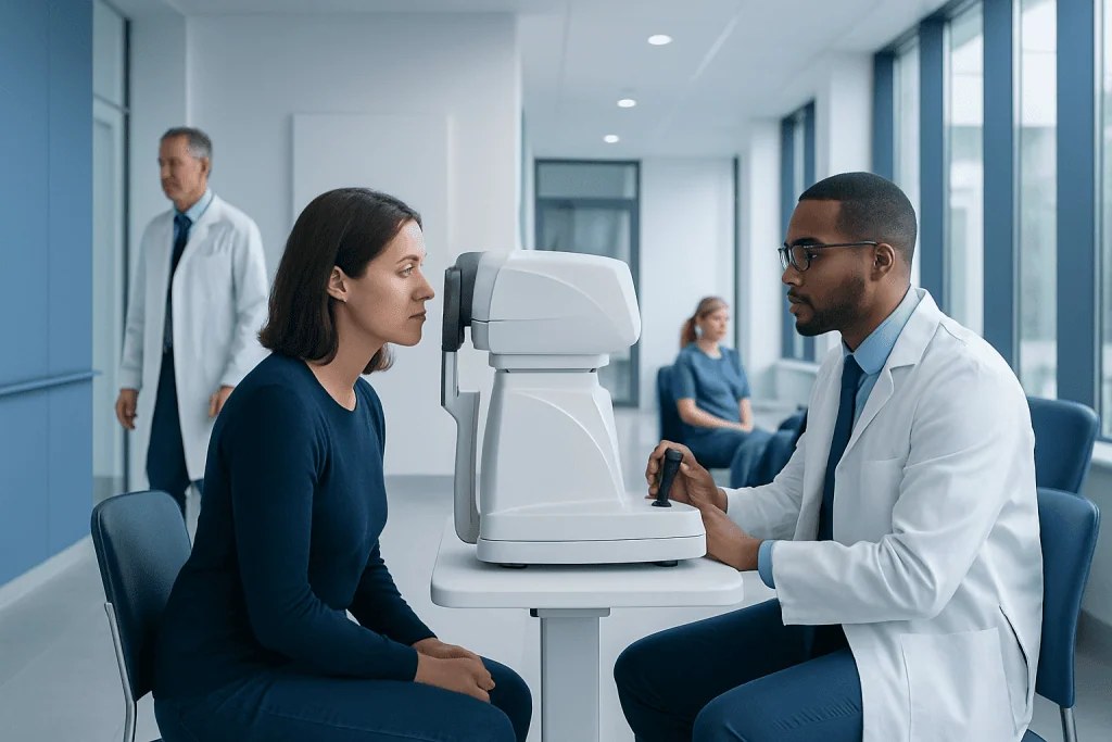
A comprehensive eye exam evaluates more than eyesight; it can identify early eye disease before symptoms develop. The exam commonly includes visual acuity, refraction, eye-pressure measurement, and an assessment of the front and back of the eye.
Key Takeaways
- A comprehensive eye exam evaluates more than eyesight; it can identify early eye disease before symptoms develop.
- The exam commonly includes visual acuity, refraction, eye-pressure measurement, and an assessment of the front and back of the eye.
- Pupil-dilating drops give the clinician a wider view of the retina and optic nerve but can cause temporary blurred vision and light sensitivity.
- People with diabetes, glaucoma risk, a family history of eye disease, or vision changes may need eye examinations more often.
- Sudden vision loss, severe eye pain, flashes of light, or a curtain-like shadow require urgent medical assessment.
An eye exam procedure is a structured assessment of vision, eye function, and eye health performed by an eye-care professional. Most tests are quick and painless, while dilating drops may temporarily blur near vision and increase sensitivity to light for several hours.
Overview: What Is an Eye Exam Procedure?
An eye exam procedure is a series of tests used to measure how clearly a person sees and to assess the health of the eyes. It may be performed by an optometrist or ophthalmologist. An ophthalmologist is a medical doctor who can diagnose and treat eye diseases and perform eye surgery, while an optometrist provides vision testing and eye-care services within their professional scope.
A comprehensive examination does not only determine whether glasses or contact lenses may help. It also examines structures such as the cornea, lens, retina, optic nerve, and eye muscles. This can help detect problems including glaucoma, cataracts, retinal conditions, and changes related to diabetes or high blood pressure, sometimes before noticeable symptoms appear.
The exact tests vary according to age, symptoms, medical history, and findings during the appointment. For most people, the process is comfortable and does not involve needles or radiation. If dilation is needed, the visit may take longer because the drops require time to work.
Who May Need a Comprehensive Eye Examination?
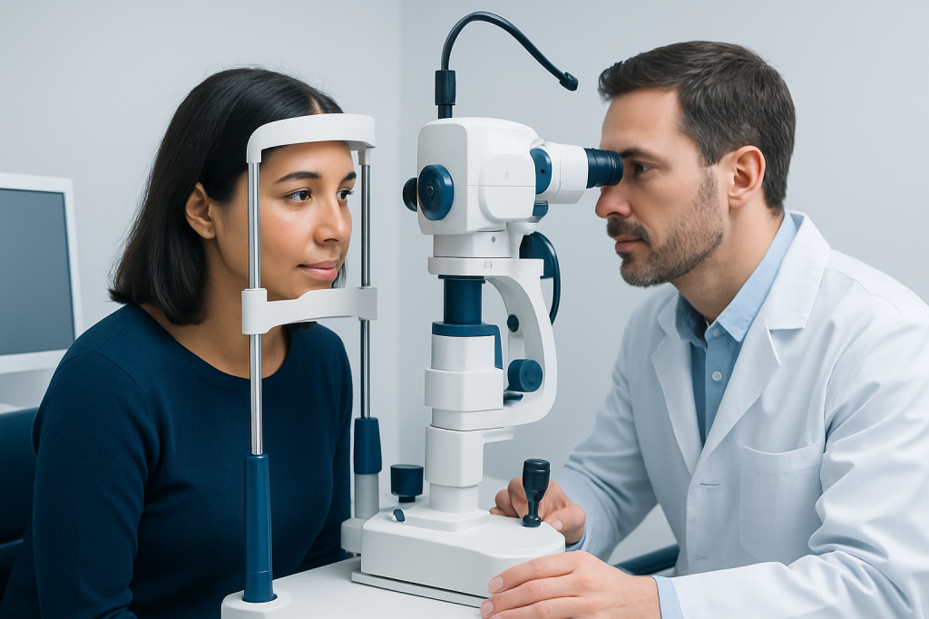
Eye examinations are useful for people of all ages, including those who feel their vision is normal. The appropriate timing depends on individual needs. Children need age-appropriate vision checks because uncorrected vision problems can affect learning and visual development. Adults may need regular reviews to update prescriptions and monitor eye health over time.
More frequent assessment may be recommended for people with diabetes, high blood pressure, autoimmune disease, a previous eye injury, high short-sightedness, or a personal or family history of glaucoma, retinal disease, or early cataracts. People using medicines that may affect the eyes should also ask their prescribing clinician whether monitoring is appropriate.
A comprehensive visit is particularly important when a person notices blurred vision, double vision, eye strain, difficulty driving at night, increasing trouble reading, new floaters, or a change in how colors appear. Regular assessment may also help identify glaucoma before permanent vision loss occurs, as early stages often cause no obvious symptoms.
How the Eye Exam Procedure Works
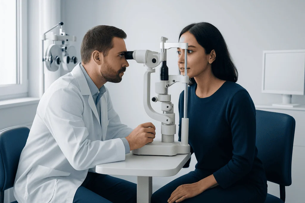
An eye-care professional begins by taking a focused history. They may ask about current symptoms, general health conditions, medicines, prior eye procedures, allergies, and family history. Bringing current glasses, contact lens information, and a medication list can help make the assessment more accurate.
The examination usually combines vision measurements with observation of the eye’s structures and function. The clinician may use charts, lenses, bright lights, magnification, imaging equipment, and eye drops. These tests help distinguish refractive errors, such as short-sightedness or astigmatism, from eye-health concerns that require additional investigation or treatment.
Many people receive a preliminary assessment from a technician before seeing the clinician. This may include visual acuity, automated refraction, eye pressure, photographs of the retina, or scans of the optic nerve. Results are interpreted in the context of the full examination; an individual screening measurement alone does not establish a diagnosis.
Step by Step: Common Tests During an Eye Exam
Visual acuity testing is often the first step. The person reads letters, symbols, or pictures at a measured distance, with each eye tested separately and together. Near-vision testing may also be performed. If vision is not as clear as expected, refraction testing uses a phoropter or trial lenses to identify whether a glasses or contact lens prescription could improve focus.
The clinician may assess how the pupils react to light, how the eyes move and work together, and whether side vision is affected. Color-vision tests may be used when relevant. A slit-lamp examination uses a microscope and narrow beam of light to inspect the eyelids, tear film, cornea, iris, and lens in detail. Fluorescein dye may occasionally be placed in the eye to highlight surface irritation or damage; it is generally painless and washes away with tears.
Eye pressure can be measured using a gentle air puff or an instrument that briefly touches the eye after numbing drops are applied. Pressure is only one part of evaluating glaucoma risk. The clinician may also perform visual-field testing, optic-nerve imaging, or retinal photography when there is a clinical reason to do so.
A dilated eye examination uses drops to widen the pupils. Once the pupils are enlarged, the clinician can examine the retina, macula, blood vessels, and optic nerve more thoroughly. This detailed review can support the evaluation of conditions such as diabetic retinopathy and retinal tears or detachments.
Dilation, Imaging, and What Recovery Is Like
Not every eye exam requires dilation, but it is often advised for a complete view of the back of the eye or when symptoms or risk factors are present. The drops may sting briefly when applied. They commonly take about 15 to 30 minutes to reach full effect, although this varies between individuals and products.
After dilation, near vision is often blurred and bright light may feel uncomfortable. These effects usually improve over several hours, but can occasionally last longer. Sunglasses can improve comfort outdoors. People should ask the clinic whether they can safely drive after their appointment; arranging transport may be sensible, especially if dilation is planned or vision is already affected.
Retinal photographs and optical coherence tomography, often called OCT, are noninvasive imaging tests that provide detailed images of eye structures. They may be used to monitor changes over time. If a problem is identified, the clinician may explain options ranging from updated glasses to specialist monitoring, medication, laser treatment, or procedures such as laser eye surgery when appropriate for the diagnosed condition.
Benefits, Limitations, and Possible Risks
The main benefit of an eye exam procedure is early recognition of correctable vision problems and eye conditions that may not initially cause discomfort. Earlier identification can allow timely monitoring or treatment and may help protect vision. The appointment can also identify signs that warrant assessment of general health conditions, although an eye examination does not replace routine medical care.
Most components of an eye examination carry little risk. Some tests may feel mildly uncomfortable because of bright lights, a brief air puff, or the sensation of drops. Dilating drops can temporarily reduce focusing ability and increase glare sensitivity. Rarely, dilation can trigger a sudden rise in eye pressure in susceptible people with narrow drainage angles.
Patients should tell the clinician about prior reactions to eye drops, known narrow-angle glaucoma, eye surgery, pregnancy, and relevant medical conditions. They should also understand that a normal examination does not prevent future changes. New symptoms between routine appointments still deserve prompt evaluation.
Preparing for an Eye Exam and Protecting Eye Health
Before the appointment, a person can note when symptoms began, whether they affect one or both eyes, and what makes them better or worse. Current glasses and contact lenses should be brought along, as well as information about previous eye treatments and family eye-health history. Contact lens wearers may be instructed to remove lenses before some measurements, particularly when a new prescription or corneal assessment is planned.
Long-term eye health also benefits from practical habits. These include using suitable protective eyewear for work, sports, or ultraviolet exposure; managing conditions such as diabetes and high blood pressure; avoiding smoking; and seeking care for persistent discomfort rather than relying repeatedly on non-prescription drops. Screen use does not generally damage the eyes, but regular breaks and blinking can reduce dryness and strain.
At Acibadem International, multidisciplinary specialists in JCI-accredited hospitals assess vision concerns and eye conditions for international patients, with investigations and treatment plans tailored to clinical findings. Patients should discuss any test result, proposed treatment, and follow-up interval with a qualified eye-care professional.
When to Seek Medical Care
Routine eye care should be arranged when vision gradually changes, glasses no longer seem effective, or a person has ongoing eye strain, redness, dryness, discomfort, headaches related to visual tasks, or difficulty seeing at night. A timely appointment is also advisable after an eye injury, even if symptoms seem minor, because some injuries are not immediately visible.
Urgent medical care is needed for sudden loss of vision, a sudden marked decrease in vision, severe eye pain, a painful red eye with nausea, new flashes of light with many floaters, or a dark curtain or shadow across the visual field. These symptoms can have several causes, including conditions that need rapid evaluation to protect sight.
People who experience chemical exposure to the eye should immediately rinse the eye continuously with clean water or saline and seek emergency advice. They should not try to remove embedded objects or use leftover prescription eye drops unless instructed by a clinician.
Frequently asked questions
How long does an eye exam procedure take?
A routine eye examination often takes around 30 to 60 minutes, depending on the tests needed and whether dilation is used. A visit may take longer if there are symptoms, complex medical history, contact lens fitting needs, or additional imaging and visual-field testing.
Does an eye exam hurt?
Most eye exam tests are painless. Bright lights, air-puff pressure testing, and eye drops may cause short-lived discomfort, but they should not be painful. A person should tell the clinician immediately if a test causes significant pain.
Can I drive after my eyes are dilated?
Dilating drops can blur near vision and make bright light uncomfortable for several hours. Some people can drive safely, while others should avoid it, particularly in strong sunlight or at night. It is best to ask the clinic in advance and consider arranging a ride.
How often should adults have an eye exam?
The ideal interval varies with age, vision needs, eye-health risks, and previous findings. Adults with diabetes, glaucoma risk, high short-sightedness, or eye symptoms may need more frequent monitoring. An eye-care professional can recommend a schedule suited to the individual.
Can an eye exam detect diabetes or high blood pressure?
An examination can reveal changes in retinal blood vessels that may be associated with diabetes or high blood pressure. However, it does not replace blood tests, blood pressure checks, or assessment by a primary-care clinician. Abnormal retinal findings may prompt referral for broader medical evaluation.
Should contact lenses be worn to an eye exam?
It depends on the reason for the visit and the tests planned. Contact lenses may be useful to assess vision with the current correction, but they may need to be removed for some measurements or evaluations. The clinic can provide specific instructions before the appointment.
References
- American Academy of Ophthalmology
- National Eye Institute
- Centers for Disease Control and Prevention
- World Health Organization
This article is for general information only and is not a substitute for professional medical advice. Please consult a qualified doctor about your individual situation.
Explore treatments in Turkey — costs, top hospitals & a free quote
JCI-accredited · board-certified surgeons · reply within 24h
Add us as a Preferred Source to see more of our trusted health content across Google Search, AI Overviews and Discover.
Check your numbers in seconds
BMI, calories, due date, blood pressure and 30+ more clinical calculators — free, instant, doctor-reviewed ranges.

More from the Health Library



Related Specialists

Ezgi Nur Çiçek
Physical Medicine & Rehabilitation
Assoc. Prof. Dr. Ahmet Özveren
Medical Oncology
Dr. Gülistan Yeşilyurt
Emergency Service
Dr. Ezel Aydoğ
Urology