Fibroadenoma of the Breast: When a Lump Needs Imaging, Biopsy, or Removal

A fibroadenoma is a benign solid breast lump that often feels smooth, firm, and movable. Breast imaging helps confirm whether a lump looks typical for fibroadenoma and whether more testing is needed.
Key Takeaways
- A fibroadenoma is a benign solid breast lump that often feels smooth, firm, and movable.
- Breast imaging helps confirm whether a lump looks typical for fibroadenoma and whether more testing is needed.
- Biopsy may be recommended if the lump is new, growing, atypical on imaging, or present in an older patient.
- Treatment is individualized and may include observation, core needle biopsy, or surgical removal.
- Any new breast lump should be assessed by a qualified clinician rather than assumed to be harmless.
Fibroadenoma of the breast is a common, usually noncancerous breast lump, especially in younger women. Many fibroadenomas can be monitored safely, but some need imaging, biopsy, or removal based on their features, growth, symptoms, and a person’s age.
Overview
Fibroadenoma of the breast is one of the most common benign breast lumps. It is made of both glandular and fibrous breast tissue and often appears in adolescents and younger adults, although it can occur at other ages as well. Many people first notice it while bathing, dressing, or doing routine breast self-awareness.
A typical fibroadenoma often feels round or oval, smooth, rubbery or firm, and easy to move under the skin. These features can be reassuring, but a lump cannot be identified by touch alone with certainty. That is why a medical evaluation is important, even when the lump seems harmless.
In many cases, fibroadenomas do not cause pain and do not turn into cancer. Still, some lumps need closer assessment because other benign conditions and, less commonly, breast cancer can sometimes look similar on exam or imaging. The main question is not simply whether a lump is present, but whether it has the typical features of a fibroadenoma and whether follow-up, biopsy, or removal is the safest next step.
Symptoms and How a Fibroadenoma May Feel
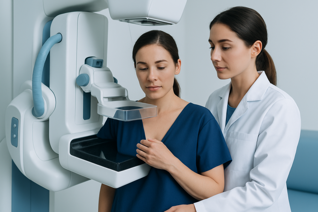
Many fibroadenomas are found as a painless breast lump. A person may notice a small, well-defined mass that moves slightly when pressed. Some are discovered during a routine breast examination by a clinician or on imaging done for another reason.
Although fibroadenomas are often symptom-free, they can sometimes feel tender or uncomfortable, especially around hormonal changes such as the menstrual cycle. Larger fibroadenomas may create a sense of fullness, visible asymmetry, or concern because they are easier to feel.
Features that can occur with fibroadenoma include:
- A smooth, solid-feeling lump
- A round or oval shape
- A movable mass rather than one fixed in place
- One lump or, less commonly, multiple lumps
- Little or no pain
Symptoms alone cannot confirm the diagnosis. A new lump, a lump that is enlarging, or any breast change associated with skin dimpling, nipple changes, or spontaneous bloody nipple discharge should be assessed promptly to rule out other conditions.
Causes and Risk Factors
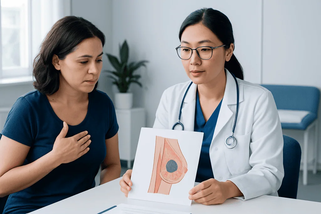
The exact cause of fibroadenoma is not fully understood, but hormones are thought to play an important role. These lumps are most common during the years when breast tissue is more hormonally responsive. They may change somewhat in size over time and can occasionally become more noticeable during pregnancy or with hormone-related changes.
Fibroadenomas are not caused by an injury to the breast, and having one does not mean a person has done anything wrong. Most are sporadic, meaning they occur without a single clear cause. Some people may develop more than one fibroadenoma, and some may have fibroadenomas in both breasts.
Risk can be influenced by age and hormonal environment. Fibroadenomas are generally more common in younger patients than in older adults. A clinician will also consider whether the lump could represent another benign breast condition, such as breast cysts, or whether a different diagnosis should be excluded based on exam findings and imaging.
When Imaging Is Needed
Any new breast lump should be medically evaluated. The first step is usually a clinical breast exam, followed by imaging chosen according to age, breast density, symptoms, and pregnancy status. In younger patients, ultrasound is often the preferred initial test because it can distinguish a solid lump from a fluid-filled cyst without using radiation.
Mammography may also be used, particularly in older patients or when the clinical picture is less straightforward. In some situations, both mammography and ultrasound are recommended. Imaging helps the radiologist assess the lump’s shape, margins, density, and internal pattern to see whether it looks typical of a benign fibroadenoma or whether additional testing is advisable.
If the lump appears classic for fibroadenoma and matches the clinical exam, short-term imaging follow-up may be enough. If it is larger than expected, growing, irregular, difficult to classify, or discordant with the exam, a biopsy may be recommended. Imaging is not just about detecting cancer; it also helps avoid unnecessary surgery when a lump looks clearly benign.
People who have a strong family history of breast cancer, prior abnormal imaging, or other risk factors may need a more individualized imaging plan. In selected cases, additional breast imaging such as mammography or breast ultrasound helps guide the next step with more confidence.
When a Biopsy Is Recommended
A biopsy is not needed for every fibroadenoma, but it is appropriate when the diagnosis is uncertain or when the lump has features that deserve tissue confirmation. The most common method is a core needle biopsy, in which small tissue samples are taken using imaging guidance and then examined by a pathologist.
Doctors may recommend biopsy if a lump is new in an older patient, grows over time, has suspicious or atypical imaging features, feels different from a typical fibroadenoma, or causes ongoing concern despite reassuring imaging. Biopsy can also help distinguish a fibroadenoma from related but less common lesions, including a phyllodes tumor, which may also present as a firm, growing breast mass.
In general, a core needle biopsy provides more information than a fine needle aspiration for a solid breast lump. The goal is to match the pathology result with the clinical and imaging findings. If all three are consistent with fibroadenoma, observation may be appropriate. If they do not match, the care team may suggest further sampling or removal.
For many patients, a breast biopsy offers reassurance and helps avoid prolonged uncertainty. Results are interpreted in the wider context of symptoms, imaging, age, and whether the lump is stable or changing.
Treatment Options: Observe, Remove, or Follow Up
Treatment for fibroadenoma of the breast depends on the size of the lump, the patient’s age, symptoms, imaging appearance, biopsy results, and personal preference. Many fibroadenomas do not need immediate treatment. If the lump has typical benign features and remains stable, a doctor may recommend observation with repeat exams or imaging after a defined interval.
Removal may be considered if the fibroadenoma is large, painful, enlarging, cosmetically bothersome, or uncertain in diagnosis even after imaging and biopsy. Surgical excision may also be advised if pathology shows atypical features or if the lesion cannot be confidently separated from another tumor type. Some centers may offer minimally invasive image-guided removal for selected benign lesions.
It is helpful for patients to know that observation is an active medical plan, not neglect. The purpose is to avoid unnecessary procedures while still watching for change. During follow-up, the clinician looks for stability in size and appearance. If the lump changes, the plan can be updated.
When an operation is chosen, the aim is to remove the lump while preserving as much normal breast tissue as possible. In centers with coordinated breast care, treatment planning may involve specialists in general surgery and breast imaging so that diagnosis and management are aligned with the individual case.
Prevention, Self-Care, and Living With a Fibroadenoma
There is no proven way to prevent fibroadenomas, because they are not linked to a single avoidable cause. Still, regular breast self-awareness is valuable. This means being familiar with how the breasts usually look and feel and reporting any new lump or change to a healthcare professional rather than trying to diagnose it at home.
For someone already diagnosed with fibroadenoma, self-care mainly involves keeping follow-up appointments and noting any changes in size, tenderness, or shape. If the lump becomes harder to move, increases noticeably, or new lumps appear, a repeat assessment is sensible. Wearing a supportive bra may help if there is discomfort, especially around the menstrual cycle.
General breast health habits can also support overall well-being:
- Attend recommended breast screening based on age and risk
- Discuss family history of breast or ovarian cancer with a doctor
- Maintain a healthy lifestyle with regular physical activity and balanced nutrition
- Limit delays in evaluating new breast symptoms
For international patients who need evaluation or treatment, Acibadem International’s multidisciplinary specialists in JCI-accredited hospitals diagnose and treat benign and suspicious breast lumps using coordinated imaging, biopsy, and surgical care when needed.
When to See a Doctor
A doctor should assess any new breast lump, even if it feels smooth and movable. Many breast lumps are benign, but it is not possible to confirm that by touch alone. Prompt evaluation helps determine whether the lump is a fibroadenoma, another benign condition, or something that needs further investigation.
Medical review is especially important if the lump is growing, firm or fixed, associated with skin changes, nipple inversion that is new, spontaneous nipple discharge, persistent focal pain, or swelling in the underarm. People with a personal history of breast disease or a strong family history of breast cancer should not delay assessment.
If a fibroadenoma has already been diagnosed, follow-up is still important when advised. Returning for review is sensible if symptoms change, anxiety remains high, or the lump interferes with comfort or body confidence. The best plan is individualized and should be guided by a qualified breast specialist or surgeon.
Frequently asked questions
Is fibroadenoma of the breast cancer?
Fibroadenoma is usually a benign, meaning noncancerous, breast lump. It does not mean a person has breast cancer. However, a new lump should still be evaluated because not every lump is a fibroadenoma.
Can a fibroadenoma go away on its own?
Some fibroadenomas stay the same size for years, some shrink, and some become less noticeable over time. Others may grow, especially during periods of hormonal change. That is why follow-up advice should be tailored to the individual case.
Does every fibroadenoma need a biopsy?
No. If the lump has a very typical appearance on exam and imaging, a doctor may recommend monitoring instead of biopsy. A biopsy is more likely if the lump is growing, atypical, or seen in a patient whose age or risk profile makes tissue confirmation more important.
When is removal recommended?
Removal may be advised if the lump is large, painful, growing, cosmetically bothersome, or uncertain in diagnosis. It may also be recommended when biopsy results do not fully explain the imaging findings. The decision is based on symptoms, imaging, pathology, and patient preference.
Can fibroadenoma come back after removal?
The exact lump that is removed does not usually return in the same way, but some people can develop new fibroadenomas in the future. This is more likely in people who tend to form multiple benign breast lumps. Ongoing breast awareness remains important.
What imaging test is usually used first?
That depends partly on age and clinical context. Ultrasound is often used first in younger patients, while mammography may be added or used first in older patients. Sometimes both are needed to characterize the lump fully.
References
- World Health Organization
- American Cancer Society
- National Cancer Institute
- American College of Radiology
- Breast Cancer Now
This article is for general information only and is not a substitute for professional medical advice. Please consult a qualified doctor about your individual situation.
Oncology care in Turkey — second opinion and treatment plan
JCI-accredited · board-certified surgeons · reply within 24h
Add us as a Preferred Source to see more of our trusted health content across Google Search, AI Overviews and Discover.

More from the Health Library


Related Specialists
Prof. Dr. Zübeyde Gündüz
Pediatrics
Assoc. Prof. Dr. Ahmet Küçükçelebi
Aesthetic Plastic & Reconstructive Surgery
Dr. Ezgi Darıcı
Gynecology & Obstetrics
Prof. Dr. Namik Demir
Gynecology & Obstetrics