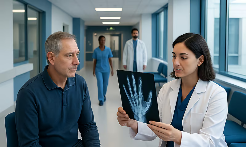
Fibrous Dysplasia Treatment: How It Works, Results and What to Expect

Fibrous dysplasia is a noncancerous condition in which normal bone is replaced by fibrous, less-strong tissue. Observation is appropriate for many stable cases with no major symptoms or functional problems.
Key Takeaways
- Fibrous dysplasia is a noncancerous condition in which normal bone is replaced by fibrous, less-strong tissue.
- Observation is appropriate for many stable cases with no major symptoms or functional problems.
- Pain management, physical rehabilitation and treatment of vitamin or hormone abnormalities can support bone health.
- Surgery may stabilize a fracture, correct deformity, remove pressure from important structures or improve function.
- The condition usually becomes less active after skeletal maturity, but follow-up remains important.
Fibrous dysplasia treatment is individualized: many people need monitoring and symptom relief, while surgery may be recommended for fractures, significant deformity, nerve compression or persistent pain. Care aims to protect bone function, improve comfort and help each person remain active safely.
Fibrous Dysplasia Treatment: How It Works
Fibrous dysplasia treatment is based on the bone involved, the person’s age, symptoms and whether the affected bone is stable. The condition occurs when an early genetic change causes an area of normal bone to be replaced with fibrous tissue. This tissue may enlarge the bone, alter its shape or make it more vulnerable to pain, deformity and fracture.
There is no treatment that removes the underlying genetic change from all affected cells. Instead, care focuses on protecting the bone, controlling symptoms and addressing complications early. A stable, painless area may only need periodic clinical review and imaging, whereas a painful lesion, progressive deformity or fracture may require more active treatment.
Fibrous dysplasia is not cancer, and it does not spread from one bone to another. It can affect one bone (monostotic fibrous dysplasia) or multiple bones (polyostotic fibrous dysplasia). In some people it occurs with hormone-related features and skin pigment changes as part of McCune-Albright syndrome, which requires coordinated specialist care.
Symptoms, Pain and Everyday Life
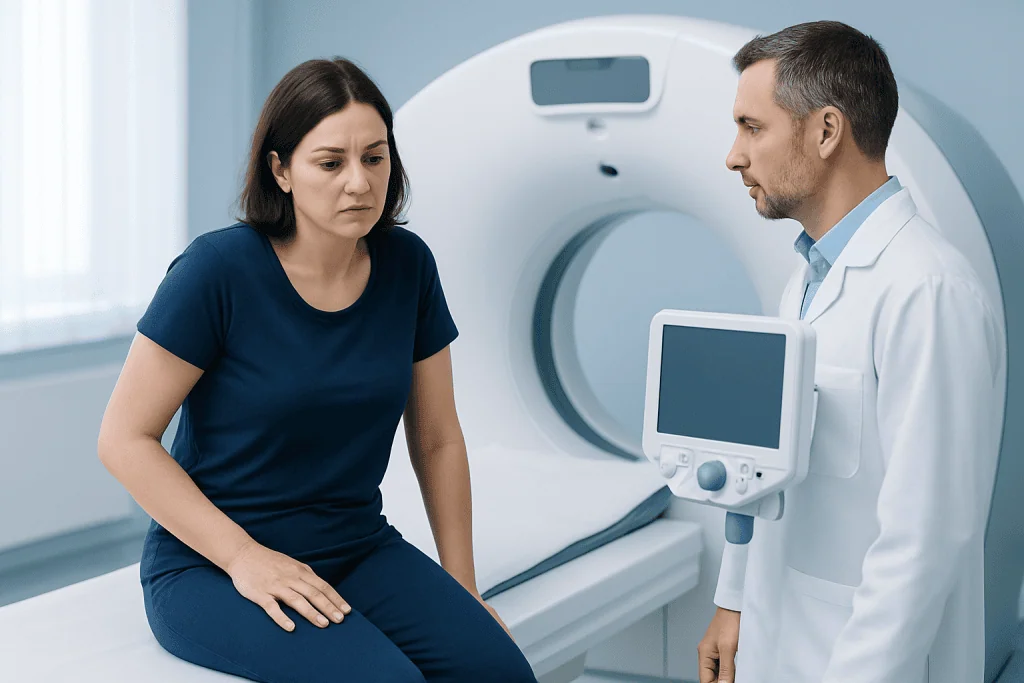
Can you live a normal life with fibrous dysplasia?
Many people with fibrous dysplasia live active, independent lives. A small, stable area found incidentally on an X-ray may never cause meaningful limitations. The outlook depends mainly on the location and extent of bone involvement, the presence of fractures or deformity, and whether associated endocrine conditions are present.
Regular follow-up helps clinicians identify changes before they interfere with mobility, vision, hearing, dental health or other functions. A person’s care team may include an orthopedic surgeon, endocrinologist, radiologist, rehabilitation specialist, dentist or craniofacial surgeon, depending on the affected area.
How painful is fibrous dysplasia?
Pain varies considerably. Some people have no pain, while others experience intermittent aching, activity-related discomfort or persistent pain in an affected bone. New, severe or rapidly increasing pain deserves assessment because it can indicate a stress injury, fracture, cyst-like change within the lesion or another treatable cause.
Pain should not simply be accepted as unavoidable. A clinician can review activity patterns, imaging findings, bone health, sleep and other contributors, then develop a plan that may include targeted exercise, non-opioid pain relief when suitable and treatment of any underlying metabolic concern.
What Causes It and What Triggers Symptoms?

What triggers fibrous dysplasia?
Fibrous dysplasia is caused by a genetic change that happens after conception during early development. It is not usually inherited from a parent and cannot be passed on in the typical inherited pattern. The change affects only some cells, which explains why the condition may involve one region of the skeleton or several areas.
Everyday activities, foods, stress and minor injuries do not cause fibrous dysplasia. However, symptoms can become more noticeable when an affected bone is placed under repeated load, when a fracture develops or when the bone changes shape. Hormonal activity can also influence the condition in people with associated endocrine disorders.
Factors that may increase the impact of the condition include low vitamin D, inadequate calcium intake, smoking, reduced muscle strength and untreated hormone abnormalities. These do not cause the disorder, but addressing them can support overall bone health and may help reduce fracture risk.
Assessment and Candidacy for Active Treatment
Diagnosis typically combines a medical history, physical examination and imaging. X-rays often show characteristic changes, and CT or MRI may be used when more anatomical detail is needed, particularly in the skull, face, spine or pelvis. Blood tests may help assess calcium, phosphate, vitamin D and hormone function when clinically appropriate.
A biopsy is not required in every case. It may be considered when imaging is not typical, when the diagnosis remains uncertain or when there are concerning changes that need clarification. The care team also evaluates whether pain comes directly from fibrous dysplasia or from another musculoskeletal condition.
Active treatment is more likely to be considered for a fracture, high risk of fracture, progressive deformity, loss of function, persistent pain despite conservative measures, or compression of a nerve or other important structure. The decision is individualized; the appearance of a lesion on an image alone does not always mean surgery is needed.
Treatment Options and What a Procedure Involves
For stable disease, treatment may begin with observation, scheduled imaging when needed, healthy activity guidance and pain management. Clinicians may recommend physical therapy to maintain joint movement, muscle strength, balance and confidence with walking or other daily activities. Medicines that affect bone turnover may be considered in selected people with persistent bone pain, although they do not reliably reshape affected bone or eliminate the disorder.
Surgery is considered when the expected benefits outweigh the risks. Depending on the location, it may involve stabilizing a weakened bone with rods, plates, screws or other fixation; correcting a deformity; treating a fracture; or carefully reshaping bone that is affecting function. In craniofacial disease, surgery is generally planned around symptoms and function rather than cosmetic imaging changes alone.
Before surgery, the team reviews imaging, bone strength, alignment, medical conditions and rehabilitation needs. During the procedure, the surgeon uses the planned approach to stabilize, realign or remove problematic tissue while protecting nearby nerves, blood vessels and joints. A bone graft may occasionally be used, but grafted bone can be affected by fibrous dysplasia over time, so the surgical plan is tailored to the individual.
The main potential benefits are improved stability, reduced fracture risk, better alignment, relief of pressure-related symptoms and improved function. Risks depend on the site and extent of surgery and can include bleeding, infection, blood clots, nerve injury, delayed healing, persistent pain, hardware problems, recurrence of deformity and the need for further surgery. These should be discussed clearly with the surgical team before consent.
Recovery Timeline, Follow-Up and Self-Care
Recovery after fibrous dysplasia surgery varies widely. A minor procedure may allow a return to light activities relatively soon, while fixation or corrective surgery in a weight-bearing bone can require protected weight bearing, mobility aids and rehabilitation over weeks to months. The exact timeline is set by the operation performed, bone healing, imaging results and the person’s overall health.
Follow-up appointments are used to monitor wounds, pain control, movement, bone alignment and healing. Physical therapy may progress from safe movement and swelling control to strengthening, balance work and a gradual return to daily, work or sporting activities. It is important not to advance weight-bearing or high-impact exercise before the surgeon advises it.
Helpful self-care includes following the rehabilitation plan, eating a balanced diet with adequate protein and nutrients, avoiding smoking and discussing vitamin D or calcium needs with a clinician. Low-impact activity may be appropriate for many people, but exercise should be adapted if there is pain, an unstable bone or a recent procedure.
Acibadem International’s multidisciplinary specialists in JCI-accredited hospitals evaluate and treat fibrous dysplasia for international patients, coordinating orthopedic, endocrine, imaging and rehabilitation care when needed.
When Does Fibrous Dysplasia Stop Growing?
Fibrous dysplasia is generally most active during childhood and adolescence, when the skeleton is growing. In many people, lesion growth slows substantially after skeletal maturity. This means that a stable condition in adulthood may remain unchanged for long periods.
However, symptoms or complications can still arise later in life. Pain, deformity or functional concerns may be related to prior structural changes, increased stress on an affected bone, fracture or coexisting bone-health issues. Adults should therefore continue follow-up at intervals recommended by their clinician, especially if the skull, spine or a weight-bearing bone is involved.
People with polyostotic disease or associated endocrine conditions may need closer monitoring. Treating hormone excess or other metabolic abnormalities can be an important part of reducing ongoing skeletal effects and supporting long-term health.
When to Seek Medical Care
Medical review is appropriate for persistent bone pain, a limp, reduced range of motion, a noticeable change in bone shape or a lesion found on imaging. A specialist can determine whether symptoms are related to fibrous dysplasia and whether observation, rehabilitation, medication or surgery is most suitable.
Prompt assessment is important for sudden severe pain, inability to bear weight, a suspected fracture, rapidly worsening swelling, new weakness or numbness, or changes in vision, hearing or facial sensation. These symptoms do not always indicate a serious complication, but they should be evaluated without delay.
People already diagnosed with fibrous dysplasia should contact their care team if their pain pattern changes, function declines or they are planning a new high-impact activity. Shared decision-making and regular review help ensure treatment remains aligned with their symptoms and goals.
Frequently asked questions
What is the best treatment for fibrous dysplasia?
There is no single best treatment for everyone. Stable, symptom-free disease may only need monitoring, while pain, fractures, deformity or pressure on important structures may require rehabilitation, medicines or surgery. The best plan depends on the affected bone, symptoms and risk of complications.
Is surgery always needed for fibrous dysplasia?
No. Many people never need surgery, particularly when the affected bone is stable and does not cause pain, deformity or loss of function. Surgery is usually reserved for specific problems such as a fracture, substantial weakening, progressive deformity or nerve compression.
Can fibrous dysplasia become cancer?
Fibrous dysplasia is a benign, noncancerous condition. Malignant transformation is considered very rare, but any rapid change in pain, swelling or imaging appearance should be assessed by a qualified clinician. Regular review helps investigate unusual changes appropriately.
Does fibrous dysplasia get worse with age?
The condition is often more active while bones are growing and tends to stabilize after skeletal maturity. Existing deformity, arthritis around an affected joint or a fracture can still cause symptoms in adulthood. Individual follow-up plans are therefore important.
Can exercise make fibrous dysplasia worse?
Appropriate exercise is often beneficial for strength, balance and general bone health. The type and intensity should be tailored to the affected bone, pain level and fracture risk, especially after surgery. A clinician or physical therapist can advise on safe activities.
How long does recovery take after fibrous dysplasia surgery?
Recovery depends on the location and complexity of surgery, whether the bone bears weight and how healing progresses. Some people resume lighter activities within weeks, while major stabilization or corrective surgery may require rehabilitation over several months. The surgical team provides individualized activity and follow-up guidance.
References
- National Institute of Arthritis and Musculoskeletal and Skin Diseases
- National Organization for Rare Disorders
- Orphanet
- International Consortium for Fibrous Dysplasia and McCune-Albright Syndrome
This article is for general information only and is not a substitute for professional medical advice. Please consult a qualified doctor about your individual situation.
Joint and spine care in Turkey — expert assessment & treatment
JCI-accredited · board-certified surgeons · reply within 24h
Add us as a Preferred Source to see more of our trusted health content across Google Search, AI Overviews and Discover.