Hemianopia Treatment: How It Works, Results and What to Expect

Hemianopia is loss of vision in one half of the visual field, usually affecting both eyes on the same side. Stroke is a common cause, but head injury, brain tumors and other neurological conditions can also lead to hemianopia.
Key Takeaways
- Hemianopia is loss of vision in one half of the visual field, usually affecting both eyes on the same side.
- Stroke is a common cause, but head injury, brain tumors and other neurological conditions can also lead to hemianopia.
- Treatment may include urgent care for the underlying cause, neuro-ophthalmology assessment, occupational therapy and visual scanning rehabilitation.
- Some people regain part of their visual field, especially in the first months after a recent brain injury or stroke, while others benefit most from adaptation strategies.
- Driving rules vary by country and depend on formal visual-field testing; people should not resume driving until cleared by the appropriate clinician and licensing authority.
Hemianopia treatment focuses on addressing the condition causing the visual field loss and helping the person adapt safely through rehabilitation. Recovery varies with the cause, the area of brain or visual pathway affected, and the time since symptoms began.
Hemianopia treatment: how it works
Hemianopia treatment involves two connected goals: identifying and treating the medical cause of the visual field loss, and improving day-to-day function through rehabilitation. Hemianopia is not usually a problem within the eye itself. It commonly results from injury or disease affecting the visual pathways in the brain, so treatment is tailored to the underlying diagnosis.
For sudden hemianopia, the first priority is urgent medical assessment because it may be a sign of stroke or another acute neurological condition. Once the cause has been evaluated and treated, a neuro-ophthalmologist, neurologist, ophthalmologist and rehabilitation team may help the person understand the visual loss and develop practical strategies for reading, walking, navigating and returning to daily activities.
There is no single procedure that restores vision for every person with hemianopia. Rehabilitation may use compensatory eye-movement training, visual-search practice, reading strategies, mobility support and selected optical aids. The aim is to make the most effective use of the remaining visual field while supporting safety and independence.
What does someone with hemianopia see?
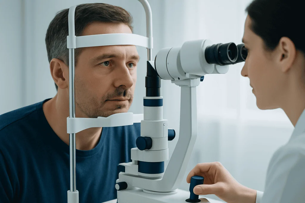
A person with hemianopia has reduced or absent vision in half of their visual field. In homonymous hemianopia, the missing half is the same side in both eyes: for example, the right side of vision is missing in both eyes, or the left side is missing in both eyes. This differs from simply losing vision in one eye.
The visual loss may feel like objects, people or parts of words suddenly disappear on one side. Someone may bump into doorframes, miss food on one side of a plate, overlook approaching pedestrians or lose their place while reading. The brain can sometimes make the missing area less noticeable, so people may not immediately recognize the extent of the problem.
Symptoms can vary according to the location and size of the affected visual pathway. Some people have a complete half-field loss, while others have a partial deficit, such as loss in one upper or lower quadrant. A formal visual-field test is needed to define the pattern accurately.
Causes, candidacy and assessment

Hemianopia most often develops after a stroke affecting the brain’s visual pathways. Other possible causes include traumatic brain injury, brain tumors, bleeding in or around the brain, inflammation, infection, seizures, migraine-related neurological symptoms and certain degenerative conditions. The onset, associated symptoms and medical history help clinicians determine the likely cause.
People who may benefit from hemianopia rehabilitation include those with a confirmed visual-field defect that affects mobility, reading, work, self-care or confidence outdoors. Rehabilitation can be appropriate whether the loss is recent or longstanding, although the plan differs depending on whether the underlying condition is stable and whether there are other difficulties, such as weakness, poor balance, language problems or reduced attention.
Assessment commonly includes a detailed eye examination, visual acuity testing and computerized perimetry, also called visual-field testing. Brain imaging such as CT or MRI may be needed, particularly when symptoms are new or the cause is uncertain. When stroke is suspected or confirmed, coordinated stroke care and prevention planning are important parts of the overall approach.
- Neuro-ophthalmology evaluation helps distinguish brain-related field loss from eye disease.
- Occupational therapy assesses the effect on everyday tasks and home safety.
- Physical therapy may support balance, walking and community mobility when needed.
- Speech and cognitive rehabilitation may be included if brain injury has affected communication, memory or attention.
Treatment plan: step by step
There is no standard surgical procedure for hemianopia itself. Instead, the treatment plan is usually built in stages. First, clinicians address the underlying cause. For example, treatment after stroke focuses on acute stroke care when appropriate, control of vascular risk factors and neurological rehabilitation. If imaging identifies a tumor, bleeding or another structural problem, the relevant specialist discusses treatment options based on that diagnosis.
Second, the visual field loss is measured and its functional effects are reviewed. This allows the team to set realistic goals, such as safer walking in busy areas, improved reading speed, greater awareness of the affected side or a supported return to work. A specialist may also assess related issues including double vision, eye movement problems, visual neglect and reduced visual attention.
Third, rehabilitation begins. Compensatory scanning training teaches purposeful, repeated eye and head movements toward the side with reduced vision. During mobility practice, a person may learn to pause, scan systematically and check the affected side before crossing roads, turning or moving through crowded environments. Reading training can include using line guides, anchors at the start or end of lines, and structured scanning patterns.
Some people may be considered for prism glasses. These lenses do not restore the missing visual field, but they can shift selected images from the blind side into the seeing side for some individuals. They require careful fitting, training and follow-up because they are not suitable for everyone. Structured neurological rehabilitation can integrate visual, physical and occupational therapy goals.
Benefits, limitations and possible risks
The potential benefit of hemianopia rehabilitation is better functional vision rather than a guaranteed return of the missing field. People may become more efficient at scanning, notice hazards earlier, read more comfortably and feel more confident managing familiar activities. Progress often depends on regular practice, attention, fatigue levels and the presence of other neurological symptoms.
Visual restoration therapies are sometimes offered as computer-based training programs intended to stimulate areas near the border of vision loss. Research into these approaches continues, but results are variable and they should not be presented as a proven way to restore vision for all patients. A qualified clinician can help interpret claims about a program and decide whether it is reasonable within an individualized rehabilitation plan.
Risks are usually related to safety rather than to invasive treatment. A person may remain vulnerable to falls, collisions, missed obstacles or road hazards if they overestimate their visual awareness. Prism lenses can initially cause visual confusion, distortion, dizziness or difficulty judging distances, which is why professional fitting and training are important. Rehabilitation should be adapted if it causes excessive fatigue, headaches or worsening symptoms.
Recovery timeline and what to expect
The recovery timeline depends strongly on the cause. When hemianopia follows a stroke or traumatic brain injury, spontaneous improvement is most likely in the early weeks and months, as swelling settles and the brain recovers from the initial injury. However, improvement can continue more gradually over time, particularly in the ability to compensate for the deficit through learned scanning and adaptive techniques.
Follow-up visual-field testing helps document changes and guide rehabilitation. Even if the field defect remains stable, a person’s practical function can improve significantly with targeted therapy, environmental changes and experience. Useful adaptations may include keeping walkways clear, improving lighting, placing frequently used items consistently and asking family members to approach from the seeing side when appropriate.
Recovery may also involve emotional adjustment. Frustration, anxiety about going outdoors or concern about employment are understandable responses to a sudden change in vision. Discussing these concerns with the care team can help identify practical support, counseling or rehabilitation resources. For international patients, Acibadem International’s multidisciplinary specialists and JCI-accredited hospitals can assess hemianopia and coordinate neurological and visual rehabilitation when indicated.
Do people recover from hemianopia?
Some people recover part of their visual field, especially when hemianopia is caused by a recent stroke, brain injury or a temporary condition that can be treated. The degree of recovery is unpredictable and depends on the cause, the location and extent of injury, overall health and associated neurological problems.
Not everyone regains the missing visual field. Even in that situation, many people improve their safety and independence through scanning practice, occupational therapy, mobility training and carefully selected visual aids. A specialist can explain whether changes on visual-field testing suggest recovery and which rehabilitation goals are most realistic.
New visual-field loss should never be assumed to be permanent or harmless. Prompt assessment is important because early treatment of the cause may affect both recovery and future health.
Can you drive if you have homonymous hemianopia?
Driving with homonymous hemianopia may be restricted or not permitted, depending on the country or region, the extent of field loss and the results of formal vision testing. A person should stop driving after new visual-field loss and should not resume until a qualified eye specialist or treating clinician has assessed them and the relevant licensing authority’s requirements have been met.
Some jurisdictions allow limited exceptions after specialized assessment, while others do not permit driving with a homonymous visual-field defect. Passing a standard eyesight chart test alone does not establish driving fitness, because safe driving also requires sufficient peripheral vision, visual attention, reaction time and awareness of hazards.
Clinicians may recommend mobility rehabilitation and alternative transport planning while fitness to drive is being clarified. This approach protects the individual, passengers and other road users without assuming that every person’s legal situation is the same.
What is the prognosis for hemianopia?
The prognosis for hemianopia is variable. It is generally linked to the cause and whether the underlying brain condition is stable, treatable or progressing. A visual-field defect caused by a completed stroke may become stable, while hemianopia caused by an untreated mass, recurrent bleeding or an inflammatory disorder may change as the condition is treated or evolves.
From a functional perspective, prognosis is often better than the visual-field test alone may suggest. With rehabilitation, many people learn reliable ways to find objects, navigate their surroundings, read and participate in meaningful daily activities. Regular reassessment is useful when symptoms change, when returning to complex tasks or when new neurological concerns arise.
Long-term care may include management of vascular risk factors, follow-up with neurology and eye specialists, and review of rehabilitation goals. When hemianopia follows stroke, healthy lifestyle measures and prescribed preventive treatment can reduce the risk of further vascular events.
When to seek medical care
Sudden loss of vision on one side, especially with facial drooping, arm weakness, numbness, speech difficulty, confusion, severe imbalance or a sudden severe headache, requires emergency medical attention. These symptoms can indicate stroke or another urgent neurological problem. Emergency services should be contacted rather than waiting to see whether symptoms improve.
A prompt medical appointment is also needed for new or worsening peripheral vision loss, repeated collisions with objects, unexplained reading difficulty, new double vision or headaches accompanied by visual changes. Even gradual symptoms deserve assessment because they can have eye, brain or systemic causes.
People with established hemianopia should seek review if their vision changes, falls increase, prism glasses are uncomfortable, new seizures occur or daily tasks become harder. Ongoing communication with the care team helps ensure that rehabilitation and safety recommendations remain appropriate.
Frequently asked questions
Is hemianopia the same as blindness in one eye?
No. Hemianopia describes loss of part of the visual field, often the same half of vision in both eyes. Blindness in one eye affects vision from one eye, whereas homonymous hemianopia usually results from a problem in the brain’s visual pathways.
Can glasses correct hemianopia?
Ordinary glasses do not restore a missing visual field caused by brain injury. In selected cases, prism lenses may help move some visual information from the affected side into the seeing side, but they require professional fitting and training.
How is hemianopia diagnosed?
Diagnosis includes an eye examination and formal visual-field testing, which maps areas of reduced or absent vision. Brain imaging and neurological assessment may be needed to identify the cause, particularly when symptoms are sudden or unexplained.
How long should visual scanning therapy continue?
The duration varies according to the person’s goals, symptoms and progress. A rehabilitation professional may provide a structured course of therapy and home practice, then review performance and adjust the plan over time.
Can hemianopia get worse?
A stable field defect after a completed stroke may remain unchanged, but new or worsening symptoms need urgent assessment. Progression can occur if the underlying cause is active, such as recurrent stroke, bleeding, inflammation or a growing lesion.
What should family members do to help someone with hemianopia?
Family members can support independence by keeping pathways clear, avoiding sudden approaches from the affected side and encouraging use of agreed scanning techniques. It is also helpful to respect the person’s pace and involve rehabilitation professionals when planning home or community safety changes.
References
- American Academy of Ophthalmology
- American Stroke Association
- National Eye Institute
- National Institute of Neurological Disorders and Stroke
- Royal College of Ophthalmologists
This article is for general information only and is not a substitute for professional medical advice. Please consult a qualified doctor about your individual situation.
Explore treatments in Turkey — costs, top hospitals & a free quote
JCI-accredited · board-certified surgeons · reply within 24h
Add us as a Preferred Source to see more of our trusted health content across Google Search, AI Overviews and Discover.