Hepatobiliary Tract Anatomy: An Evidence-Based Guide for Patients
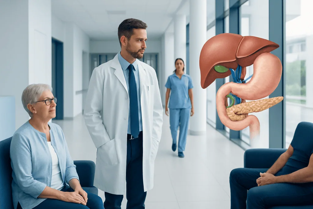
The liver makes bile continuously, while the gallbladder stores and concentrates it between meals. Bile travels through small channels in the liver into larger bile ducts and then enters the first part of the small intestine.
Key Takeaways
- The liver makes bile continuously, while the gallbladder stores and concentrates it between meals.
- Bile travels through small channels in the liver into larger bile ducts and then enters the first part of the small intestine.
- The pancreas shares an outlet area with the bile duct in many people, so conditions in one system can affect the other.
- Gallstones, inflammation, narrowing and tumors can interfere with bile flow and may cause pain, jaundice or digestive symptoms.
- Imaging tests such as ultrasound, MRCP, CT and ERCP help clinicians assess hepatobiliary anatomy and identify blockages.
- Persistent abdominal pain, yellowing of the skin or eyes, fever, or very dark urine should be assessed promptly by a healthcare professional.
The hepatobiliary tract is the system of organs and channels that produces, stores and transports bile, a fluid that supports fat digestion and helps remove certain waste products. It includes the liver, gallbladder, bile ducts and their close connection to the pancreas and small intestine.
Overview: What Is the Hepatobiliary Tract?
Hepatobiliary tract anatomy refers to the liver, gallbladder and bile ducts, along with the pathways that carry bile into the small intestine. Bile is a yellow-green digestive fluid made by the liver. It helps the body digest and absorb dietary fats and fat-soluble vitamins, and it carries substances such as bilirubin and cholesterol out of the body.
The word hepato relates to the liver, and biliary relates to bile and the structures that transport it. Although the pancreas is not technically part of the biliary tract, it is closely connected to this system because the main pancreatic duct and common bile duct usually join or open near the same area of the duodenum, the first section of the small intestine.
This anatomy is important because even a small blockage in a bile duct can affect digestion and liver function. Understanding the normal route of bile can make it easier to understand common conditions, tests and treatments involving the gallbladder, liver, pancreas and upper digestive tract.
The Main Organs and Structures
The liver is the largest internal organ and sits mainly in the upper right side of the abdomen, beneath the rib cage. It has many essential functions, including processing nutrients, making proteins, storing energy, breaking down certain substances and producing bile. The liver is supplied with oxygen-rich blood through the hepatic artery and nutrient-rich blood from the digestive organs through the portal vein.
The gallbladder is a small, pear-shaped sac located beneath the liver. It is connected to the bile ducts by the cystic duct. Between meals, the gallbladder stores bile made by the liver and concentrates it by removing some water. When a person eats, especially a meal containing fat, the gallbladder normally contracts and releases bile into the intestine.
The bile ducts are a branching network of tubes. Tiny ducts within the liver collect bile from liver cells. These join to form the right and left hepatic ducts, which drain the right and left sides of the liver. Outside the liver, these ducts form the common hepatic duct. The cystic duct from the gallbladder joins it to create the common bile duct.
The common bile duct travels downward toward the duodenum. Near its lower end, it commonly meets the pancreatic duct at a small opening called the ampulla of Vater. A circular muscle, the sphincter of Oddi, helps regulate the release of bile and pancreatic juices into the intestine.
How Bile Flows Through the System
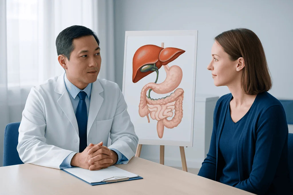
Bile is produced continuously by specialized liver cells called hepatocytes. From there, it enters microscopic channels known as bile canaliculi, then progressively larger ducts within the liver. This organized branching pattern is sometimes compared with a tree: many small branches collect into a few larger pathways.
When a person is not eating, much of the bile flowing down the common hepatic duct is directed through the cystic duct into the gallbladder. There, it is stored until food reaches the small intestine. Hormonal and nerve signals, particularly the hormone cholecystokinin, prompt the gallbladder to contract after a meal.
During digestion, bile moves from the gallbladder through the cystic duct, into the common bile duct and into the duodenum. Bile salts help break large fat droplets into smaller droplets, making them easier for digestive enzymes to process. Most bile salts are later reabsorbed farther along in the small intestine and returned to the liver for reuse; this is called enterohepatic circulation.
Bile flow can be influenced by normal anatomy, prior surgery, inflammation or a blockage. A blockage may occur within the gallbladder, cystic duct, common bile duct or the area where the duct enters the intestine. The location of a problem often helps clinicians determine which symptoms and tests are most relevant.
Why the Pancreas and Intestine Matter
The lower common bile duct lies close to the head of the pancreas. In many people, the bile duct and pancreatic duct join before entering the duodenum, although the precise arrangement varies naturally between individuals. This shared region explains why a stone in the bile duct can sometimes affect the pancreas as well as bile flow.
The duodenum receives bile, pancreatic enzymes and partially digested food from the stomach. These fluids work together: bile supports fat digestion, while pancreatic enzymes help digest fats, proteins and carbohydrates. The intestinal lining then absorbs nutrients into the bloodstream or lymphatic system.
Anatomical variation is common and does not necessarily cause health problems. For example, the cystic duct may join the common hepatic duct at different angles or positions. Some people also have accessory bile ducts. These variations are particularly important for surgeons and radiologists to recognize before or during procedures involving the gallbladder or bile ducts.
Because these organs function so closely together, doctors may assess the liver, gallbladder, bile ducts and pancreas when investigating upper abdominal symptoms. This broader approach can help distinguish a biliary concern from stomach, intestinal or pancreatic conditions.
Common Problems That Affect Hepatobiliary Anatomy
Gallstones are hardened deposits that can form in the gallbladder. Many people with gallstones have no symptoms. However, a stone can block the cystic duct and cause gallbladder inflammation, or move into the common bile duct and interfere with bile drainage. Symptoms may include pain in the upper right or upper middle abdomen, nausea or vomiting, often after meals.
Inflammation of the gallbladder is called cholecystitis. Inflammation or infection of the bile ducts is called cholangitis and requires urgent medical evaluation. Other causes of impaired bile flow include scarring or narrowing of a duct, known as a stricture; inflammation related to pancreatic disease; and, less commonly, growths affecting the bile ducts, gallbladder, liver or pancreas.
When bile cannot drain normally, bilirubin may build up in the blood. This can lead to jaundice, which causes yellowing of the skin and the whites of the eyes. Some people may also notice dark urine, pale or clay-colored stools, itching, fatigue or poor appetite. These signs do not identify a specific cause on their own, but they warrant medical assessment.
Liver diseases may also affect bile production or flow. For example, inflammation, scarring or certain inherited conditions can involve the smaller bile ducts inside the liver. A clinician considers symptoms, blood test results, imaging findings and medical history together to identify the underlying issue.
How Doctors Assess the Hepatobiliary System
A medical assessment usually begins with a discussion of symptoms, personal and family history, medicines and prior abdominal procedures. A clinician may gently examine the abdomen for tenderness, fullness or signs of discomfort. Blood tests can measure liver enzymes, bilirubin, markers of inflammation and pancreatic enzymes when appropriate.
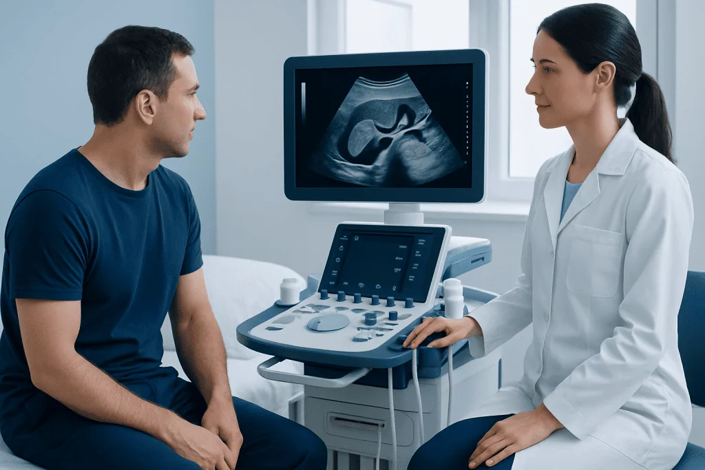
Ultrasound is often the first imaging test for suspected gallstones or gallbladder inflammation because it can show stones, gallbladder wall changes and widening of bile ducts. CT scans may provide additional detail about the abdomen. Magnetic resonance cholangiopancreatography, usually called MRCP, is a specialized MRI technique that creates detailed images of the bile and pancreatic ducts without placing an instrument into them.
Endoscopic ultrasound may help assess small stones, duct changes or nearby structures. Endoscopic retrograde cholangiopancreatography, or ERCP, combines endoscopy and X-ray imaging. It is commonly used when a doctor expects to treat a problem, such as removing a bile duct stone, placing a stent or widening a narrowed area, rather than solely for diagnosis.
Test selection depends on the person’s symptoms and likely cause. It is helpful for patients to tell their care team about pregnancy, allergies, kidney disease, implanted medical devices, blood-thinning medicines and previous reactions to contrast agents or sedation.
Supporting Hepatobiliary Health and Knowing When to Seek Care
Not every hepatobiliary condition can be prevented, and gallstones may develop even in people with healthy habits. Still, regular meals, a balanced eating pattern, physical activity and gradual weight management can support overall digestive and liver health. Rapid weight loss and repeated cycles of weight loss and regain may increase gallstone risk for some people, so individualized guidance from a clinician or dietitian can be useful.
People should avoid starting supplements or herbal products specifically for “liver cleansing” without medical advice. Some products can interact with medicines or cause liver injury. Limiting alcohol where appropriate, avoiding smoking and taking medicines only as directed can also support liver health. Vaccination and infection-prevention advice should be discussed with a healthcare professional based on individual risks.
When to seek medical care: A person should arrange a medical review for recurring upper abdominal pain, nausea after fatty meals, unexplained itching, persistent changes in stool or urine color, or yellowing of the skin or eyes. Urgent assessment is important for severe or worsening abdominal pain, fever or chills with abdominal pain, jaundice, repeated vomiting, confusion, fainting or an inability to keep fluids down.
Care is tailored to the diagnosis and may involve gastroenterologists, hepatologists, radiologists, surgeons and other specialists. Acibadem International’s multidisciplinary specialists and JCI-accredited hospitals diagnose and treat hepatobiliary conditions for international patients, with care plans based on individual clinical findings.
Frequently asked questions
What organs are included in the hepatobiliary tract?
The hepatobiliary tract includes the liver, gallbladder and bile ducts. It also has a close functional relationship with the pancreas and duodenum, where bile enters the digestive tract. Together, these structures make, store, transport and release bile.
Is the pancreas part of the hepatobiliary system?
The pancreas is generally considered a separate organ, but it is closely linked to the hepatobiliary system. Its main duct usually joins or opens near the common bile duct in the duodenum. This shared anatomy is why some bile duct problems can also affect the pancreas.
What is the difference between the common hepatic duct and common bile duct?
The common hepatic duct is formed when the right and left hepatic ducts join after leaving the liver. When the cystic duct from the gallbladder joins the common hepatic duct, the resulting channel is called the common bile duct. The common bile duct carries bile toward the small intestine.
Can a person digest food without a gallbladder?
Yes. The liver continues to make bile after gallbladder removal, and bile can flow directly into the small intestine. Some people notice temporary changes in bowel habits or difficulty tolerating very fatty meals after surgery, but many adapt well over time.
What symptoms can suggest a bile duct blockage?
Possible symptoms include abdominal pain, jaundice, dark urine, pale stools, itching, nausea and fever. These symptoms can have several causes, so they should not be used to self-diagnose a blockage. Prompt medical assessment is especially important if fever, chills, jaundice or severe pain occurs.
Which test shows the bile ducts most clearly?
MRCP is a noninvasive MRI-based test that can provide detailed pictures of the bile and pancreatic ducts. Ultrasound is often used first for gallbladder concerns, while ERCP may be chosen when treatment of a suspected duct problem is needed. The best test depends on the clinical situation.
References
- National Institute of Diabetes and Digestive and Kidney Diseases
- National Health Service
- Merck Manual Consumer Version
- American College of Gastroenterology
- RadiologyInfo.org
This article is for general information only and is not a substitute for professional medical advice. Please consult a qualified doctor about your individual situation.
Persistent digestive symptoms? Get evaluated in Turkey
JCI-accredited · board-certified surgeons · reply within 24h
Add us as a Preferred Source to see more of our trusted health content across Google Search, AI Overviews and Discover.
Check your numbers in seconds
BMI, calories, due date, blood pressure and 30+ more clinical calculators — free, instant, doctor-reviewed ranges.